J of Evolution of Med and Dent Sci/ eISSN- 2278-4802, pISSN- 2278-4748/ Vol. 4/ Issue 12/Feb 09, 2015 Page 1986
RELATIONSHIP BETWEEN TYPE 2 DIABETES AND PULMONARY
FUNCTIONS IN OBESE WOMEN
Veena C. N1, B. C. Vastrad2, Nandan T. M3HOW TO CITE THIS ARTICLE:
Veena C. N, B. C. Vastrad, Nandan T. M. Relationship between Type 2 Diabetes and Pulmonary Functions in Obese Women. Journal of Evolution of Medical and Dental Sciences 2015; Vol. 4, Issue 12, February 09;
Page: 1986-1990, DOI: 10.14260/jemds/2015/286
ABSTRACT: BACKGROUND: Type 2 diabetes mellitus (Type 2 DM) is a major metabolic disorder commonly associated with obesity. Obesity and Type 2 DM are closely associated with a state of low-grade inflammation which causes altered pulmonary function. There is faster decline in pulmonary function in obese Type 2 diabetic patients. OBJECTIVES: To study the correlation between glucose levels on pulmonary function tests in obese diabetic women. MATERIALS AND METHODS: 30 obese diabetic women aged between 30 & 65 yrs with duration of illness between 2 &10 yrs were selected for the study. Pulmonary function tests were performed using Medspiror. RESULTS AND CONCLUSION: Significant negative correlations were observed between the FVC, PEFR, mean FEF 25-75% and the raised blood glucose levels and that between the mean FEF 25-25-75% and the duration of Type 2 diabetes. This suggests that Type 2 diabetes is a risk factor for impairment of respiratory function in obese women and hence the metabolic pathways related to hyperglycemia make lungs to be potential targets.
KEYWORDS: Type 2 Diabetes, Pulmonary functions, Obese women.
INTRODUCTION: Diabetes mellitus comprises a group of metabolic disorder that share the phenotype of hyperglycemia. The metabolic dysregulation associated with it causes secondary pathophysiologic changes in multiple organ systems that impose a tremendous burden on an individual with diabetes and on the health care system.1
Type 2 diabetes mellitus (Type 2 DM) is a major metabolic disorder also strongly associated with obesity. It appears to result from a collection of multiple genetic factors including polymorphisms, each contributing their own predisposing risks that are further modified by environmental factors. Obesity, particularly visceral, is very common in Type 2 DM. The relationship between them is of such interdependence that the term diabesity has been coined. Adipocytes secrete a large number of biological products that modulate insulin secretion, insulin action and body weight, which may contribute to insulin resistance. Insulin resistance impairs glucose utilization by insulin sensitive tissues and increases hepatic glucose output; both of which contribute to hyperglycemia.2
Further, obesity and insulin resistance, the cardinal features of metabolic syndrome are closely associated with a state of low-grade inflammation.3 In adipose tissues, chronic over nutrition
leads to macrophage infiltration resulting in local inflammation that aggravates insulin resistance. Moreover, systemic inflammation is also thought to play a role in the association between reduced pulmonary function and cardiovascular mortality.4 Thus, obesity and diabetes associated with
J of Evolution of Med and Dent Sci/ eISSN- 2278-4802, pISSN- 2278-4748/ Vol. 4/ Issue 12/Feb 09, 2015 Page 1987 function, there are only a few studies designed to investigate whether poor glycemic control in obese Type 2 diabetic women is an independent determinant of reduced pulmonary function. Thus the present study was undertaken to study the correlation between glucose levels on respiratory function in obese diabetic women using the most important variables of pulmonary function tests.
MATERIALS AND METHODS: Thirty obese diabetic women aged between 30 & 65 yrs attending outpatient department in a tertiary care medical centre in Kuppam, Southern Andhra Pradesh, were selected for the study. The criteria for inclusion of the subjects were the following: obese diabetic individuals with duration of illness between 2 &10 yrs (BMI >25),5 non-smokers, subjects with no
past history of chronic respiratory illness, subjects with no symptoms of respiratory illness at the time of examination, and subjects with no chronic cardiovascular disease.
A detailed health status assessment was done for all the subjects included in the study through history taking. Body mass index (BMI) was calculated after measuring height and weight. Clinical examination was done to rule out other systemic abnormalities. The ethical clearance was obtained. The subjects were briefed about the procedure and a written consent was taken. Then, every individual was subjected to pulmonary function tests.
Pulmonary function tests were performed using Medspiror, which is a PC based spirometer with a flow transducer. Tests were performed on all the subjects in sitting position. Reference values for spirometry were based on age, sex and height provided in the software.
The whole procedure was explained and demonstrated to the subjects before testing. Later the subjects were asked to perform the forced vital capacity manoeuvre. FVC was recorded after a maximal inspiration when the subject expired forcefully with maximum expiration in to the mouth piece. A minimum of 3 acceptable FVC manoeuvres were performed and the best manoeuvres were selected and accepted. Acceptability criteria were: full inhalation before the start of test, satisfactory start of exhalation (Maximal effort exerted with no hesitation), no cough during the 1st second of manoeuvre, no early termination of exhalation (A maximum exhalation time of 6 seconds was followed).
For FVC and FEV1 manoeuvres, only if the difference between the two largest values were not
less than 200 ml and for PEFR, only if the difference between the two largest values were not less than 10%, the testing was continued for 8 trials and the values recorded. Calibration was done from time to time for accuracy. After scrutinizing the flow volume curve and the time volume curve, the parameters derived were FVC, FEV1, FEV1/FVC, PEFR and FEF-25-75%. These criteria are based on American thoracic society (ATS) and European thoracic society standards.
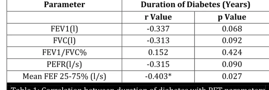
J of Evolution of Med and Dent Sci/ eISSN- 2278-4802, pISSN- 2278-4748/ Vol. 4/ Issue 12/Feb 09, 2015 Page 1988 Parameter Duration of Diabetes (Years)
r Value p Value
FEV1(l) -0.337 0.068
FVC(l) -0.313 0.092
FEV1/FVC% 0.152 0.424
PEFR(l/s) -0.315 0.090
Mean FEF 25-75% (l/s) -0.403* 0.027
Table 1: Correlation between duration of diabetes with PFT parameters
* Correlation significant at 0.05 level (2 tailed)
Parameter FBS (mg/dl) r Value p Value
FEV1(l) -0.146 0.441
FVC(l) -0.375* 0.041
FEV1/FVC% -0.269 0.151
PEFR(l/s) -0.534** 0.002
MEAN FEF 25-75 % (l/s) -0.499** 0.005
Table 2: Correlation between FBS with PFT parameters
* Correlation significant at 0.05 level (2 tailed) ** Correlation significant at 0.01 level (2 tailed)
Parameters PPBS (mg/dl)
r Value p Value
FEV1(l) -0.184 0.330
FVC(l) -0.362* 0.049
FEV1/FVC % -0.120 0.528
PEFR(l/s) -0.372* 0.043
MEAN FEF 25-75 % (l/s) -0.406* 0.026
Table 3: Correlation between PPBS with PFT parameters
* Correlation significant at 0.05 level (2 tailed) ** Correlation significant at 0.01 level (2 tailed)
DISCUSSION: Obesity and Type 2 DM are two major features of metabolic syndrome, the prevalence of which has reached epidemic proportions. Obesity mechanically restricts lung volume in addition to causing widespread lipid deposition in non-adipose tissues including the lungs, which increases pro-oxidant and pro-inflammatory cellular stress as well as alterations in lung structure.6
J of Evolution of Med and Dent Sci/ eISSN- 2278-4802, pISSN- 2278-4748/ Vol. 4/ Issue 12/Feb 09, 2015 Page 1989 respiratory function in obese women, notably, a negative correlation is obtained between blood glucose levels and the parameters of pulmonary function testing viz., FVC, FEF 25-75 and PEFR. This finding strongly suggests that the metabolic pathways related to hyperglycemia are the main factors accounting for impaired pulmonary function. The findings of the present study are in agreement with Walter. E, Robert et. al., who showed a decrease in FVC in patients with diabetes mellitus,8 Davis. M. E,
Timothy, who studied pulmonary function and its association with Type 2 DM found an average decrease of 9.5% in FVC9 and Sreeja et.al., showed a reduction in PEF and mean FEF 25-75% in the
case group.10
The present study is also in agreement with findings of Tricia M. McKeever et.al., whose findings showed that post prandial glucose levels have an inverse association with pulmonary function suggesting that impaired glucose auto regulation is associated with impaired lung function.6
On assessing the relationship between duration of diabetes with lung function, a significant negative correlation was found between the duration of diabetes and mean FEF 25-75% suggesting impairment of lung function with prolonged illness. The results of the present study is in agreement with Sultan A. Meo et.al., who studied the effect of duration of diabetes on ventilator function in ethnic Saudi group whose findings showed decreased FEF25-75% with the increased duration of disease.11
The present study is also in agreement with the studies of Shravya K.G et.al.,12 and
Kanyakumari et.al.,13 whose results showed a significant negative correlation between duration of
diabetes and lung function.
The findings of the present study is also supported by Ramirez LC whose results showed that long-term near-normoglycemia may be beneficial in preventing the deterioration of pulmonary function associated with diabetes mellitus.14
CONCLUSION: The results of the present study showed a decline in pulmonary function in obese Type 2 diabetic women with poor glycemic control. As measures of airflow limitation predict all-cause mortality in Type 2 diabetes, intensive glycemic management may reduce the risk of death through improved ventilatory function independent of other beneficial effects. Therefore, the impact of Type 2 diabetes on pulmonary function should be considered by those providing care for obese women.
REFERENCES:
1. Powers AC. Chapter 323, Diabetes mellitus. In: Klasper DL, Braunwald E et al., editors. Harrison s principles of internal medicine. 6th edition. New York: The McGraw-Hill Companies;
2005; p. 2152-2158.
2. Davis, W.A., Knuiman, M., Kendall, P., Grange, V., Davis, T.M., and Fremantle Diabetes Study. Glycemic exposure is associated with reduced pulmonary function in type 2 diabetes: the Fremantle Diabetes Study. Diabetes Care. 2004; 27: 752–757.
J of Evolution of Med and Dent Sci/ eISSN- 2278-4802, pISSN- 2278-4748/ Vol. 4/ Issue 12/Feb 09, 2015 Page 1990 4. Ochs-Balcom HM, Grant BJB, Muti P, Sempos CT, Freudenheim JL, et. al. Pulmonary Function and
Abdominal Adiposity in the General Population. CHEST 2006; 129:853–862.
5. Chiu M, Austin PC, Manuel DG, Shah BR, Tu JV. Deriving ethnic-specific BMI cutoff points for assessing diabetes risk. Diabetes Care 2011; 34: 1741–1748.
6. Yilmaz C. Ravikumar P, Bellotto DJ, Unger RH, Hsia CC. Fatty diabetic lung: functional impairment in a model of metabolic syndrome. J Appl Physiol 109: 1913-1919, 2010.
7. Phillips LK, Prins JB. The link between abdominal obesity and the metabolic syndrome; Curr Hypertens Rep. 2008 Apr; 10 (2): 156-64.
8. Walter RE, Beiser A, Givelber RJ, O'Connor GT, Gottlieb DJ; Association between glycemic state and lung function: the Framingham Heart Study; Am J Respir Crit Care Med. 2003 Mar 15; 167 (6): 911-6.
9. Davis TM, Knuiman M, Kendall P, Vu H, Davis WA; Reduced pulmonary function and its associations in Type 2 diabetes: the Fremantle Diabetes Study. Diabetes Res Clin Pract. 2000 Oct; 50 (2):153-9.
10.Sreeja CK, Samuel E, Kesavachandran C, Shashidhar S; Pulmonary function in patients with diabetes mellitus. Indian Journal of Physiology and Pharmacology 2003, 47(1):87-93.
11. Meo SA, Al DreesAM, Ahmed J, Shah SFA, Al-Regaiey K, Husain A; Effect of Duration of Disease on Ventilatory Function in an Ethnic Saudi Group of Diabetic Patients. J Diabetes Sci Technol. Sep 2007; 1 (5): 711–717.
12.Shravya KG, Bandi HK, Suresh M, Preetham JK, Reddy MN, Singh SBM. Role of Duration of Diabetes on Ventilatory Capacities and Expiratory Flow Rates in Type 2 Diabetes Mellitus..
Journal of Biology, Agriculture and Healthcare. 2012; 2 (6): 77-82.
13.Kanyakumari DH, Nataraj SM, Devaraj HS. Correlation of duration of diabetes and pulmonary function tests in Type 2 diabetes mellitus patients. Int J Biol Med Res. 2011; 2 (4): 1168 -1170. 14.Ramirez LC, Dal Nogare A, Hsia C, Arauz C, Butt I; Relationship Between Diabetes Control and
Pulmonary Function In Insulin Dependent Diabetes Mellitus; Am J Med. Oct 1991; 91 (4): 371-376.
AUTHORS:
1. Veena C. N. 2. B. C. Vastrad 3. Nandan T. M.
PARTICULARS OF CONTRIBUTORS:
1. Assistant Professor, Department of Physiology, PES Institute of Medical Sciences and Research, Kuppam. 2. Professor and HOD, Department of
Physiology, PES Institute of Medical Sciences and Research, Kuppam. 3. Assistant Professor, Department of
Microbiology, PES Institute of Medical Sciences and Research, Kuppam.
NAME ADDRESS EMAIL ID OF THE CORRESPONDING AUTHOR:
Dr. Veena C. N, Assistant Professor, Department of Physiology, PESIMSR, Kuppam,
517425.
E-mail: chinni.iyer@yahoo.co.in