Chicken Necrotic Enteritis Isolates and Comparison with
C. perfringens
Conjugative Plasmids
Valeria R. Parreira1, Marcio Costa1, Felix Eikmeyer2, Jochen Blom3, John F. Prescott1*
1Department of Pathobiology, University of Guelph, Guelph, Ontario, Canada,2Institute for Genome Research and Systems Biology, Center for Biotechnology, Bielefeld University, Bielefeld, Germany,3Bioinformatics Resource Facility, Center for Biotechnology, Bielefeld University, D-33594 Bielefeld, Germany
Abstract
Twenty-six isolates ofClostridium perfringensof different MLST types from chickens with necrotic enteritis (NE) (15netB -positive) or from healthy chickens (6netB-positive, 5netB-negative) were found to contain 1–4 large plasmids, with most netB-positive isolates containing 3 large and variably sized plasmids which were more numerous and larger than plasmids in netB-negative isolates.NetBandcpb2were found on different plasmids consistent with previous studies. The pathogenicity locus NELoc1, which includesnetB, was largely conserved in these plasmids whereas NeLoc3, present in thecpb2containing plasmids, was less well conserved. AnetB-positive and acpb2-positive plasmid were likely to be conjugative, and the plasmids were completely sequenced. Both plasmids possessed the intact tcp conjugative region characteristic of C. perfringensconjugative plasmids. Comparative genomic analysis of nineCpCPs, including the two plasmids described here, showed extensive gene rearrangements including pathogenicity locus and accessory gene insertions around rather than within the backbone region. The pattern that emerges from this analysis is that the major toxin-containing regions of the variety of virulence-associatedCpCPs are organized as complex pathogenicity loci. How these different but relatedCpCPs can co-exist in the same host has been an unanswered question. Analysis of the replication-partition region of these plasmids suggests that this region controls plasmid incompatibility, and that CpCPs can be grouped into at least four incompatibility groups.
Citation:Parreira VR, Costa M, Eikmeyer F, Blom J, Prescott JF (2012) Sequence of Two Plasmids fromClostridium perfringensChicken Necrotic Enteritis Isolates and Comparison withC. perfringensConjugative Plasmids. PLoS ONE 7(11): e49753. doi:10.1371/journal.pone.0049753
Editor:Eric A. Johnson, University of Wisconsin, Food Research Institute, United States of America
ReceivedMay 15, 2012;AcceptedOctober 15, 2012;PublishedNovember 26, 2012
Copyright:ß2012 Parreira et al. This is an open-access article distributed under the terms of the Creative Commons Attribution License, which permits unrestricted use, distribution, and reproduction in any medium, provided the original author and source are credited.
Funding:Funding provided by Canadian Poultry Research Council and the Agriculture and Agri-Food Canada Poultry Research Cluster program, by the Ontario Ministry of Agriculture and Food, and by the Natural Sciences and Engineering Research Council. FE acknowledges scholarship from the CLIB-Graduate Cluster Industrial Biotechnology co-financed by the Ministry of Innovation of North Rhine-Westphalia. JB acknowledges funding by the German Federal Ministry of Education and Research (grants 0315599A & 0315599B ‘‘GenoMik-Transfer’’). The funders had no role in study design, data collection and analysis, decision to publish, or preparation of the manuscript.
Competing Interests:The authors have declared that no competing interests exist.
* E-mail: [email protected].
Introduction
Clostridium perfringensis an important pathogen of humans and animals, and certain strains of type A isolates cause necrotic enteritis (NE) in broiler chickens. NE is a common bacterial infection of chickens that is traditionally controlled by use of antibiotics. However the removal of growth-promoting antibiotics in broiler chickens in Europe and increasing demands elsewhere for ‘‘antibiotic-free’’ chicken are focusing efforts to find alternative approaches to control of this important disease [1,2]. There has been considerable effort in recent years to understand the pathogenesis of NE in chickens, including understanding the strains involved in disease.
A major breakthrough in understanding virulence in NE isolates ofC. perfringenswas the demonstration that a new toxin, NetB, was critical for development of the disease [3]. Subsequently, our group showed thatnetBformed part of a large pathogenicity locus (PAL) present on a plasmid (NELoc1) and that a second PAL, NELoc3, containing a gene namedhdhA, which encodes 7-alpha-hydroxysteroid dehydrogenase, was also characteristic of NE isolates and was present on a separate plasmid [4]. A
chromosomally encoded locus, NELoc2, was also present in all NE isolates. Pathogenicity loci are clusters of genes that harbour a group of potential virulence genes, which may contribute to the characteristic of the diversity ofC. perfringensas a pathogen [5,6].
Clostridium perfringensproduce at least 15 potent toxins that are responsible for severe diseases in humans and animals [7]. InC. perfringens, other than the mouse-lethal alpha toxin genescpaand
Recently, three plasmids from an NE isolate have been fully sequenced (pJIR3535, pJIR3844, pJIR3537); one of these containednetBand another thecpb2gene; the third was a smaller tetracycline-resistance plasmid that did not contain virulence-associated genes [10].
In the study reported here, we sequenced two different plasmids containing NELoc1 and NELoc3 and analysed the diversity of plasmid profiles of a group ofC. perfringens strains isolated from chickens. Further, comparative genomics tools were used to analyse DNA sequences. We found that both plasmids contained multiple genes which shared high similarity to well-known C. perfringensconjugative plasmids (CpCPs).
Materials and Methods
Bacterial Strains and Media
Twenty sixC. perfringensstrains belonging to different multi-locus sequence types (ST) were examined (Table 1). NE strains, field isolates from NE cases, and healthy isolates from the same farm in Ontario as the outbreak flock, were obtained from Patrick Boerlin, Department of Pathobiology, University of Guelph [16]. Each isolate was grown overnight at 37uC under anaerobic conditions (80% N2, 10% H2, 10% CO2) on TGY medium (3% Tryptic Soy
Broth (Difco Laboratories, Detroit, MI) containing 2% D-glucose (Difco ), 1% yeast extract (Difco), and 0.1% L-cysteine (Sigma-Aldrich Co., St. Louis, MO). All isolates were also cultivated in blood agar (Trypticase Soy Agar (Fisher) with 5% sheep blood) plates aerobically to confirm purity.E. colistrains were grown on Luria-Bertani agar plates (Difco) at 37uC in aerobic conditions.
Genomic and Plasmid DNA isolation
Genomic DNA was isolated from 5 ml of overnight culture in Brain Heart Infusion broth (Difco) at 37uC under anaerobic conditions [17]. After precipitation, DNA pellets were washed twice with 70% ethanol and resuspended in TE buffer (10 mM Tris-Cl, pH 7.5 1 mM EDTA). Plasmid DNA was purified using midi-Qiagen columns (Qiagen, Mississauga, Canada) following the manufacturer’s instructions.
Construction ofnetBandcpb2Mutants
The generation of C. perfringens mutants was conducted as described in Heap et al. [18]. ClosTron intron targeting and design tool (http://clostron.com) identified possible intron target sites. The insertion sites in the sense strands at positions 461/ 462 bp in thenetB open reading frame (orf) and 390/391 bp in the cpb2 orf were preferentially chosen to generate ClosTron-intron modifications, which were obtained by PCR from primers (IBS, EBS2, EBS1 and EBS universal) designed by the ClosTron website (Table S1). The 350-bp products and pMTL007 ClosTron-shuttle vector were both digested with
HindIII and BsrGI, ligated and then transformed by heat shock into E. coli DH5a. Recombinant plasmids were isolated and sequenced in order to verify sequences of the retargeted intron specific for netB and cpb2 insertions. The recombinants pMTLnetB and pMTLcpb2 containing the modified netB and
cpb2 intron were then electroporated into electrocompetent C. perfringens strain CP1. Recombinant colonies were selected and restreaked onto BHI agar containing 2.5mg/ml of erythromycin to select for bacteria in which the intron had been inserted. Insertions were confirmed by PCR using the EBS universal primer and target gene specific reverse primers. ErmRAM-F and ErmRAM-R primers were used to demonstrate the ErmRAM splicing, primers used are shown in Table S1.
Conjugation Experiments
Plasmid transfer experiments were carried out withC. perfringens
CW504 used as recipient strain. Overnight cultures ofC. perfringens
donor and recipient strains grown in TGY were mixed with a donor:recipient ratio of 2:1 and a total of 200ml of both cultures were seeded onto BHI agar without antibiotics and incubated anaerobically at 37uC overnight. Subsequently, the bacterial growth was removed and resuspended in 3 ml of BHI broth. Transconjugants were selected on BHI agar plates supplemented with rifampicin (20mg/ml), nalidixic acid (20mg/ml) and erythromycin (2.5mg/ml). Transconjugants were initially screened by PCR amplifications of specific genes (netBandcpb2) followed by Pulsed Field Gel Electrophoresis (PFGE) to analyze the presence of plasmids.
Pulsed Field Gel Electrophoresis
PFGE was performed to analyze the presence of plasmids in 26 poultryC. perfringensisolates, as described by Leppet al. [4]. Briefly, DNA plugs for PFGE were prepared from overnight cultures ofC. perfringensgrown in TGY and the bacterial pellets incorporated into a final agarose concentration of 1% in PFGE certified agarose (Bio-Rad Laboratories, Hercules, CA). Plugs were incubated overnight with gentle shaking at 37uC in lysis buffer (0.5M EDTA pH 8.0, 2.5% of 20% sarkosyl (Sigma-Aldrich), 0.25% lysozyme (Sigma-Aldrich) and subsequently incubated in 2% proteinase K (Roche Applied Science, Laval, QC) buffer for 2 days at 55uC. One-third of a plug per isolate was equilibrated in 200mL of restriction buffer at room temperature for 20 min and then digested with 10 U ofNotI (New England Biolabs, Pickering, ON) at 37uC overnight. Electrophoresis was performed in a 1% PFGE-certified gel and separated with the CHEF-III PFGE system (Bio-Rad) in 0.56 Tris-borate-EDTA buffer at 14uC at 6 V for 19 h with a ramped pulsed time of 1 to 12 s. Gels were stained in ethidium bromide and visualized by UV light. Mid-Range II PFG markers (New England Biolabs) were used as molecular DNA ladder.
Preparation of DIG Probes and PFGE Southern Blotting
DNA probes for all PFGE Southern blot steps were labelled by PCR amplification in the presence of digoxigenin-11-dUTP (DIG; Roche Applied Science) according to the manufacturer’s recom-mendation. DNA probes were amplified fromC. perfringensstrain CP1. DNA probes fornetB and hdhA genes were prepared with specific primers (Table S1). DNA from PFGE gels was transferred to nylon membranes (Roche Applied Science, Mannheim, Germany). DNA hybridizations and detection were performed by using the DIG labelling and CSPD substrate according to the manufacturer’s recommendation (Roche). For Southern blot hybridizations, nylon membranes were prehybridized for at least 2 h at 42uC in hybridization solution without labelled probe and then hybridized separately at 42uC with specific DNA probes for 16 h. The membranes were washed at 68uC under high-stringency conditions. For each different DIG labelled probe, the membrane was first stripped with 0.2 N NaOH and 0.1% sodium dodecyl sulfate, incubated with prehybridization solution, and then reprobed.
Overlapping PCR Analysis of NE Locus 1–3
each primer. A touchdown PCR program was used: 94uC for 3 min, 35 cycles of 94uC for 15 s, 65uC to 50uC for 15 s/cycle (the annealing temperature is decreased by 1uC every cycle until 50uC),
extension at 68uC for 5 min, and finally, 68uC for 10 min. For longer-range fragments the extension time was increased to 15 min. All primers used are described in Table S1. PCR product Table 1.General features of bacterial strains and plasmids.
Strains/Plasmids
Sequence
Type1 Characteristics/Clinical signs Source
E. coli
DH5a F-W80 lacZDM15D(lacZYA-argF)U169 endA1 recA1
hsdr17(rK2mK2) deoR thi-1 supE44 gyrA96 relA1
Invitrogen
C.perfringens
CW504 RifR2NalRconjugation recipient J.I.Rood, Monash University
CP1 Necrotic enteritis [34]
CP1DnetB::ErmRAM ClosTron insertion innetBgene This study
CP1Dcpb2::ErmRAM ClosTron insertion incpb2gene This study
T98 CW504 derived transconjugant RifRNalRErmR
with plasmid pCpb2 from CP1Dcpb2::ErmRAM
This study
NE01 01 Necrotic enteritis [16]
NE04 10 Necrotic enteritis [16]
NE06 02 Necrotic enteritis [16]
NE09 04 Necrotic enteritis [16]
NE10 03 Necrotic enteritis [16]
NE14 05 Necrotic enteritis [16]
NE15 06 Necrotic enteritis [16]
NE19 08 Necrotic enteritis [16]
NE20 09 Necrotic enteritis [16]
NE23 10 Necrotic enteritis [16]
NE28 13 Necrotic enteritis [16]
NE30 14 Necrotic enteritis [16]
NE32 15 Necrotic enteritis [16]
NE42 16 Necrotic enteritis [16]
NE57 22 Necrotic enteritis [16]
H+18 08 Healthy [16]
H+22 01 Healthy [16]
H+26 11 Healthy [16]
H+27 12 Healthy [16]
H+34 10 Healthy [16]
H+60 06 Healthy [16]
H-16 07 Healthy [16]
H-45 17 Healthy [16]
H-46 19 Healthy [16]
H-47 18 Healthy [16]
H-54 20 Healthy [16]
Plasmids [16]
pMTL007 Clostridial vector for expression of ClosTron, containing
ErmRAM, ColE1, CmR
[19]
pMTL-netB pMTL007 containing intron retargeted toC. perfringens
netB
(sense insertion at 461–462 bp)
This study
pMTL-cpb2 pMTL007 containing intron retargeted toC. perfringens
cpb2
(sense insertion at 390–391 bp)
This study
1Chalmerset al. 2008 [16].
sizes were determined by agarose gel electrophoresis and visualized by ethidium bromide staining and photographed under UV light.
Plasmids Sequencing and Sequence Assemblies
The complete nucleotide sequence of two plasmids from C. perfringenspNetB and pCpb2 were determined using the 454 GS Junior Titanium platform (Roche Applied Science, Indianapolis, IN, USA). In brief, plasmid DNA (10mg) was nebulized at 45 psi for 1 min to shear the DNA into fragments smaller than 400 bp. Sheared DNA was end repaired by incubating with 15 U of T4 polynucleotide kinase and 15 U of T4 DNA polymerase in the presence of buffer and a dNTP mix (10 mM each) at 12uC for 15 min and 25uC for 15 min. DNA was then purified by MinElute PCR Purification Kit (Qiagen). The 454-sequencing adaptors were ligated to the DNA fragments according to the GS Junior Titanium shotgun DNA Library Preparation Method (Roche) by incubating with 104 U of ligase in the presence of ligase buffer at 25uC for 15 min. The reaction was purified by MinElute PCR Purification Kit (Qiagen). The nucleotide sequence reads obtained were assembled using the Newbler (version 2.5p1)de novosequence assembly software (Roche). Gaps between contigs were closed by DNA amplification using conventional PCR techniques and Sanger sequencing.
Sequencing and Annotation
Complete sequences were automatically annotated by Rapid Annotation using Subsystem Technology (RAST) and manually rectified. BLASTN and BLASTX analyzes were performed to compare the established sequences to knownC. perfringensplasmids in the NCBI database.
Comparative Analyses
To analyse the similarity and the phylogeny of theC. perfringens
conjugative plasmids (CpCPs) the sequences of the sequenced plasmids pNetB-NE10 and pCpb2-CP1 were aligned with sequences of plasmids pCPF5603 (AB236337), pCPF4969 (NC_007772), pJIR3535 (JN689219), pJIR3844 (JN689217), pCPPB-1 (AB604032), pCP8533etx (NC_011412) and pCW3 (NC_010937) using the tool M-GCAT with default settings [19]. A custom PerlScript was used to visualize the alignment mapped on the respective GenBank files [20]. The computation of the core genome (predicted gene products encoded on every plasmid in this study) of the C.perfringens plasmids was carried out using the bioinformatics tool EDGAR [21]. The phylogenetic tree was built by the Neighbor-joining algorithm using MEGA5 software [22].
Nucleotide Sequence Accession Numbers
The CpCP sequences were assigned GenBank accession numbers JQ655731 and JQ655732 for pNetB-NE10 and pCpb2-CP1, respectively.
Results
Pulsed-field Gel Electrophoresis and Southern Blot
To determine the presence of large plasmids in NE and healthy chicken isolates, DNA from 26 poultry isolates (Table 1) were subjected to PFGE.In silicorestriction endonuclease analysis of the genomes ofC. perfringensstrains SM101 and ATCC13124, as well as of plasmids pCPF5603, pCPF4969 and pCP8533etx, revealed that NotI cleaved the genomes at no more than one location, whereas all plasmids were cleaved once; this restriction enzyme was therefore chosen to linearize the plasmids prior to PFGE. The PFGE profiles of the virulent NE type A strains digested withNotI
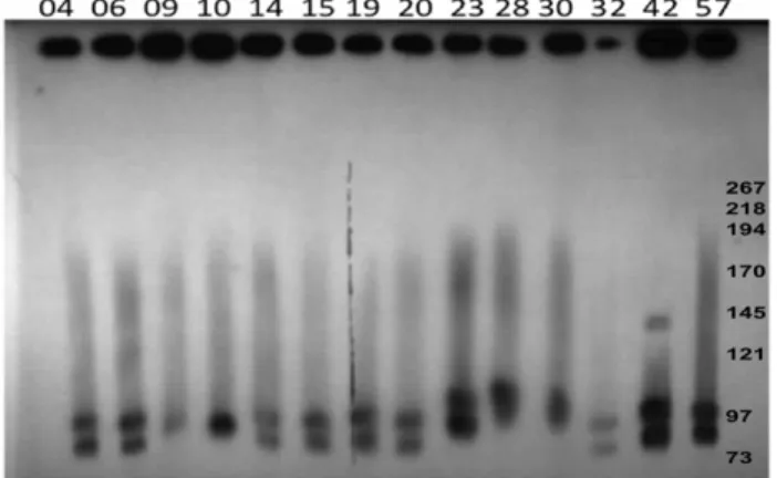
revealed the presence of one to four large plasmids ranging in size from 45 kb–150 kb in all strains (Figure 1, Table 2). PFGE analysis (Figure 1) showed the diversity of plasmids among the type AC. perfringens poultry isolates and their sometimes marked size variations, which were confirmed by PFGE/Southern blotting experiments (Figure 2). C. perfringens strains NE09 and NE10 carried just a single largenetB-positive plasmid (Table 2) but most NE isolates carried at least 3 large plasmids, in which thenetBand
cpb2-hdhAgenes were on distinct plasmids (Figure 2). Interestingly, a group of six netB-positive isolates from healthy chickens also showed three to four large plasmids, whereas five netB-negative healthy chicken isolates had fewer and smaller large plasmids (Figure S4A).
Southern blotting showed the presence ofcpb2in two plasmids in the same isolate (Figure S4B). When NotI-digested genomic DNA was probed withnetB,a hallmark of NELoc-1, it hybridized as a single large band in NE isolates as well as innetB-positive healthy chicken isolates (Figure S4C). Hybridization to,80 kb to 100 kb bands confirmed the plasmid identity of these PFGE bands and showed that the netBgene was always located in the larger plasmids (Figure 2). The hdhA probe (NELoc-3) hybridized to different and smaller plasmids than the netB-probe (Table 2, Figure 2), in 13 of 15 virulent NE isolates as well as in allnetB -positive healthy chicken isolates.
Mutants and Conjugation
The netB and cpb2 genes were insertionally inactivated in the strain CP1, resulting in the mutant strains CP1DnetB::ErmRAM
Figure 1. PFGE analyses of plasmids from NE C. perfringens
poultry strains.Agarose plugs containing DNA from each specified isolate were digested withNotI and subjected to PFGE and staining with ethidium bromide. Line numbers indicate isolate numbers M: Mid-Range II PFG molecular DNA ladder (Kb).
and CP1Dcpb2::ErmRAM. The insertion of ErmB-carrying introns into the target genes was confirmed by PCR using primers flanking the insertion site (data not shown). The netB and cpb2
genes located in different plasmids in CP1 strain were thus marked with erythromycin-cassette resistance (ermB) and this resistance could subsequently be used as a selective marker.
Conjugation assays were performed usingC. perfringensstrains CW504 RifRNalRas the recipient and CP1Dcpb2::ErmRAM and CP1DnetB::ErmRAM as donor strains in plate matings. Both plasmids (pNetB and pCpb2) transferred to the recipient strain, however we were unable to find one transconjugant harbouring only pNetB. Erythromycin-resistant transconjugants were con-firmed by specific PCR amplifications of DnetB::ErmRAM and
Dcpb2::ErmRAM; the ermB gene was amplified from the transconjugants but not from the wild-type or donor strains.
Conservation of NELoc-1, 2 and 3 in Poultry Isolates
Overlapping PCR assays were used to check the diversity of the three loci and their sites of insertion in nine virulent NE strains which represented different ST and plasmid profiles (classified by number and sizes) and two netB-positive isolates from healthy
chickens (Figure S1). NELoc-1 showed a general uniformity and conservation. For NELoc-2 just one isolate (NE 30) showed no PCR amplification for reaction #5 and two healthy chicken isolates (H26, H34) showed slightly smaller products. Most differences were found in the NELoc-3 (Figure S1).
Sequencing of Plasmids
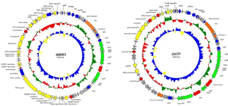
The two plasmids pNetB-NE10 and pCpb2-CP1 were isolated from wild-type NE10 and transconjugant T98 (Table 1 and Figure S5) respectively and sequenced on the Roche 454 GS Junior system. The complete nucleotide sequences of the plasmids pNetB-NE10 and pCpb2-CP1 were assembled into circular DNA sequences of 81,693 bp and 65,875 bp with an average depth of coverage of 200, respectively (Figure 3). The average G+C content is 25.7% for pNetB-NE10 and 26.8% for pCpb2-CP1, which is very similar to the G+C content of mostC. perfringensplasmids [23].
pNetB-NE10 and pCpb2-CP1
Sequence annotation of pNetB-NE10 showed the presence of 82 open read frames (orfs) whereas pCpb2-CP1 contained 73 orfs (Figure 3). Both plasmids are organized in the typical plasmid backbone of otherC. perfringensplasmids [10,11,24]. Of the fully sequenced CpCPs, the sequences of plasmids pNetB-NE10 and pCpb2-CP1 have identical gene organizations to plasmids pJIR3535 and pJIR3844 [10], respectively. The pNetB-NE10 and pCpb2-CP1 plasmids sequenced in this study are 99.1% and 97.9% similar to previous published plasmids pJIR3535 and pJIR3844, respectively. All these plasmids share a high degree of similarity with a major difference at the orfs 4 and 5 (Table S2).
ComparativeC. perfringensConjugative Plasmid Analyses
The DNA sequences of plasmids pNetB-NE10 (JQ655731) and pCpb2-CP1 (JQ655732) were compared to those of plasmids pCPF5603 (AB236337), pCPF4969 (NC_007772), pJIR3535 (JN689219), pJIR3844 (JN689217), pCPPB-1 (AB604032), pCP8533etx (NC_011412) and pCW3 (NC_010937). Figure 4 shows a diagrammatic representation of the organization among these differentCpCPs. The software tool EDGAR [21] was used for the assessment of genes that are present on all nineCpCPs and definition of a conserved backbone structure for these plasmids. A total of 24 core genes were identified (Table 3). From 24 core genes, 22 genes belong to the conserved backbone, encoding the Table 2.Properties ofClostridium perfringensstrains.
Strains Plasmids1 PCR3 Southern blot
netB cpb2 netB cpb2
CP1 4 + + 90 80
NE01 3 + + 90 80, 70
NE04 3 + + 85 75
NE06 4 + + 85 75
NE09 1 + 2 85 –
NE10 1 + 2 82 –
NE14 4 + + 88 77
NE15 3 + + 87 77
NE19 3 + + 90 77
NE20 3 + + 87 75
NE23 4 + + 95 75
NE28 3 + + 85 80, 70
NE30 3 + + 85 80, 70
NE32 3 + + 85 74
NE42 4 + + 94 81
NE57 3 + + 93 82
H+18 3 + + 90 80, 50
H+22 3 + + 95 85
H+26 3 + + 95 90, 60
H+27 3 + + 95 90, 60
H+34 4 + + 90 80
H+60 3 + + 90 85
H-16 1 2 + – 60
H-45 3 2 + – 65
H-46 3 2 + – 65
H-47 2 2 + – 73
H-54 3 2 + – 60
1Number of plasmids showed by PFGE analysis. Numbers indicate the approximate size of the plasmid (in kb).
2Genes detected by PCR amplification. (2) negative; doi:10.1371/journal.pone.0049753.t002
Figure 2. PFGE Southern blot of plasmids from NEC. perfringens
poultry strains.Southern blotting of PFGE (Figure 1) was performed with DIG-labelled probes fornetBandhdhA. Results from bothnetBand hdhAprobes are shown overlayed. In all lanes with two bands, the upper band representsnetBand the lower bandhdhA. M: Mid-Range II PFG molecular DNA ladder (Kb).
plasmid replication protein (rep), a DNA-binding transcriptional repressor (regD), the PemK protein, a sortase family protein, proteins required for conjugative transfer (tcpACDEFGHIJ), a DNA adenine-specific methyltransferase (dam), a tyrosine integrase (intP)
and seven hypothetical proteins, for a total of around 35 kb of the plasmid.
A second comparative analysis that considered only plasmids from necrotic enteritis isolates showed that 39 common genes Figure 3. Genetic maps of the sequenced NE plasmids pNetB-NE10 and pCpb2-CP1.The circles represent (from inner to outer most): (i) G+
C skew; (ii) G+C content and (iii) open reading frames; arrows indicate the direction of transcription. doi:10.1371/journal.pone.0049753.g003
Figure 4. Comparative analysisof C. perfringensconjugative plasmids.Comparative analysis of the sequenced NE plasmids pNetB-NE10 and pCpb2-CP1 and the published Cp plasmids pCPF5609, pCPF4969, pJIR3535, pJIR3844, pCPPB1, p8533etx and pCW3. Conserved regions within the analysed plasmids, pNetB (JQ655731), pCpb2 (JQ655732), pCPF5603 (AB236337), pCPF4969 (NC_007772), pJIR3535 (JN689219), pJIR3844 (JN689217), pCPPB-1 (AB604032), pCP8533etx (NC_011412) and pCW3 (NC_010937) are highlighted by grey boxes. Similarities between plasmids were calculated using the M-GCAT tool and visualised using PerlScript.
among those plasmids (pNetB-NE10, pJIR3535, pCpb2-CP1 and pJIR3844) (Table 4) are conserved. These 39 genes additionally encode the LexA repressor (regB), replication protein (rep), DNA-binding transcriptional repressor (regD), PemK family protein, sortase protein, DNA adenine-specific methyltransferase (dam), tyrosine integrase (intP), conjugation proteins described above besides conjugation proteins TcpA and TcpI, group II intron reverse transcriptase LtrA, DNA-cytosine methyltransferase (dcm), swim zinc finger domain protein and 15 hypothetical proteins of unknown functions in a total of approximately 41 kb of the plasmid size. As expected, the analyzed plasmids differ mostly in genes located in their pathogenicity loci, labelled yellow in Figures 3 and 4.
Plasmid Central Control Region
The initial region (,6 kb sequence) of plasmids pNetB-NE10 and pCpb2-CP1 as well as the two other fully sequenced and annotated NE plasmids (pJIR3535 and pJIR3844), which harbour five genes for regulation (regB, regCBorregCC, regD), replication (rep) or putative partitioning (parM), seems to be the ‘‘central control region’’ (CCR) (Figure 5). All nineC.perfringensplasmids compared in this study (Figure 4) carry a replication gene (rep) encoding a highly conserved replication initiation protein with about 90% identity on nucleotide sequence (Figure S2). Further genes surrounding therepgene appear to be responsible for regulation of plasmid copy number and function as transcriptional repressors.
The orf5 (parM) of the sequenced NE plasmids (pNetB-NE10, pCpb2-CP1) is found in the CCR, is transcribed divergently from therepgene, and encodes an ATPase involved in putative plasmid partitioning similar to the protein ParM of the ParMRC plasmid partitioning system.
Phylogenetic Tree
The sequences of the two sequenced NE plasmids pNetB-NE10 and pCpb2-CP1 and the seven completely sequencedCpCPs were analyzed phylogenetically (Figure 6) as described previously [18]. The phylogenetic tree suggests that these plasmids are closely related phylogenetically, and that there are closer relationships within each of thenetBand thecpb2-containing plasmids. Based on the homologous sequences of all plasmids the % identity varies
between 92,3% (pCpb2/pCPF4969) and 99,1% (pNetB/
pJIR3535).
Discussion
The current study provides complete DNA sequences of two NE
C. perfringensvirulence-associated plasmids (pNetB-NE10, pCpb2-CP1), further insight into the conjugative plasmids associated with NE, and significant new understanding of Clostridium perfringens
conjugative plasmids.
In this study, PFGE analyses revealed the presence of one to four large plasmids .45 kb in fifteen NE isolates of known virulence and different MLST type [16]. The variation in size of Table 3.Core genome genes ofC. perfringensplasmids.
pNetB-NE10 pCpb2-CP1 Gene/orf Name/Function
pNetB-NE10_1 pCpb2-CP1_1 hypothetical protein, unknown
pNetB-NE10_6 pCpb2-CP1_6 rep plasmid replication protein
pNetB-NE10_8 pCpb2-CP1_8 regD DNA-binding transcriptional repressor
pNetB-NE10_9 pCpb2-CP1_9 hypothetical protein, unknown
pNetB-NE10_10 pCpb2-CP1_10 hypothetical protein, unknown
pNetB-NE10_11 pCpb2-CP1_11 pemK PemK, growth inhibitor (COG2337)
pNetB-NE10_14 pCpb2-CP1_14 hypothetical protein, unknown
pNetB-NE10_15 pCpb2-CP1_15 srt sortase family protein
pNetB-NE10_16 pCpb2-CP1_16 hypothetical protein, unknown
pNetB-NE10_17 pCpb2-CP1_17 hypothetical protein, unknown
pNetB-NE10_18 pCpb2-CP1_18 dam DNA adenine-specific methyltransferase
pNetB-NE10_19 pCpb2-CP1_19 hypothetical protein, unknown
pNetB-NE10_21 pCpb2-CP1_21 intP tyrosine integrase
pNetB-NE10_22 pCpb2-CP1_23 tcpA conjugation protein TcpA, FtsK/SpoIIIE DNA translocase
pNetB-NE10_23 pCpb2-CP1_25 tcpC conjugation protein TcpC
pNetB-NE10_24 pCpb2-CP1_26 tcpD conjugation protein TcpD
pNetB-NE10_25 pCpb2-CP1_27 tcpE conjugation protein TcpE
pNetB-NE10_26 pCpb2-CP1_28 tcpF conjugation protein TcpF
pNetB-NE10_27 pCpb2-CP1_31 tcpG conjugation protein TcpG
pNetB-NE10_29 pCpb2-CP1_33 tcpH conjugation protein TcpH
pNetB-NE10_30 pCpb2-CP1_34 tcpI conjugation protein TcpI
pNetB-NE10_31 pCpb2-CP1_35 tcpJ conjugation protein TcpJ
pNetB-NE10_40 pCpb2-CP1_53 hypothetical protein, unknown
pNetB-NE10_42 pCpb2-CP1_54 hypothetical protein, unknown
Core genome composed of 24 genes of the nineC. perfringensplasmids pCPF4969, pCPF5603, pJIR3844, pCP8533etx, pCPPB-1, pCpb2-CP1, pNetB-NE10, pCW3 and pJIR3535. The core genome was computed with the software tool Edgar.
the plasmids reported by us here as well as previously [4] suggests that numerous rearrangements occur between and within the large conjugative plasmids, although further plasmid characterization is required to confirm this. For example, Southern blotting showed here for the first time that cpb2 can be present in two different plasmids within the same host strain (Table 2 and Figure S4). By contrast, in most type A isolates thenetBgene was found in a single variably sized plasmid (,80 kb–90 kb). The healthynetB-negative
chicken isolates lacked the NELoc1 and NELoc3 and their related plasmids, supporting the role of these plasmids in NE.
Overlapping PCR of the three pathogenicity loci [4] confirmed that NELoc-1, which containsnetB, is very conserved (Figure S1). Size variation of pNetB plasmids therefore must be the result of other changes in these plasmids. In contrast, the NELoc-3 showed greater variation (Figure S1). For example, the isolates NE28, NE30 and H26 possessed just a fragment of the NEloc-3 (hdhA
gene) and isolate NE42 seemed to harbour only the 59and 39links of this locus. This suggests that NELoc3, the smallest of the loci associated with NE isolates [4] is less important for NE than the other two loci. The chromosomal NELoc-2 was intact in all except one (NE30) of the eleven strains tested, confirming that it is an important signature for NE isolates. Conjugation assays using the erythromycin resistance-marked NE C. perfringens CP1 strain (which contains four large plasmids) as donor and the strain CW504 as recipient resulted in transconjugants with a variable number of the large plasmids (Figure S5), and suggests that all these plasmids are conjugative. Our sequencing data showed that that both pNetB-NE10 and pCpb2-CP1 possess thetcp conjuga-tion region, which has been found in all of the conjugative C. perfringensplasmids to date [10,12]. The pNetB-NE10 and pCpb2-CP1 plasmids sequenced in this study are 99.1% and 97.9% similar at the nucleotide level to previous published plasmids pJIR3535 and pJIR3844, respectively (Table S2). The presence of intacttcp-based conjugative regions suggests that pNetB-NE10 and pCpb2-CP1 plasmids are conjugative, supporting the recent work of others that showed that pJIR3535 and pJIR3844 plasmids to be conjugative [10].
Analysis of the two new genome sequences of plasmids pNetB-NE10 and pCpb2-CP1 isolated from NE isolates C. perfringens
(Figure 3) showed the high similarity with two other recently sequenced avian necrotic enteritisC. perfringensplasmids pJIR3535 and pJIR3844 [10] and confirmed the extensive conservation of the common backbone among allCpCPs [10,11,24,25,26].
Comparative genomic analysis showed that CpCPs, including the two plasmids described here, showed greater gene rearrange-ments including pathogenicity locus and accessory gene insertions around rather than within the backbone region (Figure 4). The
CpCPs have a mosaic organization in which transposons and Table 4.Core genome genes of NEC. perfringensplasmids.
pNetB-NE10 Gene/orf Name/Function
pNetB-NE10_1 hypothetical protein, unknown
pNetB-NE10_2 regB SOS-response repressor and protease LexA
pNetB-NE10_6 rep Plasmid replication protein
pNetB-NE10_8 regD DNA-binding transcriptional repressor
pNetB-NE10_9 hypothetical protein
pNetB-NE10_10 cysteine-rich hypothetical protein
pNetB-NE10_11 pemK PemK family protein
pNetB-NE10_13 hypothetical protein
pNetB-NE10_14 hypothetical protein
pNetB-NE10_15 srt Sortase family protein
pNetB-NE10_16 hypothetical protein
pNetB-NE10_17 hypothetical protein
pNetB-NE10_18 dam DNA adenine-specific methyltransferase
pNetB-NE10_19 hypothetical protein, unknown
pNetB-NE10_20 hypothetical protein, unknown
pNetB-NE10_21 intP tyrosine integrase/recombinase
pNetB-NE10_22 tcpA FtsK/SpoIIIE DNA translocase TcpA
pNetB-NE10_23 tcpC conjugation protein TcpC, putative Tn916
pNetB-NE10_24 tcpD conjugation protein TcpD
pNetB-NE10_25 tcpE conjugation protein TcpE
pNetB-NE10_26 tcpE conjugation protein TcpF
pNetB-NE10_27 tcpF conjugation protein TcpG
pNetB-NE10_28 G2 group II intron reverse transcriptase LtrA
pNetB-NE10_29 tcpH conjugation pore, membrane protein TcpH
pNetB-NE10_30 tcpI conjugation protein TcpI
pNetB-NE10_31 tcpJ conjugation protein TcpJ
pNetB-NE10_33 hypothetical protein, unknown
pNetB-NE10_34 dcm DNA-cytosine methyltransferase
pNetB-NE10_35 hypothetical protein, unknown
pNetB-NE10_36 hypothetical protein, unknown
pNetB-NE10_37 swim zinc finger domain protein
pNetB-NE10_38 conserved hypothetical protein, unknown
pNetB-NE10_40 conserved hypothetical protein, unknown
pNetB-NE10_42 hypothetical protein, unknown
pNetB-NE10_43 nuclease family transposase
pNetB-NE10_52 cell wall surface anchor family protein
pNetB-NE10_53 srt sortase A, LPXTG specific
pNetB-NE10_58 Recombinase
pNetB-NE10_66 hypothetical protein
Core genome composed of 39 genes of the five NEC. perfringensplasmids type A, pJIR3844, pCpb2-CP1, pNetB-NE10, pJIR3535. The core genome was computed with the software tool Edgar.
doi:10.1371/journal.pone.0049753.t004
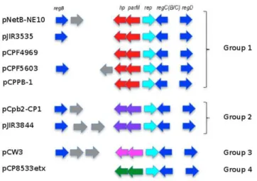
Figure 5. Comparative analysis of central control region ofC. perfringensconjugative plasmids.Comparative genomic analysis of the central control region ofC. perfringensplasmids starting fromregB regulatory gene. Identical colors designate similar function on pNetB-NE10, pCpb2-CP1, pCPF5609, pCPF4969, pJIR3535, pJIR3844, pCPPB1, pCP8533etx and pCW3.
integrases have played a role (Figure 4). The plasmids showed a conserved backbone region highlighted in gray. Hence, differences between the plasmids are related to their pathogenicity locus (yellow), also including different toxin genes (pink) (Figure 4). The nuclease family transposase recognized as orf50 in the pCW3 plasmid seems to be the insertion site of the pathogenicity loci of these conjugative plasmids leading to different organizational types (Figure 4). Inter- and intra-strain rearrangements of CpCPs are apparently responsible for the large size variation of conjugative plasmids from NEC. perfringensisolates (Table 2; Figure 1 and 2) possibly by duplications, insertions and deletions. Figure 4 shows a clear pattern of organization around the backbone region and in the pathogenicity loci of theCpCPs, and of the development of these plasmids. Thedcmregion has previously been described as a possible hot spot for insertion of iota-toxin genes in plasmids of
C. perfringens type E [9], which supports our suggestion of insertional ‘‘hot spots’’ between the regulation and partitioning genes and downstream of the tcp transfer region, where dcm is located. The pattern that emerges in the analysis shown in Figure 4 is that the major toxin-containing regions of the CpCPs are organized as pathogenicity loci. This was first described for the NELoc1 and NELoc3 of the NE-associated plasmids [4], but might be a general feature of these virulence plasmids.
The comparative analysis of all nine C. perfringens plasmids showed a core genome of 24 genes, most of them belonging to the conserved backbone structure (Table 3) which includes the transfer of a clostridial plasmid (tcp) locus. The backbone region comprises a large portion of the conjugative plasmids [10,11,25,26,27], so that for both NE virulence plasmids, as well as for other characterized major virulence plasmids, a size around 35 kb seems to be optimized for efficient replication, conjugative transfer, plasmid maintenance and stability functions. Within the backbone region, there is what we designate the central control region (CCR) consisting of the replication (rep), regulatory genes (reg) and putative partition genes (parM).
The common core genome for NE-isolate associated plasmids is larger (39 genes, around 41 kb). Comparison of the four NE-isolate-associatedC. perfringens plasmids (pNetB-NE10, pJIR3535,
pCpb2-CP1 and pJIR3844) identified not only 35 genes in the backbone region but interestingly also four genes common in the pathogenicity loci. These genes encode a cell wall surface anchor family protein, the sortase A, a resolvase/recombinase and a hypothetical protein. It is clear that C. perfringens conjugative plasmids are closely related since they show remarkable homology [8,11,27].
Plasmid partition is classified by one of three types of par
systems. Type I systems use ParA ATPase proteins with Walker-type folds and centromere-binding proteins called ParB; Walker-type II systems use actin-like ParM ATPases and centromere-binding proteins called ParR; and a recently described type III system uses a tubulin-like protein, TubZ [28]. The ParMRC operon is a well-known partition system for bacterial DNA segregation in low copy number plasmids [29]. These partition system consists of three components: two genesparMand parRlocated side-by-side, with
parM encoding a NTPase protein, and ParR, a specific centro-mere-binding protein, and acis-acting centromere-like site parC, a small non-coding plasmid region with a series of 11 bp repeats. Interestingly, the equivalent to the ParR protein was not found in any of theCpCPs, but there is a gene adjacent toparM, transcribed in the same orientation asparM, that encodes a conserved protein of unknown function (orf4 in pNetB-NE10). Sequence differences in the ParM ortholog encoded in the replication and maintenance regions of these plasmids may be involved in this process. The mechanism of segregation is presently unknown forC. perfringens
conjugative plasmids, and no Par system was described in their DNA sequence.
Six genes were found to be unique within the backbone region for the NE plasmids. These include the collagen adhesion protein (orf12) and a hypothetical protein (orf40) and four genes located in the CCR (orf3, orf4, orf5(parM),orf7(regCB),using pNetB-NE10 as reference). These differences in the CCR transcriptional regulatory genes and segregation genes suggest that these differences may allow this family of plasmids to co-exist in their
may be a function of the limited variation in the CCR. Gurjaret al
[27] had earlier suggested that only certain toxin plasmid combinations could be stably maintained within a single C. perfringenscell. Earlier analysis suggested that differences in ParM orthologs may be involved in this process [10]. It will be of interest to examine the CCR regions of otherCpCPs found in NE strains with multiple plasmids, to determine how these relate to the postulated incompatibility system described here.
The mechanism of the partitioning system incompatibility in
CpCPs proposed here is different from the well-known replication-mediated incompatibility [30]. Two different plasmids with the same partitioning system cannot coexist stably in the same host because of the competition between identical partitioning systems [31,32,33]. Based on this comparative genomic analysis, we suggest that C. perfringens conjugative plasmids can be grouped according to their types of putative partitioning genes, in particular
parM (orf5) and orf4 (hypothetical protein), which seems to be equivalent toparRin the ParMRC system.
ATPase/ParM protein showed the highest similarity (99%) in amino acid sequence in the group of plasmids pNetB-NE10 (orf5), pJIR3535 (orf00004) pCPF4969 (orf61), pCPF5603 (orf16) and pCPPB-1 (orf63) (Figure5) (Fig S2). Amino acid sequence alignments showed that the ParM proteins contain conserved domain actin-like ATPases (PRK13917) and a predicted function of a plasmid segregation protein as part of a type II Par system [28,29]. Plasmids pCpb2-CP1 (orf5) and pJIR3844 (orf6) form a second group that encodes a different ATPase with no conserved domain and just 27% protein identity with orthologues of the first plasmid group. Although ATPase/ParM proteins from plasmids pCW3 (orf13) and pCP8533etx (orf52) have low homology (27%) with each other, both proteins belong to a superfamily of StbA proteins, a family that consists of several bacterial StbA plasmid stability proteins.
The orf4 gene in pNetB-NE10 and its homologues in other
CpCPs have no conserved domain or significant similarity to other known proteins in GenBank. Speculatively, this hypothetical protein is suggested to be the potential ParR component of the partitioning system of CpCPs, primarily because it is located adjacent to parMwith the same transcriptional orientation in all
CpCPs analysed. Interestingly, this hypothetical protein is also conserved in the same plasmid groups described above, as is shown in the multiple sequence alignments (Figure S2). Another important element to complete the ParMRC system is the presence of parC, the centromeric region of the plasmid. Centromeres consist of a series of tandem DNA repeats of eight 10-bp or four 20-bp repeats typically located adjacent of theparM
gene [28,29]. However, the precise size and organization of the
parC site varies among ParMRC system [29]. The upstream sequence of parM genes of the nine CpCPs revealed several imperfect 11 bp repeats and conserved regions among the sequences which appear to be the equivalent of aparCsite (Figure S3).
In conclusion, the complete sequencing of two new conjugative plasmids from NE isolates described here, when combined with comparative analysis of previously sequenced plasmids, adds considerably to understanding the evolution of virulence-associat-ed plasmids in C. perfringens, and contributes to the unanswered question of how these different but related plasmids can co-exist in the same host. The suggestion proposed here of classifyingCpCPs into incompatibility groups, of which four are described here, based on the partitioning systems, requires confirmation by experimental data. There are important areas still to be un-derstood including the function of conserved hypothetical proteins, the presence of additional plasmid incompatibility systems, and the
basis of any limitation of specific CpCP family members to particularC. perfringenstypes. Sequencing of further large CpCPs (Figure 1) might add confirmation to our supposition about the role of the CCR in maintenance of different family members in the same host.
Supporting Information
Figure S1 Overlapping PCR analysis of NE locus in C. perfringens.PCR reactions were performed using DNA fromC. perfringens strains described on Table 1. Healthy and NE C. perfringensisolates H26, H34, NE04, NE09, NE10, NE14, NE20, NE23, NE28, NE30, NE42, respectively. Genetic organization of NE loci. (A) Overlapping PCR analysis of NE locus 1. (B)
Overlapping PCR analysis of NE locus 2.(C)Overlapping PCR analysis of NE locus 3. PCR products spanning the entire locus are represented by black bars and the PCR results for each strain tested are given below as follows:+.PCR product was of expected size;2, no PCR product produced. Where the PCR product did not match the expected size, the actual size is given.
(PPT)
Figure S2 Amino acid alignments of proteins encoded by differentC. perfringens plasmids. Plasmid names and their respective orf number (plasmid name orf#) are described for each protein. Identical residues (*), conservative amino acid substitutions (:), and semi-conservative amino acid substitutions (.) are shown below the aligned sequences. (MUSCLE23.7). (DOCX)
Figure S3 Repeats found on the upstream region of
parM gene. Possible tandem repeats found on the upstream region ofparMgene next to rep gene fromC. perfringensplasmids using etandem (http://emboss.bioinformatics.nl/cgi-bin/emboss/ etandem).
(DOCX)
Figure S4 PFGE and Southern blot analyses of plasmids from healthy C. perfringens poultry strains. (A) PFGE analyses of plasmids from healthy C. perfringens poultry strains. Agarose plugs containing DNA from each specified isolate were digested with NotI and subjected to PFGE and staining with ethidium bromide. See Table1 and 2 for isolate features. Line numbers indicate isolate numbers M: Mid-Range II PFG molecular DNA ladder (Kb).(B)PFGE Southern blot of plasmids from healthy C. perfringens poultry strains. Southern blotting of PFGE (Figure S4A) was performed with only DIG-labelled probe for cpb2 gene. M: Mid-Range II PFG molecular DNA ladder (Kb). (C) PFGE Southern blot of plasmids from healthy C. perfringenspoultry strains. Southern blotting of PFGE (FigureS 4A) was performed with only DIG-labelled probe fornetB gene. M: Mid-Range II PFG molecular DNA ladder (Kb).
(DOCX)
Figure S5 PFGE analyses of plasmids from transconju-gantsC. perfringensstrains.Agarose plugs containing DNA from each specified isolate were digested withNotI and subjected to PFGE and staining with ethidium bromide. Lines indicate: CW504 recipient strain (plasmid free); T98 (transconjugant carrying the plasmid pCpb2); T117 (transconjugant carrying three of CP1 plasmids); T118 (transconjugant carrying four of CP1 plasmids); T119 (transconjugant carrying two of CP1 plasmids); T125(transconjugant carrying two of CP1 plasmids); T128 (transconjugant carrying two of CP1 plasmids); CP1 donor strain (harbours four large plasmids); M: Mid-Range II PFG molecular DNA ladder (Kb).
Table S1 List of primers. (A) Primers used for PCR DIG labelling and mutation (B) Primers used for overlapping PCR reactions of the three Pathogenicity loci characteristic of necrotic enteritisC. perfringensisolates.
(DOCX)
Table S2 Comparison of NE C. perfringens plasmids. (A)Comparison of coding sequences pNetB-NE10 and pJIR3535 NE C.perfringensplasmids by means of BLASTn analyses. Open reading frames are labeled according to the annotation of plasmid pNetB-NE10(B)Comparison of open reading frames pCpb2-CP1 and pJIR3844 NE C. perfringensplasmids by means of BLASTn analyses. Open reading frames are labeled according to the annotation of plasmid pCpb2-CP1.
(DOCX)
Acknowledgments
The authors would like to thank Dr. Nigel P. Minton (University of Nottingham) for providing plasmid pMTL007 for the ClosTron system, Dr. Julian I. Rood (Monash University) for theC. perfringensconjugative strain CW504, Cornelius Poppe for the conjugation experiments and his support, Andrew Kropinski for his comments and suggestions on the manuscript and Patrick Boerlin forC. perfringensstrains.
Author Contributions
Conceived and designed the experiments: VRP JFP. Performed the experiments: VRP MC. Analyzed the data: VRP MC FE JB JFP. Wrote the paper: VRP JFP.
References
1. Cooper KK, Songer JG (2009) Necrotic enteritis in chickens: A paradigm of enteric infection byClostridium perfringenstype A. Anaerobe 15: 55–60. 2. Van Immerseel F, Rood JI, Moore RJ, Titball RW (2009) Rethinking our
understanding of the pathogenesis of necrotic enteritis in chickens. Trends Microbiol 17: 32–36.
3. Keyburn AL, Boyce JD, Vaz P, Bannam TL, Ford ME, et al. (2008) NetB, a new toxin that is associated with avian necrotic enteritis caused by Clostridium perfringens. PLoS Pathog. 4: e26.
4. Lepp D, Roxas B, Parreira VR, Marri PR, Rosey EL, et al. (2010) Identification of novel pathogenicity loci inClostridium perfringens strains that cause avian necrotic enteritis. PLoS One 5: e10795.
5. Songer JG (1996) Clostridial enteric diseases of domestic animals. Clin Microbiol Rev. 9: 216–34.
6. Myers GSA, Rasko DA, Cheung JK, Ravel J, Seshadri R, et al. (2006) Skewed genomic variability in strains of the toxigenic bacterial pathogen,Clostridium perfringens. Genome Res 16: 1031–1040.
7. Popoff MR, Bouvet P (2009) Clostridial toxins. Future Microbiol. 4: 1021–1064. 8. Miyamoto K, Fisher DJ, Li J, Sayeed S, Akimoto S, et al. (2006) Complete Sequencing and Diversity Analysis of the Enterotoxin-Encoding Plasmids in Clostridium perfringensType A Non-Food-Borne Human Gastrointestinal Disease Isolates. J Bacteriol 188: 1585–1598.
9. Li J, Miyamoto K, McClane BA (2007) Comparison of Virulence Plasmids amongClostridium perfringensType E Isolates. Infect Immun 75: 1811–1819. 10. Bannam TL, Yan XX, Harrison PF, Seemann T, Keyburn AL, et al. (2011)
Necrotic enteritis-derivedClostridium perfringensstrain with three closely related independently conjugative toxin and antibiotic resistance plasmids. MBio. 2. pii: e00190–11.
11. Miyamoto K, Li J, Sayeed S, Akimoto S, McClane BA (2008) Sequencing and Diversity Analyses Reveal Extensive Similarities between Some Epsilon-Toxin-Encoding Plasmids and the pCPF5603Clostridium perfringensEnterotoxin Plasmid. J Bacteriol 190: 7178–7188.
12. Bannam TL, Teng WL, Bulach D, Lyras D, Rood JI (2006) Functional identification of conjugation and replication regions of the tetracycline resistance plasmid pCW3 fromClostridium perfringens.J Bacteriol. 188: 4942–4951. 13. Porter CJ, Bantwal R, Bannam TL, Rosado CJ, Pearce MC, et al. (2012) The
conjugation protein TcpC fromClostridium perfringensis structurally related to the type IV secretion system protein VirB8 from Gram-negative bacteria. Mol Microbiol. 83: 275–88.
14. Bantwal R, Bannam TL, Porter CJ, Quinsey NS, Lyras D, et al.(2012) The peptidoglycan hydrolase TcpG is required for efficient conjugative transfer of pCW3 inClostridium perfringens. Plasmid 67: 139–147.
15. Steen JA, Bannam TL, Teng WL, Devenish RJ, Rood JI (2009) The putative coupling protein TcpA interacts with other pCW3-encoded proteins to form an essential part of the conjugation complex. J Bacteriol. 19: 2926–2933. 16. Chalmers G, Bruce HL, Hunter DB, Parreira VR, Kulkarni RR, et al. (2008)
Multilocus sequence typing analysis ofClostridium perfringensisolates from necrotic enteritis outbreaks in broiler chicken populations. J Clin Microbiol. 46: 3957– 3964.
17. Pospiech A,Neumann B (1995) A versatile quick-prep of genomic DNA from gram-positive bacteria. Trends Genet. 11: 217–8.
18. Heap JT, Pennington OJ, Cartman ST, Carter GP, Minton NP (2007) The ClosTron: a universal gene knock-out system for the genus Clostridium. J Microbiol Methods. 70: 452–464.
19. Treangen TJ, Messeguer X (2006) M-GCAT: interactively and efficiently constructing large-scale multiple genome comparison frameworks in closely related species. BMC Bioinformatics. 7: 433.
20. Eikmeyer F, Hadiati A, Szczepanowski R, Wibberg D, Schneiker-Bekel S, et al. (2012) The complete genome sequences of four new IncN plasmids from wastewater treatment plant effluent provide new insights into IncN plasmid diversity and evolution. Plasmid. Feb 2. (Epub ahead of print).
21. Blom J, Albaum SP, Doppmeier D, Pu¨hler A, Vo¨rholter FJ, et al. (2009) EDGAR: a software framework for the comparative analysis of prokaryotic genomes. BMC Bioinformatics 10, 154.
22. Kumar S, Nei M, Dudley J, Tamura K. (2008) MEGA: a biologist-centric software for evolutionary analysis of DNA and protein sequences. Bioinformatics 9: 299–306.
23. Shimizu T, Ohtani K, Hirakawa H, Ohshima K, Yamashita A, et al. (2002) Complete genome sequence ofClostridium perfringens, an anaerobic flesh-eater. PNAS 99: 996–1001.
24. Sayeed S, Li J, McClane BA (2007) Virulence Plasmid Diversity inClostridium perfringensType D Isolates. Infect Immun 75: 2391–2398.
25. Sayeed S, Li J, McClane BA (2010) Characterization of Virulence Plasmid Diversity amongClostridium perfringensType B Isolates. Infect Immun 78: 495– 504.
26. Miyamoto K, Yumine N, Mimura K, Nagahama M, Li J, et al.(2011) Identification of novelClostridium perfringenstype E strains that carry an iota toxin plasmid with a functional enterotoxin gene. PLoS One 6: e20376.
27. Gurjar A, Li J, McClane BA (2010) Characterization of toxin plasmids in Clostridium perfringenstype C isolates. Infect Immun 78: 4860–4869.
28. Schumacher MA, Glover TC, Brzoska AJ, Jensen SO, Dunham TD, et al. (2007) Segrosome structure revealed by a complex of ParR with centromere DNA. Nature 450: 1268–1271.
29. Salje J, Gayathri P, Lo¨we J. (2010) The ParMRC system: molecular mechanisms of plasmid segregation by actin-like filaments. Nat Rev Microbiol 8: 683–692. 30. Novick RP (1987) Plasmid incompatibility. Microbiol Rev 51: 381–395. 31. Funnell BE, Slavcev RA (2004) Partition systems of bacterial plasmids. In:
Phillips G, Funnell BE, eds. Plasmid biology. Washington: ASM Press. 81–103. 32. Ebersbach G, Gerdes K (2005) Plasmid segregation mechanisms. Annu Rev
Genet 39: 453–479.
33. Bouet JY, Nordstro¨m K, Lane D (2007) Plasmid partition and incompatibility-the focus shifts. Mol Microbiol. 65: 1405–1414.
34. Thompson DR, Parreira VR, Kulkarni RR, Prescott JF (2006) Live attenuated vaccine-based control of necrotic enteritis of broiler chickens. Vet Microbiol. 113: 25–34.