A role for adeno-associated viral vectors in gene therapy
Renata dos Santos Coura and Nance Beyer Nardi
Laboratório de Imunogenética, Departamento de Genética, Universidade Federal do Rio Grande do Sul,
Porto Alegre, RS, Brazil.
Abstract
Gene therapy constitutes a therapeutic intervention based on modification of the genetic material of living cells, by correcting genetic defects or overexpressing therapeutic proteins. The success of gene therapy protocols depends on the availability of therapeutically suitable genes, appropriate gene delivery systems and proof of safety and effi-cacy. Recent advances on the development of gene delivery systems, particularly on viral vectors engineering and improved gene regulatory systems, have led to marked progress in this field. Although the available vector systems can successfully transfer genes into cells, the ideal delivery vehicle has not been found. In this context, adeno-associated virus vectors (AAV) are arising as a promising tool for a wide range of applications, due to a combi-nation of characteristics such as lack of pathogenicity and immunogenicity, wide range of cell tropism and long-term gene expression. Since its isolation, the biological properties of the adeno-associated virus have been increasingly understood, improving our ability to manipulate and use it as a safe and efficient gene therapy vector of wide spec-trum. In this work, we review the bases of gene therapy, main types of gene transfer systems and basic properties and use of AAV vectors.
Key words:Adeno-associated virus, AAV-based recombinant vectors, gene therapy.
Received: August 29, 2007; Accepted: November 8, 2007.
Introduction
Gene therapy is a clinical strategy involving gene transfer with therapeutic purposes. It is based on the con-cept that an exogenous gene (transgene) is able to modify the biology and phenotype of target cells, tissues and organs. Initially designed to definitely correct monogenic disorders, such as cystic fibrosis, severe combined immu-nodeficiency or muscular dystrophy, gene therapy has evolved into a promising therapeutic modality for a diverse array of diseases. Targets are expanding and currently in-clude not only genetic, but also many acquired diseases, such as cancer, tissue degeneration or infectious diseases (Worgall, 2005; Zubler, 2006). Depending on the duration planned for the treatment, type and location of target cells, and whether they undergo division or are quiescent, differ-ent vectors may be used, involving nonviral methods, non-integrating viral vectors or non-integrating viral vectors (re-viewed by El-Aneed, 2004; Zubler, 2006).
The first gene therapy clinical trial was carried out in 1989, in patients with advanced melanoma, using tumor-infiltrating lymphocytes modified by retroviral
transduc-tion (Rosenberget al., 1990). In the early nineties, a clinical trial with children with severe combined immunodefi-ciency (SCID) was also performed, by retrovirus transfer of the deaminase adenosine gene to lymphocytes isolated from these patients (Blaeseet al., 1995). Since then, more than 5,000 patients have been treated in more than 1,000 clinical protocols all over the world (http://www.wiley.co. uk/genetherapy/clinical). Despite the initial enthusiasm, however, the efficacy of gene therapy in clinical trials has not been as high as expected; a situation further compli-cated by ethical and safety concerns (Verma and Somia, 1997; Rubanyi, 2001; El-Aneed, 2004; Edelstein et al., 2004; Zubler, 2006). Further studies are being developed to solve these limitations.
Vectors
The primary challenge in gene therapy is the develop-ment of efficient gene delivery vectors to target the selected tissues, where proper gene expression may be achieved. The ideal gene delivery method should be able to protect the transgene against degradation by nucleases, allow transport of the transgene into the nucleus of target cells, and have no potential inflammatory or cytotoxic side ef-fects. However, the choice of the vector, as well as the pro-tocol of treatment and the outcome of the therapy, are
www.sbg.org.br
Send correspondence to Nance Beyer Nardi. Laboratório de Imu-nogenética, Departamento de Genética, Universidade Federal do Rio Grande do Sul, Av. Bento Gonçalves 9500, 91501-970 Porto Alegre, RS, Brazil. E-mail: nardi@ufrgs.br.
determined mainly by the nature of the disease and by the target organ (Salaniet al., 2005; Gaoet al., 2007). There-fore, other important parameters to be considered in the definition of the best type of vector include its purity and ti-ter, efficiency, size limitations for insertion of transgenes, ability to infect dividing and/or quiescent cells, duration of expression of the transgene, integration into the host ge-nome, tropism, and toxicity or immunogenicity of the vec-tor (Goverdhanaet al., 2005).
The introduction of therapeutic genes into target cells, leading to efficient and stable expression of the trans-gene with minimal adverse effects, can be achieved using viral and nonviral vectors. Viral vectors can mediate gene transfer with high efficiency and the possibility of long-term gene expression, since they can easily enter cells and deliver their genetic material into the nucleus; therefore, they are in most cases more efficient than nonviral delivery systems. However, the risks of immunogenicity and inser-tion mutagenesis, disclosed in gene therapy clinical trials, pose serious safety concerns about some commonly used viral vectors. Limitations in the size of the transgene that re-combinant viruses can carry and issues related to the pro-duction of viral vectors present additional practical challenges.
In trying to bypass safety issues intrinsic to the use of viral vectors, several nonviral vectors are being developed (Goverdhanaet al., 2005). Although significant progress has been made in studies on the basic biology and applica-tions of various nonviral gene delivery systems, most of them are still much less efficient than viral vectors, particu-larly forin vivogene delivery (Gaoet al., 2007).
Non-viral systems
The underlying principle of nonviral vector systems is to complex the DNA vector with molecules that will fa-cilitate DNA entry into the cells of interest. Complexed DNA binds to the cell membrane, triggering either nonspe-cific or receptor-mediated endocytosis. Upon entry into the cell, these complexes remain in endosomes, and their abil-ity to escape before lysosomal enzymes destroy them is an essential characteristic of a successful nonviral vector.
Once released from the endosomes, these complexes must enter the nucleus and complexed DNA must be released from its carrier molecules and adequately transcribed.
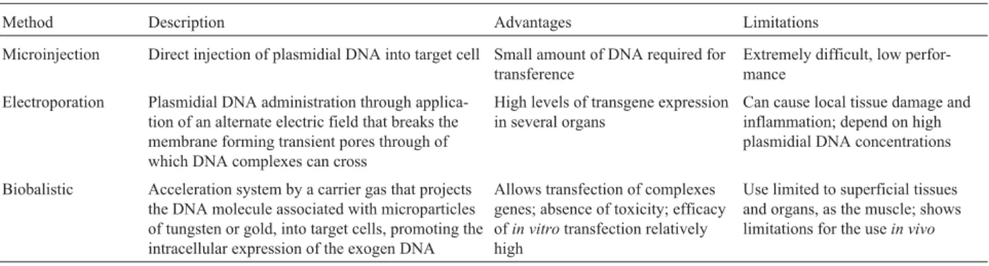
Methods of nonviral gene delivery have been ex-plored using physical (carrier-free gene delivery) and che-mical approaches (synthetic vector-based gene delivery). Physical approaches, including needle injection, electro-poration, gene gun, ultrasound, and hydrodynamic deliv-ery, employ a force that permeates the cell membrane and facilitates intracellular gene transfer (Table 1). Chemical approaches use synthetic or naturally occurring compounds as carriers to deliver the transgene into cells (Table 2).
Viral systems
For the production of efficient and safe viral vectors, essential sequences for viral particle assembly, genome package, and transgene delivery to target cells must be identified. Dispensable genes are then deleted from the vi-ral genome in order to reduce its pathogenicity and immunogenicity and, finally, the transgene is integrated into the construct. Some viral vectors are able to integrate into the host genome, whereas others remain episomal. In-tegrating viruses result in persistent transgene expression. Non-integrating vectors, such as adenoviruses whose viral DNA is maintained in episomal form in infected cells, lead to transient transgene expression. Each type of vector pres-ents specific advantages and limitations that make them ap-propriate for particular applications (Pfeifer and Verma, 2001). Most of the vectors currently used for gene transfer are derived from human pathogens, from which essential viral genes have been deleted to make them nonpathogenic. They usually have a broad tropism, so that different types of cells and/or tissues may be targeted.
The ideal vector has not been described yet, but its characteristics should include:
• easy and efficient production of high titers of the vi-ral particle;
• absence of toxicity for target cells and undesirable effects such as immune response against the vector or the transgene;
Table 1- Physical methods of gene therapy with non-viral vectors.
Method Description Advantages Limitations
Microinjection Direct injection of plasmidial DNA into target cell Small amount of DNA required for transference
Extremely difficult, low perfor-mance
Electroporation Plasmidial DNA administration through applica-tion of an alternate electric field that breaks the membrane forming transient pores through of which DNA complexes can cross
High levels of transgene expression in several organs
Can cause local tissue damage and inflammation; depend on high plasmidial DNA concentrations
Biobalistic Acceleration system by a carrier gas that projects the DNA molecule associated with microparticles of tungsten or gold, into target cells, promoting the intracellular expression of the exogen DNA
Allows transfection of complexes genes; absence of toxicity; efficacy ofin vitrotransfection relatively high
• capacity of site-specific integration, allowing long-term transgene expression, for treating diseases such as ge-netic disorders;
• capacity of transduction of specific cell types; • infection of proliferative and quiescent cells. The most commonly used viral vectors for gene ther-apy are based on adenoviruses (Ad), adeno-associated vi-ruses (AAV) and retrovirus/lentivirus vectors. Their main characteristics are summarized in Table 3.
Adeno-associated viruses - structure and life cycle
Adeno-associated viruses (AAV) are non-pathogenic parvoviruses. The Parvoviridae family includes small, ico-sahedral and non-enveloped viruses whose genome is a sin-gle stranded DNA. AAV is one of the smallest viruses, with a capsid of approximately 22 nm, and one of the most fre-quent members of the family. Despite its high prevalence,
the virus has not been associated to any human illness. Since a co-infecting helper virus is required for productive infection, adeno-associated viruses are ascribed to a sepa-rate genus (Dependovirus) in the Parvoviridae family. Nu-merous serotypes of AAV have been identified, but the majority of rAAV vectors are based on AAV serotype 2 (AAV2).
The wild AAV2 life cycle in human cells consists first in its binding to a serotype-specific receptor/co-receptor at the cell surface followed by its entry into the cell through endocytosis. Thereafter, the virus needs to escape from the endosome, uncoat and enter the nucleus. In the nu-cleus, the single-stranded (ss) viral DNA needs to be con-verted into double-stranded (ds) replicative intermediates, from which regulatory and structural viral proteins are tran-scribed, as well as the ssDNA for packaging into the new viral particles. This conversion is probably carried out by
Table 2- Chemical methods of gene therapy with non-viral vectors.
Method Description Advantages Limitations
Nude DNA Direct injection of plasmidial DNA into target cell (non-complexed to any particle)
Successful transgene expression (1%-40%) transferred through nude DNA hidrodynamically* injected in mice
Fast degradation and neutralization by en-dogenous DNAses; limited potential of electrostatic interactions with anionic lipids of the cellular membrane (negative charge of DNA molecule); uncertain clini-cal applicability and problems for large-scale use
Lipoplexes Cationic lipids complexed to DNA that neutralize its electrostatic charge, allowing DNA-cellular membrane interaction, and thus, increasing the transfection efficacy
Lipoplexes are powerful systems to intro-duce plasmids into target cells; high level transgene expression after direct adminis-tration or injection in target tissues
Interaction with plasmatic proteins and other extracellular proteins that can inacti-vate them due to their hydrophobic and cationic surface; indiscriminate transgene expression in cells at the administration site (non-specific membrane activity); low efficacy of quiescent cells transfection Poliplexes Non-particulate complexes formed by
binding of DNA to cationic polymers
Idem to lipoplexes; possibility of choice of the ideal polymer structure to target cell transfection; easy to obtain in large-scale
Idem to lipoplexes
*fast intravenous administration of a great volume (up to ~2.5 mL in mice).
Table 3- Main viral vector systems.
Viral vector Description Advantages Limitations Applications
Adenovirus (Ad) Icosahedric, non- enveloped, genome of 36 kb, non-integrative
Easy propagation in high titers, infection of most cell types;
insertion of large DNA frag-ments
High immunogenicity, in-ducing important cellular and humoral immune responses that can be fatal
Therapies that require tran-sient gene expression: cancer therapy, angiogenesis induc-tion and DNA vaccine pro-duction (due to its inflamma-tory and immunogenic properties)
Retroviruses (Retrovirus and Lentivirus)
Integrative in proliferative (retrovirus and lentivirus) and quiescent (lentivirus) cells
Low immunogenicity, possi-bility of insertion of large DNA fragments (up to 8 kb)
Insertional mutagenesis Genetic diseases of T cells and hematological diseases (Retrovirus),
HIV/AIDS Adeno-associated virus
(AAV)
Icosahedric, non- enveloped, single-stranded DNA, genome of 4.7 kb, integrative
Low immunogenicity, easy propagation in high titers, infection of most of cell types, long-term gene expression
Limited capacity for inser-tion of DNA fragments
some not yet identified cellular DNA repair mechanism (re-viewed in Stenderet al., 2007). These dsDNA intermedi-ates integrate into a specific site of human chromosome 19 (19q13.3q-ter) called AAVS1 (Kotinet al., 1990; Kotinet al., 1991; Samulski et al., 1991; McCarty et al., 2004; Philpottet al., 2004; Wang and Lieber, 2006). In the ab-sence of a co-infection by a helper virus (commonly adeno-virus or herpesadeno-virus), the integrated AAV genome remains in a latent state. When the host cell is co-infected by a helper virus, the changes of the cellular milieu lead to the activation of the integrated AAV dsDNA that initiates the transcription of the regulatory and structural viral genes, as well as the replication of viral DNA, generating the ssDNA to be packaged into novel viral particles. The viral DNA replication process relies on host cell polymerase activities, since AAV does not encode a proper polymerase (Niet al., 1998). Although the encapsidation process is still not well understood, there is evidence that it is a nuclear process (Wistuba et al., 1997). This evidence also indicate that there are specific patterns of distribution and transport of viral proteins between cellular compartments in each stage of a productive infection. Capsid assembly probably occurs in the nucleolus and is then relocalized into the nucleo-plasm, with some influence of Rep proteins. The co-localization of the empty assembled capsid, the Rep pro-teins and the ssDNA AAV genome in the nucleoplasm al-lows the encapsidation process to occur (Im and Muzyczka, 1989; Wistubaet al., 1997; Kinget al., 2001; Blekeret al., 2005). Finally, the novel viral particles exit the cell, proba-bly by an unknown exocytosis mechanism.
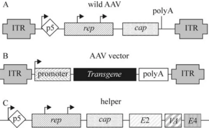
The wild AAV has a linear single-stranded DNA ge-nome of approximately 4.7 kb of either plus or minus polar-ity. The AAV DNA ends are constituted of a 145 bp inverted terminal repeat (ITR), whose terminal 125 bases are multipalindromic, leading to a characteristic T-shaped hairpin structure (Koczotet al., 1973). This structure be-comes a 3’ hydroxyl group available as a primer for the ini-tiation of viral DNA replication (Berns,1990).
The ITRs that act as DNA replication origin, packag-ing and integration signal, also act as a regulator element for wild AAV gene expression. Two viral genes located be-tween the ITRs,repandcap, encode proteins involved in viral replication and capsid formation, respectively (Fig-ure 1A). The rep gene encodes four regulatory proteins called Rep78, Rep68, Rep52 and Rep40, according to their molecular weight. This is possible due to the use of two al-ternative promoters (p5 and p19) and an internal splice do-nor and acceptor site.
Rep78 and Rep68 are site-specific DNA binding pro-teins that interact with specific sites within the ITRs (Rep-binding element - RBE - and terminal resolution site -trs), playing a crucial role in DNA replication, transcriptional control and site-specific integration processes. They also act as positive or negative regulators of AAV gene expres-sion, depending on the presence or absence of a helper
vi-rus, respectively (Pereiraet al., 1997). Moreover, these pro-teins have endonuclease (Berns and Giraud, 1996), helicase and ATPase (Im and Muzyczka,1990) activities.
The Rep52 and Rep40 proteins participate in the gen-eration and accumulation of the plus and minus ssDNA vi-ral genome from the dsDNA intermediates (Chejanovsky and Carter, 1989). The single-stranded genomes of both po-larities are packaged with equal efficiency (Berns and Adler, 1972). Similar to Rep78 and Rep68, these proteins also exhibit helicase and ATPase activities (Smith and Kotin, 1998; Collacoet al., 2003).
Thecapgene has a single promoter (p40) that tran-scribes two messenger RNAs (mRNA) through alternative splicing at two acceptor sites. The larger mRNA encodes the virion protein 1 (VP1), the largest protein subunit of the capsid (~87 kDa). The shorter transcript presents a conven-tional initiation codon (AUG), which originates the VP3 capsid protein (~62 kDa), and an upstream non-canonical start codon (ACG) that generates VP2 (~72 kDa). These three capsid proteins differ among them at the N terminus. VP1, VP2 and VP3 assemble in a molar ratio of 1:1:10, re-spectively, forming a near-spherical protein shell of 60 sub-units that leads to perfect icosahedral symmetry (Blekeret al., 2005). A conserved phospholipase A2(PLA2) motif,
located within the VP1 N-terminal region (Zádoriet al., 2001), was reported to have a biological significance in AAV2 infection (Girodet al., 2002). It seems to play a cru-cial role in the trafficking of the AAV genome from the endosome to the nucleus and in the initiation of viral gene expression (Girodet al., 2002; Blekeret al., 2005).
Adeno-associated virus biology and trafficking in host cells
As mentioned above, AAV infection is initiated by binding to cell surface receptors that are serotype-specific. AAV2, for instance, has the heparan sulfate proteoglycan (HSPG) as one of its primary attachment receptors
merford and Samulski, 1998). The binding to a receptor is not a sufficient stimulus for viral internalization, which, ad-ditionally, requires binding to co-receptors, such asαvβ5
integrin heterodimers (Summerfordet al., 1999), fibroblast growth factor receptor type 1 (Qinget al., 1999) and the hepatocyte growth factor receptor, c-Met (Kashiwakuraet al., 2005). HSPG is ubiquitously distributed in cells and tis-sues of several species. This can explain in part the broad tropism of this virus that includes human, non-human pri-mate, canine, murine and avian cell types. N- and O-linked sialic acids act as receptors for AAV5 and AAV4, respec-tively (Kaludovet al., 2001). The platelet-derived growth factor receptor has been identified as another cellular deter-minant involved in AAV5 infection (Di Pasqualeet al., 2003).
Different AAV serotypes preferentially transduce different cell types (Burgeret al., 2004). In general, the tro-pism is associated with abundance of the specific recep-tor/coreceptor, but there are exceptions (Duanet al., 1999; Duanet al., 2000), suggesting that probably there are other internalization pathways independent of known receptors (Dinget al., 2005).
The events that control rAAV transduction following receptor binding and leading to nuclear uptake have been found to be a major rate-limiting step in rAAV transduction for many cell types. Some studies in this area have also sug-gested that the rate of intracellular processing and the pro-cess of uncoating of viral vectors may critically affect host immune responses toward input capsids, in the absence of new viral protein synthesis (Campbell and Hope, 2005; Dinget al., 2005).
The current knowledge of AAV trafficking is based on a limited number of serotypes, particularly AAV2 and AAV5, and there are relatively few reports about intra-cellular trafficking for any of the other AAV serotypes. The knowledge of the intracellular processing in these and other existing serotypes is becoming even more important with the increasing identification of new AAV serotypes (Campbell and Hope, 2005; Dinget al., 2005).
Although current advances in technology have greatly increased our ability to dissect the complexities of AAV intracellular trafficking, the events and processes that regulate the trafficking of AAV particles into the nucleus are still not fully understood.In vitroexperiments have in-dicated that one of the routes this virus can use to get through the plasma membrane involves receptor-mediated endocytosis, via the formation of clathrin-coated pits (Duan
et al., 1999; Bartlettet al., 2000). Other studies have sug-gested that internalized virions escape from endosomes and are released in the cytosol by a low pH-dependent process (Bartlettet al., 2000). In addition, experiments employing a powerful new imaging technique that enables real-time monitoring of the trajectories of individual virions (Seisen-bergeret al., 2001) have shown that each endosome carries a single AAV particle. There are reports that support the
in-volvement of microtubule assembly and motor proteins in active AAV intracellular transport. It has also been sug-gested that AAV particles can access the nucleus through the nuclear pore complex (NPC) due to their very small size. However, recent research points to a nuclear entry process that is not dependent on NPC activity (Hansenet al., 2001; Xiaoet al., 2002), whereas the issue of whether AAV capsids enter nuclei intact or remodeled seems to de-pend on the presence or absence, respectively, of co-infec-ting helper Ad particles (Xiaoet al., 2002).
After reaching the nucleus, the wild-type AAV2 is able to integrate into the host genome in dividing cells and, at a lesser frequency, also in non-dividing cells. This inte-gration occurs preferentially into a specific site of the q arm of chromosome 19 (AAVS1) in a Rep-dependent manner (Kotinet al., 1990; Kotinet al., 1991; Samulskiet al., 1991; McCarty et al., 2004; Philpott et al., 2004; Wang and Lieber, 2006). Specific integration sites for the other AAV serotypes have not yet been identified.
Adeno-associated virus serotypes
Human AAV was discovered as a contaminant of adenovirus (Ad) preparations in 1965 (Atchison et al., 1965). After the establishment of the first infectious clone of AAV2 in 1982 (Samulski et al., 1982), it has rapidly gained popularity in gene therapy applications, due to char-acteristics such as lack of pathogenicity, wide range of infectivity, and ability to establish long-term transgene ex-pression.
Up to date, several AAV serotypes and over 100 AAV new isolates have been acquired from adenovirus stocks or from human/nonhuman primate tissues (Atchison
et al., 1965; Hoggan et al., 1966; Parks et al., 1967; Bantel-Schaal and zur Hausen, 1984; Rutledgeet al., 1998; Gao et al., 2002; Gao et al., 2004; Mori et al., 2004; Schmidtet al., 2006). The over 100 new AAV isolates are called AAV variants since their serology is not currently available. Alternative AAV serotypes can result in lower vector load requirements due to their potentially higher transduction efficiency. They also help to evade preexisting neutralizing antibodies generated as a result of humoral im-mune responses to natural infection. Moreover, as more serotypes are characterized and the capsids of the different serotypes are combined to obtain new tropisms, it will be possible that tissues not easily infected by known AAV serotypes become susceptible to gene transfer based on this vector, expanding and complementing the current range of AAV-based vectors.
2002; Grimm and Kay,2003; Gaoet al., 2004), and sero-logical profiles of AAV10 and AAV11 are not well charac-terized yet (Moriet al., 2004).
There is evidence that thein vivotransduction effi-ciency of one AAV vector serotype is generally unaffected by preexisting neutralizing antibodies to another serotype (Xiaoet al., 1999; Halbertet al., 2000; Pedenet al., 2004). However, there are also reports that indicate that the extent of AAV serotype cross-reactivity could be species-specific or dependent on target tissue and route of administration (Wuet al., 2006).
With exemption of AAV5, which was isolated from a human penile condylomatous wart (Bantel-Schaal and Hausen, 1984), AAV serotypes 1 to 6 were isolated as con-taminants in laboratory adenovirus stocks. Since there is a relatively high prevalence of neutralizing antibodies in hu-man populations against AAV2, AAV3 and AAV5, these serotypes are thought to be of human origin (Blacklowet al., 1968; Parkset al., 1970; Georg-Frieset al., 1984; Erles
et al., 1999). In contrast, AAV4 is thought to originate from monkey samples since antibodies against it are common in nonhuman primates (Parks et al., 1970). Antibodies to AAV1 were found in monkey sera (Parkset al., 1970), whereas AAV1 viral genomes have been isolated from hu-man tissues (Gaoet al., 2004). Therefore, it still remains an open question whether AAV1 originated from humans or from nonhuman primates.
AAV6 is thought to be a hybrid recombinant between AAV1 and AAV2, since its left ITR and p5 promoter re-gions are virtually identical to those of AAV2, while the rest of its genome is nearly identical to that of AAV1 (Rutledgeet al., 1998; Xiaoet al., 1999). However, it is not clear whether recombination occurredin vivoorin vitro.
In the past 4 years, several novel AAV serotypes, in-cluding AAV7, 8 and 9, and over 100 AAV variants have been found in human or nonhuman primate tissues (Gaoet al., 2002; Gaoet al., 2004; Moriet al., 2004; Schmidtet al., 2006; Gaoet al., 2005). Different from AAV1 to 6, the new AAV serotypes and variants were not isolated as live virus forms; instead, they were isolated as DNA sequences using a specific PCR-based strategy (Gaoet al., 2004; Gaoet al., 2005).
AAV genomes have not been isolated only from pri-mates, but also from other species such as horse (Dutta, 1975), lizard (Jacobsonet al., 1996), chicken (Bossis and Chiorini, 2003), cow (Schmidtet al., 2004), snake (Farkas
et al., 2004) and goat (Olsonet al., 2004; Arbetmanet al., 2005). Among these, AAV isolates from bovine, avian and caprine species have been developed into vectors for gene transfer studies (Schmidtet al., 2004; Bossis and Chiorini, 2003; Arbetmanet al., 2005).
These AAV serotypes share a common genome struc-ture, but show variation in cell and tissue tropism due to dif-ferences in their capsid proteins that lead to recognition by different cell surface receptors. The more commonly used
serotype for vector production is still AAV2 which exceed-ingly infects the brain, retina and kidney, and is being widely used in applications involving these organs. AAV1 and AAV7 show a major tropism for skeletal muscle. AAV4 and AAV5 appear to have a stronger tropism for the central nervous system and photoreceptor cells. Finally, AAV8 shows greater tropism for heart, pancreas, liver and muscle; and AAV9, for lung, muscle and liver (Dinget al., 2005; reviewed in Wuet al., 2006).
AAV-based recombinant vectors
All AAV-based vectors are derived from a plasmid that has only the AAV 145 bp inverted terminal repeats (ITRs) flanking the cassette with the transgene of choice. Since all requiredcisfunctions for vector production are lo-cated in the ITR region and in the immediately adjacent 45 nucleotides, the two open reading frames (ORFs),repand
cap, can be completely replaced by the transgene of interest and its promoter (Figure 1B). Vectors with up to 5.2 kb size can be packaged, although the optimal size for the AAV vector genome is between 4.1 and 4.9 kb. Therepandcap
genes are provided in another plasmid, the package plasmid (Tal, 2000; Pfeifer and Verma, 2001). Acquisition of rAAV used to require the transfection of both constructs in Ad in-fected cells (Pfeifer and Verma, 2001). Since there is no homology between vector and helper sequences, rAAVs produced in this system are essentially free from wild AAV. This minimizes the possibility of undesirable gene expression that causes host immune responses, as observed for other vectors. In addition to AAV capacity to infect both mitotic and post-mitotic cells, recentin vivostudies are re-sulting in efficient and persistent gene transfer to several tissues, organs and systems, including the central nervous system, retina, muscle, lung and liver (reviewed in Xiao and Samulski, 1998; Tal, 2000; Pfeifer and Verma, 2001).
Although involved in wild AAV gene expression, ITR sequences are excluded from the helper plasmid to avoid generation of recombinant wild AAV. The AAV pro-moter region p5 also acts as an enhancer in the regulation of expression of proteins p19 and p40, Rep52/42 and Cap. Therefore, gene expression of packaged plasmid should be optimized for Rep and Cap expression in order to achieve efficient vector replication and packaging. Overexpression of AAV Rep 78/68 by the replacement of p5 promoter with strong heterologous promoters results in a considerably lower rAAV production. On the other hand, Rep 78/68 underexpression, through replacement of the initiation codon ATG by ACG and addition of a second copy of the p5 promoter 3’ fromCap, inhibiting its own transcriptional activity, results in higher rAAV production (approximately 15 fold more than by conventional package plasmids). This suggests that proper regulation of AAV gene expression has a crucial role in rAAV production, and that the handling of the package plasmid to optimize gene expression can lead to an improvement in vector production. Moreover, the proper supply of helper functions from Ad essential genes is also crucial, since these genes regulate AAV gene expression and also modify the cellular microenvironment to allow AAV spreading (Tal, 2000; Pfeifer and Verma, 2001; Smith, 2003).
As mentioned previously, a remarkable characteristic of AAV is the requirement of co-infection with a non-related virus, such as Ad, to supply essential helper func-tions for a productive viral cycle. Several adenoviral genes, includingE1a,E1b,E2a,E4andVA RNAhave these helper functions.E1aacts as a transactivator, overregulating the transcriptional activity of various Ad genes, as well as AAV repand capgenes. TheE1bgene interacts withE4 fa-cilitating viral mRNA transport in the appropriate moment. TheE4gene, especially ORF 6, is also involved in the facil-itation of AAV DNA replication.E2aandVA RNAgenes increase the stability and efficacy of AAV mRNA transla-tion, especially forCapgene transcripts (Pfeifer and Ver-ma, 2001).
Although this is the most efficient procedure to intro-duce Ad helper genes, several problems arise as a result of Ad infection. The first is the requirement to remove con-taminating Ad particles. Moreover, the inherent competi-tion between AAV and Ad for critical viral gene funccompeti-tions affects the final vector production. The complete removal of Ad is based on physical techniques that include CsCl2,
chromatography and a step of heat denaturation in order to inactivate any Ad residual particles (Vincentet al., 1997). Although the majority of these procedures is well suc-ceeded to a certain level, potential Ad contamination is still an undesirable risk and the presence of denatured Ad proteins is unacceptable for clinical purposes. Therefore, the possibility of producing rAAV in the absence of Ad, as described by Xiao and Samulski (1998), through a system of three plasmids, a rAAV vector plasmid, a package
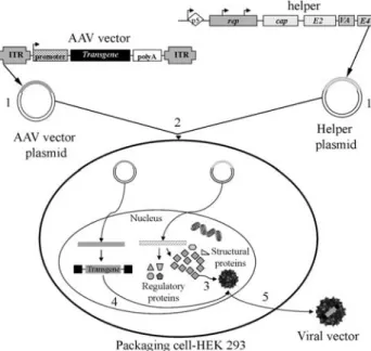
plasmid (with rep and cap genes) and a helper plasmid (containing only the Ad5 genesE2a,E4 andVA RNA), constitutes a promising alternative for the potentialization of AAV vectors in gene therapy. More recently, efficient systems with only two plasmids - a rAAV vector plasmid and the helper containing the AAV and Ad genes required for package and replication - have been described (Zolotukhinet al., 2002) (Figure 1C). In all cases, the cell packaging line used is HEK293, whose genome contains the AdE1gene (Figure 2).
Despite the currently great number of available re-combinant AAV (rAAV) serotypes and variants, several bi-ological barriers appear to limit the effectiveness of rAAVs for gene therapy. Understanding the fundamental basis of these barriers has aided in establishing methods to improve the efficiency of rAAV-mediated gene delivery. These bio-logical barriers are inherent to the AAV life cycle and to its infectious process, as already described above (Figure 3) (Dinget al., 2005). Each intracellular stage required for successful AAV infection constitutes a barrier for rAAV cell transduction. AAV entry pathways probably determine the routes by which the virus crosses the cell cytoplasm to enter into the nucleus. Consequently, viral receptors and coreceptors involved in endocytosis and movement of AAV through the endosomal compartment should be the focus of intracellular trafficking studies.
AAV-based vectors gene therapy applications
The establishment of the first infectious clone of AAV serotype 2 (AAV2) in 1982 (Samulskiet al., 1982) and the pioneering work on the successful cloning of AAV, also in the early 1980s, laid the foundation of recombinant AAV vectors capable of expressing foreign genes in mam-malian cells (Laughlin et al., 1983; Hermonat and Muzyczka, 1984). Since then AAV2 vectors have rapidly gained popularity in gene therapy applications, due to their unique and desirable biological features (reviewed by Cou-ra and Nardi, 2007). This class of viruses has adapted to co-exist with mammalian hosts in a manner that allows for long-term persistence without any detectable deleterious effect on the host. Despite the deceptively simple structure of AAV, this virus is able to use its nonstructural proteins to facilitate replication as a satellite of other DNA viruses dur-ing its productive phase, as well as to establish stable inte-grated and episomal forms during its latent phase (Bernset al., 1975; Berns, 1990; Berns and Linden, 1995). There-fore, the nonpathogenic and persistent long-term nature of AAV infection combined with its wide range of infectivity has made this virus an important candidate as a therapeutic gene transfer vector.
Recombinant adeno-associated viral (rAAV) vectors have rapidly advanced to the forefront of gene therapy in the past decade, since they have been successfully over-coming gene therapy challenges such as transgene mainte-nance, security, host immune response and target diseases, and meeting the desirable vector system features of high level of safety combined with clinical efficacy and
versatil-ity in terms of potential applications. Consequently, despite the already existing limitations and concerns to be resolved, rAAV are increasingly becoming the vectors of choice for a wide range of gene therapy approaches.
Until recently, the better studied and the most fre-quently used AAV serotype for gene therapy was AAV2. Other new serotypes, however, are gaining more and more popularity. Recombinant AAV2 vectors have been tested in preclinical studies for a variety of diseases such as genetic diseases (hemophilia,µ1-anti-trypsin deficiency, cystic fi-brosis, Duchenne muscular dystrophy and others) (Hildin-ger and Auricchio, 2004), tumors (Nget al., 2007), neuro-logical disorders (Paternaet al., 2007), ocular (Martinet al., 2002; Smithet al., 2005) and cardiovascular diseases (Jazwaet al., 2007; Liuet al., 2006; Shenoudaet al., 2006), chronic disorders (Idenoet al., 2007; Blenmanet al., 2006; Sandalonet al., 2007) and others.
Several clinical trials have already been performed for various diseases, especially monogenic diseases and tu-mors. Currently, there are more than 20 open clinical trials evaluating the use of AAV vectors for genetic and acquired diseases (Pfeifer and Verma, 2001; Worgall, 2005; Clinical trial database - http://www.wiley.com.uk/genetherapy/ clinical/).
Future directions
Since its isolation, the biological properties of the adeno-associated virus have been increasingly understood, improving our ability to manipulate and use it as a safe and efficient gene therapy vector of wide spectrum. AAV-based vectors seem to bypass the main gene therapy barri-ers, such as long-term and stable transgene expression in many tissues, safety, broad range of target diseases and lack of immunogenicity and pathogenicity. However, although some obstacles have already been overcome, others are ris-ing and need to be surpassed now, and research advances will certainly bring more challenges in the near future.
Acknowledgments
This work was supported by Conselho Nacional de Desenvolvimento Científico e Tecnológico (CNPq), through the program Instituto do Milênio - Rede de Terapia Gênica.
References
Arbetman AE, Lochrie M, Zhou S, Wellman J, Scallan C, Do-roudchi MM, Radley B, Patarroyo-White S, Liu T, Smith P
et al.(2005) Novel caprine adeno-associated virus (AAV) capsid (AAVGo1) is closely related to the primate AAV-5 and has unique tropism and neutralization properties. J Virol 79:15238-15245.
Atchison RW, Casto BC and Hammon M (1965) Adenovirus-associated defective virus particles. Science 149:754-756. Figure 3- Course of AAV productive infection. Scheme showing the
Bantel-Schaal U and zur Hausen H (1984) Characterization of the DNA of a defective human parvovirus isolated from a geni-tal site. Virology 134:52-63.
Bartlett JS, Wilcher R and Samulski RJ (2000) Infectious entry pathway of adeno-associated virus and adeno-associated vi-rus vectors. J Virol 74:2777-2785.
Berns KI, Pinkerton TC, Thomas GF and Hoggan MD (1975) De-tection of adeno-associated virus (AAV)-specific nucleotide sequences in DNA isolated from latently infected Detroit 6 cells. Virology 68:556-560.
Berns K (1990) Parvovirus replication. Microbiol Rev 54:316-329.
Berns KI and Adler S (1972) Separation of two types of adeno-associated virus particles containing complementary polynucleotide chains. J Virol 9:394-396.
Berns KI and Linden RM (1995) The cryptic life style of adeno-associated virus. Bioessays 17:237-245.
Berns KI and Giraud C (1996) Biology of adeno-associated virus. Curr Top Microbiol Immunol 218:1-23.
Blacklow NR, Hoggan MD and Rowe WP (1968) Serologic edence for human infection with adenovirus-associated vi-ruses. J Natl Cancer Inst 40:319-327.
Blaese RM, Culver KC, Miller AD, Carter CS, Fleisher T, Sheare G, Chang L, Chiang Y, Tolstoshev P, Greenblatt JJet al.
(1995) T lymphocyte-directed gene therapy for ADA-SCID: Initial trial results after 4 years. Science 270:475-480. Bleker S, Sonntag F and Kleinschmidt JA (2005) Mutational anal-ysis of narrow pores at the fivefold symmetry axes of adeno-associated virus type 2 capsids reveals a dual role in genome packaging and activation of phospholipase A2 ac-tivity. J Virol 79:2528-2540.
Blenman KR, Duan B, Xu Z, Wan S, Atkinson MA, Flotte TR, Croker BP and Morel L (2006) IL-10 regulation of lupus in the NZM2410 murine model. Lab Invest 86:1136-1148. Bossis I and Chiorini JA (2003) Cloning of an avian
adeno-associated virus (AAV) and generation of recombinant AAV particles. J Virol 77:6799-6810.
Burger C, Gorbatyuk OS, Velardo MJ, Peden CS, Williams P, Zolotukhin S, Reier PJ, Mandel RJ and Muzyczka N (2004) Recombinant AAV viral vectors pseudotyped with viral capsids from serotypes 1, 2, and 5 display differential effi-ciency and cell tropism after delivery to different regions of the central nervous system. Mol Ther 10:302-317.
Campbell EM and Hope TJ (2005) Gene therapy progress and prospects: Viral trafficking during infection. Gene Ther 12:1353-1359.
Chejanovsky N and Carter BJ (1989) Mutagenesis of an AUG codon in the adeno-associated virus rep gene: Effects on vi-ral DNA replication. Virology 173:120-128.
Collaco RF, Kalman-Maltese V, Smith AD, Dignam JD and Trempe JP (2003) A biochemical characterization of the adeno-associated virus rep40 helicase. J Biol Chem 278:34011-34017.
Coura RD and Nardi NB (2007) The state of the art of adeno-associated virus-based vectors in gene therapy. Virol J 4:99. Di Pasquale G, Davidson BL, Stein CS, Martins I, Scudiero D,
Monks A and Chiorini JA (2003) Identification of PDGFR as a receptor for AAV-5 transduction. Nat Med 9:1306-1312.
Ding W, Zhang L, Yan Z and Engelhardt JF (2005) Intracellular trafficking of adeno-associated viral vectors. Gene Ther 12:873-880.
Duan D, Li Q, Kao AW, Yue Y, Pessin JE and Engelhardt JF (1999) Dynamin is required for recombinant adeno-associated virus type 2 infection. J Virol 73:10371-10376. Duan D, Yue Y, Yan Z, Yang J and Engelhardt JF (2000)
Endo-somal processing limits gene transfer to polarized airway epithelia by adeno-associated virus. J Clin Invest 105:1573-1587.
Dutta SK (1975) Isolation and characterization of an adenovirus and isolation of its adenovirus-associated virus in cell cul-ture from foals with respiratory tract disease. Am J Vet Res 36:247-250.
Edelstein ML, Abedi MR, Wixon J and Edelstein RM (2004) Gene therapy clinical trials worldwide 1989-2004-an over-view. J Gene Med 6:597-602.
El-Aneed A (2004) Current strategies in cancer gene therapy. Eur J Pharmacol 498:1-8.
Erles K, Sebokova P and Schlehofer JR (1999) Update on the prevalence of serum antibodies (IgG and IgM) to adeno-associated virus (AAV). J Med Virol 59:406-411.
Farkas SL, Zadori Z, Benko M, Essbauer S, Harrach B and Tijssen P (2004) A parvovirus isolated from royal python (Python regius) is a member of the genus Dependovirus. J Gen Virol 85:555-561.
Gao G, Alvira MR, Wang L, Calcedo R, Johnston J and Wilson JM (2002) Novel adeno-associated viruses from rhesus monkeys as vectors for human gene therapy. Proc Natl Acad Sci USA 99:11854-11859.
Gao G, Vandenberghe LH, Alvira MR, Lu Y, Calcedo R, Zhou X and Wilson JM (2004) Clades of adeno-associated viruses are widely disseminated in human tissues. J Virol 78:6381-6388.
Gao G, Vandenberghe LH and Wilson JM (2005) New recombi-nant serotypes of AAV vectors. Curr Gene Ther 5:285-297. Gao X, Kim K-S and Liu D (2007) Nonviral gene delivery: What
we know and what is next. AAPS J 9:E92-E104.
Georg-Fries B, Biederlack S, Wolf J and zur Hausen H (1984) Analysis of proteins, helper dependence, and seroepide-miology of a new human parvovirus. Virology 134:64-71. Girod A, Wobus CE, Zádori Z, Ried M, Leike K, Tijssen P,
Kleinschmidt JA and Hallek M (2002) The VP1 capsid pro-tein of adeno-associated virus type 2 is carrying a phospho-lipase A2 domain required for virus infectivity. J Gen Virol 83:973-978.
Goverdhana S, Puntel M, Xiong W, Zirger JM, Barcia C, Curtin JF, Soffer EB, Mondkar S, King GD, Hu J et al.(2005) Regulatable gene expression systems for gene therapy appli-cations: Progress and future challenges. Mol Ther 12:189-211.
Grimm D and Kay MA (2003) From virus evolution to vector rev-olution: Use of naturally occurring serotypes of adeno-associated virus (AAV) as novel vectors for human gene therapy. Curr Gene Ther 3:281-304.
Halbert CL, Rutledge EA, Allen JM, Russell DW and Miller AD (2000) Repeat transduction in the mouse lung by using adeno-associated virus vectors with different serotypes. J Virol 74:1524-1532.
pro-cessing enhances transduction efficiency in murine fibro-blasts. J Virol 75:4080-4090.
Hermonat PL and Muzyczka N (1984) Use of adeno-associated virus as a mammalian DNA cloning vector: Transduction of neomycin resistance into mammalian tissue cultured cells. Proc Natl Acad Sci USA 81:6466-6470.
Hildinger M and Auricchio A (2004) Advances in AAV-mediated gene transfer for the treatment of inherited disorders. Eur J Hum Genet 12:263-271.
Hoggan MD, Blacklow NR and Rowe WP (1966) Studies of small DNA viruses found in various adenovirus preparations: Phy-sical, biological and immunological characteristics. Proc Natl Acad Sci USA 55:1467-1472.
Ideno J, Mizukami H, Kakehashi A, Saito Y, Okada T, Urabe M, Kume A, Kuroki M, Kawakami M, Ishibashi Set al.(2007) Prevention of diabetic retinopathy by intraocular soluble flt-1 gene transfer in a spontaneously diabetic rat model. Int J Mol Med19:75-79.
Im DS and Muzyczka N (1989) Factors that bind to adeno-associated virus terminal repeats. J Virol 63:3095-3104. Im DS and Muzyczka N (1990) The AAV origin binding protein
Rep68 is an ATP-dependent site-specific endonuclease with DNA helicase activity. Cell 61:447-457.
Jacobson ER, Kopit W, Kennedy FA and Funk RS (1996) Coin-fection of a bearded dragon, Pogona vitticeps, with adeno-virus- and dependoadeno-virus-like viruses. Vet Pathol 33:343-346.
Jazwa A, Jozkowicz A and Dulak J (2007) New vectors and strate-gies for cardiovascular gene therapy. Curr Gene Ther 7:7-23.
Kaludov N, Brown KE, Walters RW, Zabner J and Chiorini JA (2001) Adeno-associated virus serotype 4 (AAV4) and AAV5 both require sialic acid binding for hemagglutination and efficient transduction but differ in sialic acid linkage specificity. J Virol 75:6884-6893.
Kashiwakura Y, Tamayose K, Iwabuchi K, Hirai Y, Shimada T, Matsumoto K, Nakamura T, Watanabe M, Oshimi K and Daida H (2005) Hepatocyte growth factor receptor is a coreceptor for adeno-associated virus type 2 infection. J Virol 79:609-614.
King JA, Dubielzig R, Grimm D and Kleinschmidt JA (2001) DNA helicasemediated packaging of adeno-associated virus type 2 genomes into preformed capsids. EMBO J 20:3282-3291.
Koczot FJ, Carter BJ, Garon CF and Rose JA (1973) Self-complementarity of terminal sequences within plus or minus strands of adenovirus-associated virus DNA. Proc Natl Acad Sci USA 70:215-219.
Kotin RM, Siniscalco M, Samulski RJ, Zhu XD, Hunter L, Lau-ghlin CA, McLauLau-ghlin S, Muzyczka N, Rocchi M and Berns KI (1990) Site-specific integration by adeno-associated vi-rus. Proc Natl Acad Sci USA 87:2211-2215.
Kotin RM, Menninger JC, Ward DC and Berns KI (1991) Map-ping and direct visualization of a region-specific viral DNA integration site on chromosome 19q13-qter. Genomics 10:831-834.
Laughlin CA, Tratschin JD, Coon H and Carter BJ (1983) Cloning of infectious adeno-associated virus genomes in bacterial plasmids. Gene 23:65-73.
Linden RM, Ward P, Giraud C, Winocour E and Berns KI (1996) Site-specific integration by adeno-associated virus. Proc Natl Acad Sci USA 93:11288-11294.
Liu Y, Li D, Chen J, Xie J, Bandyopadhyay S, Zhang D, Nemar-kommula AR, Liu H, Mehta JL and Hermonat PL (2006) In-hibition of atherogenesis in LDLR knockout mice by sys-temic delivery of adeno-associated virus type 2-hIL-10. Atherosclerosis 188:19-27.
Martin KR, Klein RL and Quigley HA (2002) Gene delivery to the eye using adeno-associated viral vectors. Methods 28:267-275.
McCarty DM, Young Jr SM and Samulski RJ (2004) Integration of adeno-associated virus (AAV) and recombinant AAV vectors. Annu Rev Genet 38:819-845.
Mori S, Wang L, Takeuchi T and Kanda T (2004) Two novel adeno-associated viruses from cynomolgus monkey: Pseudotyping characterization of capsid protein. Virology 330:375-383.
Nakai H, Montini E, Fuess S, Storm TA, Grompe M and Kay MA (2003) AAV serotype 2 vectors preferentially integrate into active genes in mice. Nat Genet 34:297-302.
Ng SS, Gao Y, Chau DH, Li GH, Lai LH, Huang PT, Huang CF, Huang JJ, Chen YC, Kung HFet al.(2007) A novel glio-blastoma cancer gene therapy using AAV-mediated long-term expression of human TERT C-long-terminal polypeptide. Cancer Gene Ther 14:561-572.
Ni T-H, McDonald WF, Zolotukhin I, Melendy T, Waga S, Stillman B and Muzyczka N (1998) Cellular proteins re-quired for adeno-associated virus DNA replication in the ab-sence of adenovirus coinfection. J Virol 72:2777-2787. Olson EJ, Haskell SR, Frank RK, Lehmkuhl HD, Hobbs LA,
Warg JV, Landgraf JG and Wunschmann A (2004) Isolation of an adenovirus and an adeno-associated virus from goat kids with enteritis. J Vet Diagn Invest 16:461-464. Parks WP, Green M, Pina M and Melnick JL (1967)
Physi-cochemical characterization of adeno-associated satellite vi-rus type 4 and its nucleic acid. J Virol 5:980-987.
Parks WP, Boucger DW, Melnich JL, Taber LH and Yow MD (1970) Seroepidemiological and ecological studies of the adeno-associated satellite viruses. Infect Immun 2:716-722. Paterna JC, Leng A, Weber E, Feldon J and Büeler H (2007) DJ-1
and Parkin modulate dopamine-dependent behavior and in-hibit MPTP-induced nigral dopamine neuron loss in mice. Mol Ther 15:698-704.
Peden CS, Burger C, Muzyczka N and Mandel RJ (2004) Circu-lating anti-wildtype adeno-associated virus type 2 (AAV2) antibodies inhibit recombinant AAV2 (rAAV2)-mediated, but not rAAV5-mediated, gene transfer in the brain. J Virol 78:6344-6359.
Pereira DJ, McCarty DM and Muzyczka N (1997) The adeno-associated virus (AAV) Rep protein acts as both a repressor and an activator to regulate AAV transcription during a pro-ductive infection. J Virol 71:1079-1088.
Pfeifer A and Verma IM (2001) Gene therapy: Promises and prob-lems. Annu Rev Genomics Hum Genet 2:177-211. Philpott NJ, Gomos J and Falck-Pedersen E (2004) Transgene
ex-pression after rep-mediated site-specific integration into chromosome 19. Hum Gene Ther 15:47-61.
co-receptor for infection by adeno-associated virus 2. Nat Med 5:71-77.
Rosenberg SA, Aebersold P, Cornetta K, Kasid A, Morgan RA, Moen R, Karson EM, Lotze MT, Yang JC and Topalian SL (1990) Gene transfer into humans - Immunotherapy of pa-tients with advanced melanoma, using tumor-infiltrating lymphocytes modified by retroviral gene transduction. N Engl J Med 323:570-578.
Rubanyi GM (2001) The future of human gene therapy. Mol As-pects Med 22:113-142.
Russell DW (2003) AAV loves an active genome. Nat Genet 34:241-242.
Rutledge EA, Halbert CL and Russell DW (1998) Infectious clones and vectors derived from adeno-associated virus (AAV) serotypes other than AAV type 2. J Virol 72:309-319.
Salani B, Damonte P, Zingone A, Barbieri O, Chou JY, D’Costa J, Arya SK, Eva A and Varesio L (2005) Newborn liver gene transfer by an HIV-2-based lentiviral vector. Gene Ther 12:803-814.
Samulski RJ, Berns KI, Tan M and Muzyczka N (1982) Cloning of infectious adeno-associated virus into pBR322: Rescue of intact virus from the recombinant plasmid in human cells. Proc Natl Acad Sci USA 79:2077-2081.
Samulski RJ, Zhu X, Xiao X, Brook JD, Housman DE, Epstein N and Hunter LA (1991) Targeted integration of adeno-associated virus (AAV) into human chromosome 19. EMBO J 10:3941-3950.
Sandalon Z, Bruckheimer EM, Lustig KH and Burstein H (2007) Long-term suppression of experimental arthritis following intramuscular administration of a pseudotyped AAV2/1-TNFR:Fc vector. Mol Ther 15:264-269.
Schmidt M, Katano H, Bossis I and Chiorini JA (2004) Cloning and characterization of a bovine adeno-associated virus. J Virol 78:6509-6516.
Schmidt M, Grot E, Cervenka P, Wainer S, Buck C and Chiorini JA (2006) Identification and characterization of novel adeno-associated virus isolates in ATCC virus stocks. J Virol 80:5082-5085.
Seisenberger G, Ried MU, Endress T, Buning H, Hallek M and Brauchle C (2001) Real-time single-molecule imaging of the infection pathway of an adenoassociated virus. Science 294:1929-1932.
Shenouda SM, Johns C, Kintsurashvili E, Gavras I and Gavras H (2006) Long-term inhibition of the central alpha(2B)-adre-nergic receptor gene via recombinant AAV-delivered anti-sense in hypertensive rats. Am J Hypertens 19:1135-1143. Smith KR (2003) Gene therapy: Theoretical and bioethical
con-cepts. Arch Med Res 34:247-268.
Smith RH and Kotin RM (1998) The Rep52 gene product of adeno-associated virus is a DNA helicase with 3’-to-5’ po-larity. J Virol 72:4874-4881.
Smith JR, Verwaerde C, Rolling F, Naud MC, Delanoye A, Thillaye-Goldenberg B, Apparailly F and De Kozak Y (2005) Tetracycline-inducible viral interleukin-10 intrao-cular gene transfer, using adeno-associated virus in
experi-mental autoimmune uveoretinitis. Hum Gene Ther 16:1037-1046.
Stender S, Murphy M, O’Brien T, Stengaard C, Ulrich-Vinther M, Soballe K and Barry F (2007) Adeno-associated viral vector transduction of human mesenchymal stem cells. Eur Cell Mater 13:93-99.
Summerford C and Samulski RJ (1998) Membrane-associated heparan sulfate proteoglycan is a receptor for adeno-associa-ted virus type 2 virions. J Virol 72:1438-1445.
Summerford C, Bartlett JS and Samulski RJ (1999) AlphaVbeta5 integrin: A coreceptors for adeno-associated virus type 2 in-fection. Nat Med 5:78-82.
Tal J (2000) Adeno-associated virus-based vectors in gene ther-apy. J Biomed Sci 7:279-291.
Verma IM and Somia N (1997) Gene therapy - Promises, prob-lems and prospects. Nature 389:239-242.
Vincent KA, Piraino ST and Wadsworth SC (1997) Analysis of recombinant adeno-associated virus packaging and require-ments for rep and cap gene products. J Virol 71:1897-1905. Wang H and Lieber A (2006) A helper-dependent
capsid-mo-dified adenovirus vector expressing adeno-associated virus rep78 mediates site-specific integration of a 27-kilobase transgene cassette. J Virol 80:11699-11709.
Wiley (2007) Gene Therapy Clinical Trials Worldwide. http:// www.wiley.co.uk/genetherapy/clinical/ (June, 2007). Wistuba A, Kern A, Weger S, Grimm D and Kleinschmidt J
(1997) Subcellular compartmentalization of adeno-associated virus type 2 assembly. J Virol 71:1341-1352. Worgall S (2005) A realistic chance for gene therapy in the near
future. Pediatr Nephrol 20:118-124.
Wu Z, Asokan A and Samulski RJ (2006) Adeno-associated virus serotypes: Vector toolkit for human gene therapy. Mol Ther 14:316-327.
Xiao X, Li J and Samulski RJ (1998) Production of high-titer re-combinant adenoassociated virus vectors in the absence of helper adenovirus. J Virol 72:2224-2232.
Xiao W, Chirmule N, Berta SC, McCullough B, Gao G and Wil-son JM (1999) Gene therapy vectors based on adeno-associated virus type 1. J Virol 73:3994-4003.
Xiao W, Warrington Jr KH, Hearing P, Hughes J and Muzyczka N (2002) Adenovirus-facilitated nuclear translocation of adeno-associated virus type 2. J Virol 76:11505-11517. Zádori Z, Szelei J, Lacoste M-C, Li Y, Gariépy S, Raymond P,
Allaire M, Nabi IR and Tijssen P (2001) A viral phospho-lipase A2 is required for parvovirus infectivity. Dev Cell 1:291-302.
Zolotukhin S, Potter M, Zolotukhin I, Sakai Y, Loiler S, Fraites TJ Jr, Chiodo VA, Phillipsberg T, Muzyczka N, Hauswirth WWet al.(2002) Production and purification of serotype 1, 2, and 5 recombinant adeno-associated viral vectors. Methods 28:158-67.
Zubler RH (2006)Ex vivoexpansion of haematopoietic stem cells and gene therapy development. Swiss Med Wkly 136:795-799.