Studies on the mosquito immune response:
Effect of antimalarial drugs and
Plasmodium
sporozoites
Universidade Nova de Lisboa
Instituto de Higiene e Medicina Tropical
Centro de Malária e outras Doenças Tropicais, LA
Studies on the mosquito immune response:
Effect of antimalarial drugs and
Plasmodium sporozoites
Susana Filipa Garcia Ramos
A dissertation submmited to obtain a Doctor of
Philosophy degreee in Biomedical Sciences,
Parasitology speciality
Supervisor: Doctor Henrique Silveira
Instituto de Higiene e Medicina Tropical, Centro de Malária e outras Doenças Tropicais
Co-Supervisor: Doctor Jean-Marc Reichhart
Institut de Biologie Moléculaire et Cellulaire,
Réponse immunitaire et développment chez les insects
This work was performed at the Centro de Malária e outras Doenças Tropicais LA (CMDT), in the Instituto de Higiene e Medicina Tropical (IHMT), Lisbon, at the Institut de Biologie Moléculaire et Cellulaire (IBMC), Strasbourg, and also at the Division of Cell and Molecular Biology, Imperial College, London. This work was financed by a PhD grant (SFRH/BD/12210/2003) awarded by the Fundação para a Ciência e Tecnologia.
This work would not have been completed without the help and support given to me by the people I had the chance of having by my side, both professionally and personally. To these people I here express my truthful acknowledgments.
First I would like to thank to Prof. Dr. Henrique Silveira, my supervisor, for taking me as a student and giving me the opportunity to perform this work. I thank your supervision, discussion and ideas that helped me build this work. I thank you for trusting and for your endless support. Thank you for making these years of learning so rewarding.
To Prof. Dr. Jean-Marc Reichhart, from the IBMC, Strasbourg, for accepting me as a student and to co-supervise this project. Thank you for the support and the opportunity you conceded me. Thank you for teaching me about flies. Thank you for providing me the chance to perform the proteomic analysis vital to the conclusion of this work. The time spent in your lab was a great learning and growing experience, both professionally and personally, for which I will always be grateful.
To Prof. Dr. Virgílio E. do Rosário, Director of the Unidade de Malária of IHMT, and member of my tutorial commission, for accepting me in his lab, for his availability and for always providing valuable suggestions for the development of this work.
work.
To Laurence Sabatier, Philippe Hamman and Christelle Guilier, from IBMC/IGBMC, Strasbourg, for teaching me and allowing me to perform the proteomic analysis vital to this work.
To Dr. Hans Michael Muller, for kindly providing me the mosquito hemocyte-like cell lines and the anti-PO2 antibodies used in this work.
To Fátima Nogueira, my devil’s advocat, for the companionship and the scientific discussions we engaged on. You were very important to me in this step of my life. Thank you for being there all the time, for sharing late hours at the lab, for teaching me and for all the discussions we had (and all the caipirinhas we shared while doing it), making me more critical about my work.
To Catarina Alves, for her friendship and for maintaining the An. sthephensi and An.
gambiae insectaries and for taking care of our mosquitoes, allowing me to perform my
experiences.
To my colleagues and friends Rute Félix, Patrícia Machado, Cristina Mendes, Ana Gabriel, Luís Filipe Lopes, José Luís Vicente, Patrícia Abrantes, Isabel Ferreira, Cláudia Marques, Vera Pinto e Sónia Cardoso for the everyday friendship and for helping me in my work. To all, thank you for making my days in the lab such a wonderful time, full of joy and companionship.
To Akira Goto, Alexey Matskevich, Nadege Pelte, Stephan Wyder and Rie Tajima, my friends, for making my staying in Strasbourg wonderful and unforgettable. Thank you all for all you taught me, for accompanying me all the time, for your availability and care. Thank you for the lunch times, the walks, the friendship and all the moments we shared.
visiting me and keep me company when I was away.
To Kika, for the company during the time I wrote this thesis. Thank you for not letting me feel alone.
To Bernardo Raposo, Rita Stilwell and Paulo Ramunni, my dearest friends, for being a lighthouse in my life. Thank you for the light you shine and for teaching me how to shine mine.
To the best parents, my parents, without whom I could never have done this. Thank you for your unwavering faith in me, for your love, support and care. I can only hope I make you proud.
To David, for the love we shared. Thank you for believing in me, for giving me strength, for being my strength, for not letting me give up, for pushing me to the finish line. Thank you for being there every moment we had, for the moments when I was confident and for the ones when I stopped believing. You never did… thank you.
Este trabalho pretende contribuir para o conhecimento geral da resposta imunológica do mosquito ao parasita da malária, uma vez que a elucidação das interacções entre vector e parasita poderão facilitar o desenvolvimento de medidas eficientes para bloquear a transmissão. As experiências realizadas neste trabalho incluíram o uso de
Drosophila melanogaster como modelo de estudo das respostas imunológicas do
mosquito e a avaliação do impacto da presença de esporozoítos de Plasmodium na hemolinfa do mosquito através da determinação de alterações no número de hemócitos, activação da reacção de melanização e do padrão de expressão de proteínas na hemolinfa do mosquito.
O fármaco antimalárico cloroquina promove a transmissão no mosquito e tem sido relacionado com a expressão diferencial de péptidos antimicrobianos no mosquito. Para avaliar o efeito da cloroquina na sua produção usámos o modelo Drosophila, uma vez que a expressão e síntese de péptidos antimicrobianos na mosca está bem caracterizada, assim como as vias de sinalização da resposta imunológica. Os resultados deste trabalho não conseguiram provar algum efeito do fármaco na expressão e/ou síntese de péptidos antimicrobianos de Drosophila. O tratamento com cloroquina in vivo não afectou as vias de sinalização Toll e Imd, avaliado pela expressão de drosomicina e diptericina em moscas infectadas. Experiências in vitro em que se utilizaram linhas celulares derivadas de hemócitos de moscas produziram os mesmos resultados para a síntese de Drosomicina e Atacina. Experiências de sobrevivência de moscas infectadas e tratadas com cloroquina também não evidenciaram qualquer efeito do fármaco na resposta imunitária de Drosophila. Como este fármaco antimalárico tem um efeito conhecido na resposta imune do mosquito, propomos que a cloroquina tenha uma acção sobre moléculas específicas dos mosquito ou sobre diferentes vias de activação da sinalização que possam estar presentes apenas no mosquito. Por outro lado, a informação conhecida acerca do efeito da cloroquina na imunidade foi obtida após tratamento de humanos, ratinhos ou linhas celulares de mamíferos, implicando a metabolização do fármaco. Como tal, não é claro se o efeito observado resulta da acção do própria fármaco ou de um metabolito específico.
Neste trabalho pretendeu-se também determinar as respostas do mosquito aos esporozoítos de Plasmodium na hemolinfa, uma vez que nesta fase da infecção no mosquito o parasita sofre uma grande redução no seu número. No hemocélio do
o oocisto rompe e os esporozoítos são libertados, esta proteína pode ser reconhecida pelas moléculas de reconhecimento presentes na hemolinfa levando à activação de respostas imunes. A activação de respostas imunitárias celulares contra os esporozoítos foi testada com base na determinação de variação do número de hemócitos quando estimulados com a proteína circumsporozoítica de P. falciparum. Apenas uma das doses (5ng) de proteína utilizadas para estimular linhas celulares de hemóctios causou uma redução significativa no números de hemócitos. Isto pode ser um reflexo de uma cinética de divisão celular mais lenta ou de destruição celular, apoptose, que poderia ser despoletada pela fagocitose de parasitas, por exemplo. Não foi possível obter uma resposta correlacionada com a dose usada para estimulação. No entanto, os hemócitos do mosquito parecem reconhcer a proteína do parasita e responder à sua presença.
A activação da reacção de melanização durante a invasão da hemolinfa por esporozoítos foi testada, através da determinação da activação da enzima profenoloxidase e da actividade da fenoloxidase. Verificou-se que a actividade enzimática da fenoloxidase varia com o tempo em mosquitos submetidos a uma refeição sanguínea não infectante. A infecção por P. berhgei não pareceu impor variações na actividade da fenoloxidase. Diferenças subtis foram observadas aos dias 9, 12 e 15 pós-infecção, sendo a actividade enzimática mais elevada em mosquitos infectados. Os esporozoítos foram detectados na hemolinfa de mosquitos a partir do dia 9 pós-infecção, indicando que o parasita pode induzir um aumento subtil na activação da melanização. A actividade da fenoloxidase parece ser mantida constitutivamente num nível baixo, mesmo em mosquitos não infectados, o que pode explicar que apenas pequenas diferenças sejam observadas em mosquitos infectados. Injecções da proteína circumsporozoítica de P. falciparum em mosquitos não revelaram indução da actividade da enzima fenoloxidase. Apesar de não ter sido possível demonstrar conclusivamente a melanização de esporozoítos na hemolinfa, experiências de inibição da fenoloxidase mostraram que a actividade desta enzima é necessária para controlar o número de esporozoítos na hemolinfa e nas glândulas salivares.
A hemolinfa é extremamente rica em proteínas, e conhecida por albergar a maior parte das moléculas do sistema imunológico necessárias ao reconhecimento, sinalização e à resposta efectora. Como tal, de modo a caracterizar o proteoma da hemolinfa durante a infecção por P. berhgei ao dia 13 pós-infecção, usámos uma
poderão estar envolvidas em processos fisiológicos como metabolismo de ácidos gordos, glicólise e transporte de iões. Estes resultados indicam que o parasita impõe alterações no metabolismo do mosquito, quer directamente quer levando o mosquito a alterar o seu próprio metabolismo como forma de conter a infecção. De facto, não há evidência se as alterações observadas são danosas ou necessárias para o desenvolvimento do parasita. No entanto, os nossos resultados sugerem que mecanismos fisiológicos do mosquito podem ter um papel na resposta imune. Um dado interessante obtido neste trabalho foi a inexistência de correlação entre a regulação a nível proteico e a nível do RNA na hemolinfa. Isto pode derivar de uma janela de tempo diferente entre expressão génica e síntese proteica, uma vez que as amostras foram recolhidas ao mesmo tempo, ou pode reflectir um fonte diferente de RNA e proteína. O RNA amplificado para avaliar a expressão génica era originário dos hemócitos presentes na hemolinfa, enquanto que as proteínas podem ter sido produzidas quer pelos hemócitos quer pelo corpo gordo, que sintetiza a maior parte das moléculas imunes que são secretadas para a hemolinfa. No entanto, é importante tem em atenção que a informação resultante da análise de expressão génica ter de ser avaliada cuidadosamente, pois pode não ter uma regulação correspondente ao nível da proteína. A biosíntese de eicosanóides teve dois impactos distintos e opostos no desenvolvimento do parasita, promovendo e bloqueando a transmissão. Os eicosanóides parecem ser importantes para o desenvolvimento do parasita numa fase da infecção em que os esporozoítos se desenvolvem nos oocistos, enquanto que numa fasemais tardia, estas moléculas parecem ser importantes para controlar o número de parasitas na hemolinfa. Os nossos resultados sugerem que o parasita possa imunosuprimir o mosquito.
A resposta do mosquito ao Plasmodium parece ser muito complexa, envolvendo acções de ambos os organismos. Para responderà invasão da hemolinfa pelos esporozoítos, o mosquito parece depender de diferentes mecanismos, como a fagocitose e a melanização. Para além destes, moléculas envolvidas em processos fisiológicos do mosquito são também afectadas pela infecção. Os nossos resultados sugerem que a resposta imunológica do mosquito possa envolver mecanismos para além daqueles que são tradicionalmente relacionados com a imunidade, como a biosíntese de eicosanóides. Verificámos também que pode não existir correlação entre a expressão génica e a síntese de proteínas, e como tal, a resposta imune deveria ser analisada em primeiro lugar por uma abordagem proteómica.
This work aimed at contributing to the general knowledge of the mosquito immune responses to the malaria parasite, in hope that the elucidation of vector/parasite interactions will facilitate the development of effective transmission blocking measures. Experiments performed here include the use of Drosophila melanogaster as a model for immunity studies in the mosquito and the evaluation of Plasmodium sporozoites presence in mosquito hemolymph impact on hemocyte numbers, melanization reaction responses and protein expression pattern.
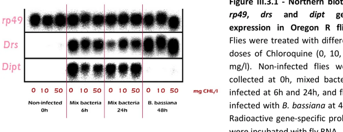
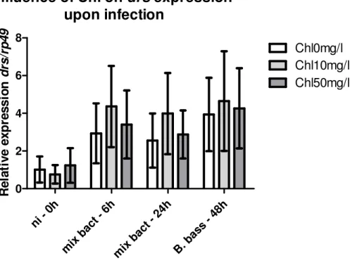
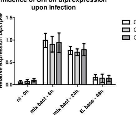
Chloroquine promotes malaria transmission in mosquito and it has been linked to differential AMP gene expression in mosquitoes. As Drosophila AMP expression and synthesis is well understood and as we have a good knowledge about immune signaling pathways, we chose this model to evaluate chloroquine effect on AMP production upon infection. Our results failed to show any drug effect on Drosophila AMP expression and/or synthesis. Drug treatment in vivo did not affect either the Toll or Imd immune signaling pathways, as shown when accessing drosomycin and
diptericin expression in infected flies, and in vitro experiments using hemocyte-like cell
lines produced the same results for Drosomycin and Attacin synthesis. Survival experiments were also performed in drug treated flies and failed to indicate any effect. For all the mechanisms tested, chloroquine did not seem to have any effect on
Drosophila immunity. As this antimalarial drug has a known effect on mosquito
immunity we propose that chloroquine may act on particular mosquito immune molecules or on different routes for pathway activation operating in the mosquito. Also, data obtained for chloroquine action on immunity were collected from treatment of humans, mice or mammalian cell-lines, implying that the drug is metabolized. Thus it is not clear if the observed effect results from an action of the drug itself, or from a specific metabolite. This would explain why direct drug feeding to flies would fail to produce an effect on Drosophila immunity.
Another purpose of this work was to determine the mosquito responses to
Plasmodium sporozoites in the hemolymph, as parasite development in the mosquito
suffers a major bottleneck at this stage of infection. Both cellular and humoral responses may be triggered in the mosquito hemocel. Upon development inside the oocysts, sporozoites are covered with a layer of circumsporozoite protein, its major
based on the evaluation of hemocyte number variation upon stimulation with the circumsporozoite protein of P. falciparum. Only one dose (5ng) stimulated hemocyte-like cell lines and led to a significant reduction in cell numbers. This may reflect a slower cell-division kinetics or cell destruction, by apoptosis, following phagocytosis. We failed to show any dose-dependent response. Nevertheless, it seems that the CS protein is recognized by mosquito hemocytes that respond to its presence only in specific conditions.
We also tested for activation of melanization reaction upon sporozoite invasion of the hemolymph by accessing PPO activation and PO activity. PO activity was found to vary over time in blood fed mosquitoes. P. berghei infection did not seem to impose variations in PO activity. Subtle differences were observed at D9, 12 and 15pi, when PO activity was higher in infected mosquitoes. Sporozoites were first detected in the hemolymph at D9pi indicating that parasite recognition may induce subtle increases in melanization activation. PO activity seems to be maintained at a low level even in non-infected mosquitoes. This may explain the fact that no great variations were observed upon infection. Pf-CS protein injections in mosquitoes failed to show PO activity induction. Although we could not conclusively determine sporozoite melanization, PO inhibition experiments showed that its activity is necessary for control of sporozoite load in the hemolymph and salivary glands.
Hemolymph is an extremely protein rich environment, and known to harbor most of the immune molecules necessary for recognition, signaling and effector mechanisms. As such, we used a two dimensional electrophoresis approach coupled with MALDI-TOF mass spectrometry to compare the hemolymph proteome of P. berghei infected and non-infected An. gambiae mosquitoes at D13pi, aiming at the identification of differentially regulated protein in infected mosquitoes. Proteins found to have altered levels in the hemolymph of infected mosquitoes are predicted to be involved in physiological processes such as fatty acid metabolism, aminoacid synthesis, glycolysis and ion transport. This indicated that the parasite imposes alterations in the overall mosquito metabolism, either directly, or secondarly to combat infection. Actually we have no evidence if the alterations observed are harmful or necessary for parasite development. Yet, the results suggest that mechanisms operating in mosquito physiology may have a role on immune responses. An interesting fact is that protein regulation in the hemolymph did not correlate at any level with gene transcription. This may reflect a different time frame between transcription and protein synthesis, as
hemocytes, but also by fat body cells, that synthesize the majority of immune-related molecules secreted into the hemolymph. Nevertheless, it is important to bear in mind that data resulting from gene expression analysis have to be carefully analyzed as it may not indicate direct protein synthesis. Eicosanoid biosynthesis was found to have two distinctive and opposite impacts in parasite development: both promoting and blocking transmission. Eicosanoids seem to be important for parasite biology and development, at a time when sporozoites are developing inside oocysts. At a later stage, these molecules seem to restrain sporozoite infection in the hemolymph. Evidence also point to mosquito immunosuppression by the parasite.
Mosquito responses to Plasmodium seem to be highly complex, involving actions from both organisms. To respond to hemolymph invasion by sporozoites, the mosquito seems to rely on different mechanisms, such as phagocytosis and melanization. Additionally, molecules involved in physiological processes are affected by hemolymph infection. Data obtained by this work suggests that immune responses may include mechanisms other than those traditionally related to immunity, as in the case of eicosanoid biosynthesis. Also, our results indicate that correlation between transcription and protein synthesis is not sure to exist and thus, immune responses should be analyzed by proteomics in a first approach.
2D – Two dimensional
2DE - Two dimensional Electrophoresis AA – Arachidonic Acid
ACN - Acetonitrile
Ae. - Aedes
AMP – Antimicrobial Peptide
An. – Anopheles
APO II/I – Apolipophorin II/I Att – Attacin
B. – Beauveria
BSA – Bovine Serum Albumin CBB – Colloidal Brilliant Blue
cDNA - complementary Deoxyribonucleic Acid CEC – Cecropin CEC1 – Cecropin 1 CEC2 – Cecropin 2 CEC3 – Cecropin 3 CECA – Cecropin A Chl – Chloroquine COX - Ciclooxygenase CSP – Circumsporozoite Protein CTL4 – C-Type Lectin 4
CTLSE – C-type Lectin Selectin
CTRP – Circumsporozoite and TRAP Related Protein Cys - Cysteine D. – Drosophila dCTP – Deoxycytidine Triphosphate DEF1 – Defensin 1 DEF2 – Defensin 2 DEFA – Defensin A DEPC - Diethylpyrocarbinate DEX - Dexamethasone Dipt –Diptericin
DNA - Deoxyribonucleic Acid
dNTP - Deoxyribonucleotide Triphosphate Dome - Domeless Drs – Drosomycin DTT - Dithiothreitol E. - Escherichia EM – Electron Microscopy En. - Enterococcus
EST – Expressed Sequence Tag FA – Fatty Acid
FCS – Fetal Calf Serum FBN9 – Fibronectin 9 FBN23 – Fibronectin 23
GALE5 – Galectin 5 GALE8 – Galectin 8 GAM – Gambicin
GFP – Green Fluorescent Protein GNBP – Gram Negative Binding Protein GNBP1 - Gram Negative Binding Protein 1 GNBP3 – Gram Negative Binding Protein 3 GNBPA1 – Gram Negative Binding Protein A1 GNBPB1 – Gram Negative Binding Protein B1 GNBPB4 - Gram Negative Binding Protein B4 GPI - Glycosylphosphatidylinositol
h - hours
HIV/AIDS – Human Immunodeficiency Virus / Acquired immune deficiency Syndrome HK – Heat-Killed
hpi – hours post-infection HRP – Horseradish Peroxidase IEF – Isoelectric Focusing IFN-γ – Interferon-γ
IGALE20 – Infection-responsive Galactose Lectin 20
IMCR14 - Immune-responsive alpha-macroglobulin and complement C3- related protein 14
IMD – Immunodeficiency IN – Indomethacin
IPG – Immobilized pH gradient
KD – Knock-Down
L-DOPA – 3,4-dihydroxy-L-phenylalanine LOX - Lipooxygenase
LPS – Lipopolysaccharide
LRIM – Leucine-rich Repeat Immune Protein LRIM1 – Leucine-rich Repeat Immune Protein1 LRR – Leucine Rich Repeats
M. – Micrococcus Ma. - Manduca
MALDI-TOF MS – Matrix Assisted Laser Desorption Ionization – Time Of Flight Mass Spectrometry.
MAPK – MAP Kinase
MDL1 – MD2-like receptor 1 min – minutes
Mix bact – Mixed bacteria
MMLV-RT – Moloney Murine Leukemia Virus Reverse Transcriptase mRNA – messenger Ribonucleic Acid
MS – Mass Spectrometry
NDGA - Nordihydroguaiaretic acid NF-KB – Nuclear Factor kB
ni – non-infected NL – Non-Linear NO – Nitric Oxide
NOI – Nitric Oxide Intermediates NOS – Nitric Oxide Synthetase
P. – Plasmodium
PAE – Prophenoloxidase Activating Enzyme PAMP – Pathogen Associated Molecular Pattern PBS - Phosphate-buffered saline
PbS21 – Plasmodium berghei Surface Protein 21 PbS28 – Plasmodium berghei Surface Protein 28 PCR – Polymerase Chain Reaction
Pf-CS – Plasmodium falciparum Circumsporozoite Protein PG – Prostaglandin
PGE2 – Prostaglandin E2 PGN – Peptidoglycan
PGRP – Peptidoglycan Recognition Protein
PGRP-L – Peptidoglycan Recognition Protein Long PGRP-LB - Peptidoglycan Recognition Protein Long B PGRPL-C – Peptidoglycan Recognition Protein Long C PGRPL-Ca – Peptidoglycan Recognition Protein Long Ca PGRPL-Cx – Peptidoglycan Recognition Protein Long Cx PGRP-LE - Peptidoglycan Recognition Protein Long E PGRP-S – Peptidoglycan Recognition Protein Short PGRP-SA - Peptidoglycan Recognition Protein Short A PRGP-SD - Peptidoglycan Recognition Protein Short D pI – Isoelectric point
PLA2 – Phospholipase A2 PO – Phenoloxidase
PPO1 – Prophenoloxidase 1 PPO2 – Prophenoloxidase 2 PPO3 – Prophenoloxidase 3 PPO4 – Prophenoloxidase 4 PPO6 – Prophenoloxidase 6 PPO9 – Prophenoloxidase 9
PRR – Pattern Recognition Receptor Psh – Persephone
PTU – Phenylthiourea
PUFA – Polyunsaturated Fatty Acid QTL – Quantitative Trait Loci
R. – Rhodnius
RNA – Ribonucleic Acid
RNAi – Ribonucleic Acid interference ROI – Reactive Oxygen Intermediates RP49 – Ribosomal Protein 49
rt – room temperature
RT-PCR – Reverse-Transcriptase PCR
S. - Serratia
SAGE – Serial Analysis of Gene Expression
SDS-PAGE – Sodium Dodecyl Sulfate Polyacrylamide Gel Electrophoresis sec - second
SM1 – Short Secreted Peptide 1
SP14D – Serine Protease 14D SP24D – Serine Protease 24D SPH – Serine Protease Homologue SPZ – Spaetzle SRPN1 – Serpin 1 SRPN2 – Serpin 2 SRPN3 – Serpin 3 SRPN6 – Serpin 6 SRPN10 – Serpin 10 SRPN27A – Serpin 27A SRPN43AC – Serpin 43Ac SSC - Saline Sodium Citrate TEP1 – Thioester-like Protein 1 TEP3 - Thioester-like Protein 3 TEP4 - Thioester-like Protein 4 TLR – Toll-like Receptor Tm – melting Temperature TNFα – Tumor Necrosis Factor α
TRAP – Thrombospondin-Related Adhesive Protein TYR – Tyrosine
UPD3 – Unpaired 3
WARP – Willebrand Factor A Related Protein WHO – World Health Organization
Acknowledgments I
Sumário V
Abstract IX
List of abbreviations XIII
General index XXI
Figure index XXIX
Table index XXXI
I – INTRODUCTION 1
I.1 – Malaria 3
I.1.1 – The parasite 3
I.1.2 – The disease 4
I.2.3 – The mosquito vector 5
I.2 – The parasite in the mosquito 7
I.3 – The insect immune response 9
I.3.1 – The Drosophila immune response 13
I.4 – The mosquito immune response to malaria 16
I.4.1 – Immune gene expression studies 17 I.4.2 – Laboratory models for mosquito refractoriness to malaria 18 I.4.3 – The mosquito response to the parasite 19
I.4.3.1 – Recognition 19
I.4.3.1.1 – Peptidoglycan Recognition Proteins 19 I.4.3.1.2 – Gram Negative Binding Proteins 20 1.4.3.1.3 – Thioester Proteins 20
I.4.3.1.7 – Fibronectins 23 I.4.3.2 – Signal modulation by extracellular protease cascades 23 I.4.3.2.1 – CLIP domain serine proteases 23
I.4.3.2.2 – Serpins 24
I.4.3.3 – Intracellular signal transduction pathways 25
I.4.3.3.1 – Toll pathway 25
I.4.3.3.2 – IMD pathway 25
I.4.3.4 – Immune effector mechanisms for parasite clearance 26
I.4.3.4.1 – AMP synthesis 26
I.4.3.4.2 – Melanization 27
I.4.3.4.3 – Lysis 30
I.4.3.4.4 – Phagocytosis 30
I.4.3.4.5 – Other killing mechanisms 31 I.4.3.5 – Immune responses after the midgut stages 32 I.4.4 – Studying rodent vs. human malaria 35 I.4.5 – Vector control by mosquito manipulation 38
I.5 – External factors influence on mosquito malaria infection 41
I.5.1 - Chloroquine 42
II – OBJECTIVES 45
II.1 – General objectives 47
II.2 – Specific objectives 47
II.2.1 – Objective 1 – Evaluation of Chloroquine effect on mosquito immune responses using the Drosophila model
47
II.2.2 – Objective 2 – Determination of mosquito immune effector mechanisms involved in the hemolymph response to Plasmodium
III.1 – Introduction 53
III.2 – Methodology 55
III.2.1 – Fly stocks 55
III.2.2 – Chloroquie solutions and administration 55 III.2.3 – Chloroquine impact on AMP production 56 III.2.3.1 – In vivo AMP expression 57 III.2.3.1.1 – Drosophila infections 57 III.2.3.1.2 – Northern blot analysis 57 III.2.3.1.3 – Real-Time analysis 59 III.2.3.2 – In vitro AMP synthesis 60 III.2.4 – Chloroquine impact on local immune responses 60 III.2.5- Chloroquine impact on fly’s survival to infection 61
III.3 – Results 61
III.3.1 – Chloroquine toxicity to flies 61 III.3.2 – AMP expression and synthesis 62 III.3.2.1 – In vivo AMP expression 62 III.3.2.1.1 – Northern blot analysis 62 III.3.2.1.2 – Real-Time analysis 65 III.3.2.2 – In vitro AMP synthesis 69 III.3.3 – Chloroquine impact on local immune responses 70 III.3.4 – Chloroquine impact on fly’s survival to infection 71
III.4 – Discussion 72
IV – MOSQUITO HEMOCYTE PROLIFERATION 77
IV.2.2 – LPS contamination of recombinant Pf-CS 82 IV.2.3 – Hemocyte proliferation assay 83
IV.3 – Results 84
IV.4 – Discussion 86
V – MOSQUITO HEMOLYMPH MELANIZATION 89
V.1 – Introduction 91
V.2 – Methodology 93
V.2.1 – Mosquitoes and parasites 93
V.2.2 – Melanization activation during mosquito infection 93 V.2.2.1 – L-DOPA assay validation in melanizing mosquitoes 93 V.2.2.1.1 – Mosquito infection and hemolymph collection 93
V.2.2.1.2 – L-DOPA assay 94
V.2.2.2 – PPO activation during mosquito infection 94 V.2.2.2.1 – Mosquito infection and hemolymph collection 95 V.2.2.2.2 – PO activity determination 95
V.2.2.2.2.1 – L-DOPA assay 95
V.2.2.2.2.2 – Western blot 95
V.2.2.2.3 – Sporozoite detection in collected hemolymph 96 V.2.3 – Melanization activation by Pf-CS 97
V.2.3.1 – Mosquito injections 97
V.2.3.2 – PO activity determination 97 V.2.4 – Effect of PPO inhibition on mosquito infection 98
V.2.4.1 – Mosquito infections 98
V.3.1 – Melanization activation during mosquito infection 100 V.3.1.1 – L-DOPA assay validation in melanizing mosquitoes 100 V.3.1.2 – PPO activation during mosquito infection 101 V.3.2 – Melanization activation by Pf-CS 103 V.3.3 – Effect of PPO inhibition on mosquito infection 105
V.4 – Discussion 106
VI – MOSQUITO HEMOLYMPH PROTEOME DURING INFECTION 111
VI.1 – Introduction 113
VI.2 – Methodology 116
VI.2.1 – Mosquitoes and parasites 116
VI.2.2 – Hemolymph collection 117
VI.2.3 – Hemolymph proteome analysis 119 VI.2.3.1 – Two dimensional electrophoresis 119 VI.2.3.1.1 – Sample preparation 119 VI.2.3.1.2 – Isoelectric focusing 120 VI.2.3.1.3 – Protein reduction/alkylation 121
VI.2.3.1.4 – SDS-PAGE 121
VI.2.3.2 – Gel imaging and analysis 122
VI.2.3.2.1 – Gel staining 122
VI.2.3.2.2 – Image analysis 123
VI.2.3.3 – Protein identification by MS 124 VI.2.3.3.1 – Tryptic digestion 124 VI.2.3.3.2 – Mass spectrometry analysis 125 VI.2.4 – Hemolymph gene expression analysis 126
VI.2.4.3 – Real-Time PCR 128 VI.2.5 – Eicosanoid biosynthesis impact on infection 131 VI.2.5.1 – Solution preparation and toxicity 131 VI.2.5.2 – Mosquito infection and injections 131 VI.2.5.3 – Sporozoite load determination 132
VI.3 – Results 134
VI.3.1 – Mosquito infections 134
VI.3.2 – Hemolymph proteome analysis 136 VI.3.2.1 – Two dimensional electrophoresis 136 VI.3.2.2 – Protein identification by MS 140 VI.3.3 – Hemolymph gene expression analysis 143 VI.3.4 – Eicosanoid biosynthesis impact on infection 147
VI.3.4.1 – Solutions toxicity 147
VI.3.4.2 – Indomethacin and arachidonic acid injections 148
VI.4 - Discussion 150
VI.4.1 – Hemolymph proteome analysis 150 VI.4.2 – Hemolymph gene expression analysis 151 VI.4.3 – Eicosanoid biosynthesis impact on infection 153
VII – GENERAL DISCUSSION 157
VIII – REFERENCES 163
IX – ANNEXES 193
Annex 1 195
Annex 2 203
I.1.1 – Plasmodium life cycle 4
I.2.1 – The sporogonic cycle of malaria parasites development in the mosquito 8
I.3.1 – Insect immune responses to pathogen infection 10
I.3.2 – Immune signaling transduction pathways in Drosophila immunity 15
I.4.1 – Interactions between Plasmodium and An. gambiae 34
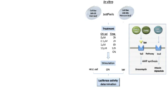
III.2.1 – Experimental design for Chloroquine effect on AMP synthesis response
of Drosophila to infection
56
III.3.1 – Northern blot of rp49, drs and dipt gene expression in Oregon R flies 62
III.3.2 – Nortern blot evaluation of the influence of Chloroquine on drs
expression upon infection
64
III.3.3 – Nortern blot evaluation of the influence of Chloroquine on dipt
expression upon infection
65
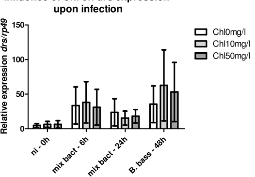
III.3.4 – Real-Time evaluation of the influence of Chloroquine on drs expression
upon infection
66
III.3.5 – Real-Time evaluation of the influence of Chloroquine on dipt expression
upon infection
68
IV.2.1 – Experimental design for hemocyte proliferation 84
IV.3.1 – Hemocyte proliferation 85
V.1.1 – PPO activation and melanin synthesis 92
V.2.1 – Experimental design to determine melanization role in the response to
hemolymph sporozoites
99
V.3.1 – Melanization activation in the mosquito hemolymph 100
V.3.2 – PPO activation during mosquito infection 101
V.3.3 – PPO activation in injected mosquitoes 103
V.3.4 – Effect of PO activity inhibition on parasite load 104
VI.2.2 – Mosquito injections to evaluate eicosanoid biosynthesis impact on
malaria infection
133
VI.3.1 – 2D maps of CBB stained hemolymph proteins from malaria non-infected
(2.33) and infected (2.34) mosquitoes
136
VI.3.2 – 2D maps of silver stained hemolymph proteins from malaria
non-infected (2.33) and non-infected (2.24) mosquitoes
138
VI.3.3 – Selected area from a 2D map of hemolymph proteins of malaria infected
mosquitoes
139
VI.3.4 – Differential regulation of protein spots highlighted in assays A and B 139
VI.3.5 – Real-Time amplification of AgPPO5 in standard mosquito DNA curve
samples, non-infected and P. berghei infected mosquito hemolymph samples collected at D13pi
143
VI.3.6 – Relative expression of Real-Time amplified genes in the hemolymph of
non-infected and P. berghei infected An. gambiae mosquitoes at D13pi
145
VI.3.7 – Mosquito survival after medium, IN and AA injections 147
VI.3.8 – The impact of eicosanoid biosynthesis on the outcome of P. berghei
infection in An. gambiae mosquitoes
I.4.1 – Stable gene expression in mosquitoes 39
I.4.2 – Introduction of foreign agents in mosquitoes to block transmission 40
III.2.1 – Primers used for RT-PCR 58
III.2.2 – Primers used for Real-Time PCR 59
III.3.1 – drs and dipt expression regulation by chloroquine 69
IV.2.1 – Sample preparation for LPS detection 82
V.2.1 – PCR conditions for Plasmodium detection 97
VI.2.1 – Primers used for Real-Time PCR 130
VI.3.1 – Infection parameters at D13pi in P. berghei infected mosquitoes
(groups 2.34)
135
VI.3.2 – Protein spot identification by MALDI-TOF MS 140
VI.3.3 – Protein families of identified spots 142
VI.3.4 – Regulation of identified spots at genomic and protein levels in P.
berghei infected vs. non-infected mosquitoes at D13pi
I
I
I
I –
–
–
– Introduction
Introduction
Introduction
Introduction
I.1 – Malaria
Malaria is one of the most life-threatening diseases affecting the human population. Together with HIV/AIDS and tuberculosis, it has become a calamity with worldwide proportions, mainly in sub-tropical countries. The WHO reports around 500 million cases of malaria each year, resulting in one million deaths, particularly of children under 5 years old.
In old days, malaria was attributed to the fetid marshes around Rome, hence the name of “bad-air” (mal-aria, in Italian), or Roman fever. In the late 19th century, scientists realized that a single-cell organism is responsible for the illness of malaria.
I.1.1 – The Parasite
The single–cell organism is a protozoan parasite from the genus Plasmodium. Taxonomically, it belongs to the Alveolata phylum, the Apicomplexa order, the Haemosporida family, and the Plasmodium genus, Levine, 1988. Biologically, the parasite has an exceedingly complex life cycle in which it alternates between a vertebrate host and an anopheline mosquito vector. In the host, the parasite is responsible for the illness observed in malaria-infected people, while in the vector it multiplies and develops compromising the mosquito’s fitness. Figure I.1.1 shows the detailed life cycle of the Plasmodium parasite.
I.1.2 – The disease
Malaria illness occurs during erythrocytic schizogony. Successive cycles of parasite invasion result in elevated numbers of red blood cells destroyed, causing acute anemia. At the same time, erythrocyte rupture releases pyrogenic substances into the blood causing high fevers. Thus, anemia, high fivers chills and renal insufficiency are the general symptoms of malaria, and are caused by infection with any of the four species of Plasmodium that infect humans: Plasmodium malariae, Plasmodium ovale,
Plasmodium vivax and Plasmodium falciparum (Tuteja, 2007). The later has an
additional property: it induces cytoadherence in the infected red blood cells that easily adhere to the blood capillaries wall, hindering oxygen delivery to the major organs. If this occurs in the brain, it leads to coma, and inescapably to death. Cerebral malaria is the worst form of malaria known, and P. falciparum is the deadliest of the malaria parasites.
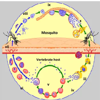
Figure I.1.1 – Plasmodium life cycle. (Adapted from www.who.int/tdr/diseases/malaria/lifecycle) Parasites are passed on to the vertebrate host through the mosquito bite (i). The parasite rapidly infects the liver (ii) where it multiplies forming new invasive forms (merozoites) – hepatic schizogony. These are released into the blood stream (iii) and invade red blood cells (iv). Merozoites mature into schizonts and undergo a series of divisions forming new invasive merozoites (v), initiating cycles of erythrocyte invasion, maturation and rupture, causing the disease – erythrocytic schizogony. Some merozoites differentiate into gametocytes (vi) that are taken up by the mosquito in a blood meal (vii). In the mosquito midgut the parasite activates gametocytes maturation into gametes. Fertilization occurs (viii) and a motile zygote is formed (ookinete). It traverses the midgut epithelia and settles down underneath the basal lamina, forming a cyst (oocyst) (ix). Inside, thousands of new invasive forms develop (sporozoites). Upon oocyst burst, these are released into the mosquito hemolymph (x) and flow to the salivary glands, where they accumulate until the next bite – sporogonic development, initiating a new cycle (i).
Mosquito Vertebrate host i ii iii iv v vi vii viii ix x
In the last few decades, the number of malaria-ill people has been rising, and the situation is becoming even more serious, since it is estimated that the numbers will (at least) double by 2020 (Breman, 2001). Several factors are contributing for this increase: i) the increasing development of parasites resistant to antimalarial drugs; ii) the existence of different Plasmodium species and their inherent antigenic variation that amplifies parasite variability; iii) the geographical and socio-economical conditions of endemic countries, and the development of other so called poverty diseases, like HIV/AIDS and Tuberculosis; iv) the hot and humid weather, the poor health and sanitary system, and insecticide resistance that contribute for the large scale development and reproduction of the mosquito vector, and v) the breakdown of control programs.
Therefore, to efficiently combat malaria, an integrated action will have to be created, so that all these factors are restrained and controlled. This will include: i) the development of new reliable drugs (or a combination of drugs to reduce resistance emergence), ii) a new efficient and highly protective vaccine, iii) the improvement of sanitary and health conditions, iv) vector control to decrease transmission (removal of mosquitoes breeding places, use of insecticide impregnated bed nets and the control of parasite development inside the mosquito), and vii) sustainability of malaria control programs.
I.1.3 – The mosquito vector
The parasite depends on its ability to develop in the mosquito in order to infect a human host. Thus, controlling transmission through the mosquito greatly reduces malaria outspread.
Transmission can be impaired by several strategies, as reducing the number of mosquitoes (using insecticides and destroying potential breeding sites), avoiding the contact between mosquitoes and humans (using insecticide-impregnated bednets) or blocking the development of the parasite in its vector. For this, it is vital to understand
the biology of parasite and mosquito, their interactions, mosquito permissiveness to parasite, and factors in parasite and mosquito necessary for an efficient transmission. The mosquito responsible for malaria transmission belongs to the genus Anopheles, and is widespread through the major temperate and tropical areas. Even though the
Anopheles genus is composed by 400 species only about 60 are able to efficiently
transmit malaria. This reveals a high degree of specificity for the parasite/mosquito combination. In fact, each mosquito species has a specific permissiveness to a particular species of Plasmodium. Mosquito and parasite factors and interactions that may account for this specificity are not yet known. It is clear, however, that both organisms engage in a series of interactions in which they are able to recognize and respond to each other (Sinden, 2002).
For instance, the blood meal alone triggers the transcriptional regulation of a set of mosquito genes, required for functions such as digestion and immunity against bacteria infected blood. A Plasmodium infected blood meal however, triggers a different set of genes, suggesting that the mosquito recognizes the parasite and mounts a response to its presence through the activation of specific genes. Moreover, the parasite itself has to undergo several morphological changes that are also regulated at the genomic level and activated with precision both temporally and spatially in the mosquito (Dimopoulos et al., 1998, 2002; Richman et al., 1997; Sinden, 2002).
Additionally, there are factors external to both organisms able to influence the outcome of infection, such as temperature, humidity and blood meal constituents. The impact of these factors on parasite development is generally characterized by an increase/decrease of parasite load in the midgut.
Thus, to be able to control transmission it is essential to fully understand which and how factors (from parasite, mosquito or external) are determinant for a successive infection.
I.2 – The parasite in the mosquito
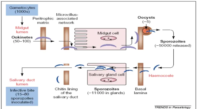
There is considerable amount of information regarding the sporogonic development of the parasite. Figure I.2.1 summarizes mosquito infection by malaria parasites and the successive bottlenecks that the parasite encounters during its development.
All interactions between parasite and mosquito, and the fact that mosquito directed antibodies are able to prevent infection by Plasmodium (Dinglasan et al., 2003), come as hope for new targets for transmission control. As malaria infection is able to compromise mosquito’s fitness, it is imperative that the mosquito controls parasite numbers for survival. Actually, the mosquito is capable of mounting efficient bottlenecks to the development of the parasite, illustrated by the dramatic losses in number suffered by the parasite along the sporogonic cycle (Figure I.2.1; Alavi et al., 2003, Sinden & Billingsley, 2001; Sinden, 2002). What mechanism(s) are responsible for the constraint in parasite number, and how they are activated remains to be fully elucidated. Nevertheless, it has become clearer that the mosquito immune system plays an important role.
Figure I.2.1 – The sporogonic cycle of malaria parasites development in the mosquito. Source: Sinden & Billingsley, 2001.
Upon a blood meal the parasite enters the mosquito midgut, where it senses changes in environment, as temperature drop, pH increase, and the presence of a mosquito factor, xanturenic acid. These cause an increase of cytoplasmic calcium release in Plasmodium gametocytes (Billker et al., 2004), triggering the differentiation into gametes. In a few minutes fertilization occurs, and a motile ookinete develops. Of the tens of thousands of gametocytes ingested by the mosquito only 50-100 ookinetes are formed. These have to evade from the midgut, traversing the peritrophic matrix, and the midgut epithelium, until settle on the basal lamina. This process involves midgut epithelium recognition and some parasite proteins have been implicated, as the secreted ookinete adhesive protein (SOAP), and the circumsporozoite- and TRAP-related protein (CTRP). Without these, the parasite’s ability to invade the midgut epithelium and progress in infection is reduced (Dessens et al., 1999, 2003). PbS21 has also been shown to be necessary for its binding to the basal lamina itself, and maturation into oocyst (Arrighi & Hurd, 2002). How the parasite recognizes the midgut cells, if it actually targets a specific type of cells, and how it reaches the basal lamina is in centre of debate and seems to differ with parasite/vector combinations. In some combinations ookinetes take an intracellular route of invasion, passing through several cells in the midgut epithelium that undergo apoptosis, and are released into the midgut lumen. This calls for a tissue repair system, directed from the surrounding cells and involving the actin cytoskeleton and microtubule remodelling that build a cover for the ookinete, in the hemocel side of the basal lamina (Han et al., 2000, Vlachou et al., 2004, 2005). Of the 50-100 ookinetes only 5 successfully develop into an oocyst that matures, producing within sporozoites that are released into the hemocel in numbers that reach tens of thousands. It is not known if the oocyst burst is a mechanical result of the growth and development of the cells within, or a result from a specific action of sporozoites. In the hemocel, sporozoites encounter a new environment, with different cells and organs of the mosquito, ultimately reaching the salivary glands. Again, only a small proportion of sporozoites (15-80) invade the salivary glands with success. How sporozoites recognize salivary glands and differentiate them from the other organs is still unknown. But it is sure that there is a specific recognition of the salivary glands, invasion of the distal lateral and medial lobes (Ghosh et al., 2001, 2002), and that gliding is essential for cell invasion, parasite locomotion in host tissues, and possibly, a target to control transmission (Frischknecht et al., 2004; Matuschewski et al., 2002). Some studies also refer to chemotatic attraction that could help to bring the parasite closer to the binding site for invasion (Akaki & Dvorak, 2005). Several proteins have been identified as essential for sporozoite invasion of the salivary glands, like the parasite proteins CSP and TRAP (Myung et al., 2004; Sultan et al., 1997) and the mosquito SGS1. The later is a salivary gland specific protein, which localizes at the region that is preferentially invaded by the sporozoites. Antibodies anti-SGS1 inhibit sporozoites invasion in Aedes aegypti, making it a good candidate for the sporozoites receptor in the salivary gland epithelium (Korochkina et al., 2006).
I.3 – The insect immune response
Numerous studies have shown that insects have the ability to recognize pathogens and trigger the activation of effective mechanisms to control or clear an infection. The current knowledge about insect response to pathogens is summarized in Figure I.3.1. Insects show several lines of defense against pathogens: i) the external cuticle avoids massive infections hindering pathogen entry into the hemocel, ii) chitinous membranes, acids and enzymes in the digestive tract help restrain infection by ingested microorganisms, iii) if pathogens manage to enter the hemocel the insect is able to mount a complex immune response to clear the infection.
Insects do not possess an adaptative immune system as vertebrates, relying solely upon an innate immune response. Nevertheless, this can be highly complex, presenting outstanding specificity and effectiveness.
Insects have several tissues able to engage in immunity: hemocytes, fat body and epithelium. Upon an infection, one or more tissues may be stimulated to participate in the response. How they signal to each other is not very clear, but it may require cytokine-like molecules.
Hemocytes (or blood cells) are the immune-responsive cells by excellence. They move freely through the hemolymph and possess the arsenal to combat infections, and are probably the first to be activated upon an infection in the hemocel.
Fat body is the equivalent to the mammalian liver. This organ is responsible for the production in large scale of pathogen killing molecules such as antimicrobial peptides, when a systemic response is in order.
Epithelium encloses the local immune responses. Epithelium responses are critical, as localized immunity avoids massive spread of infection, facilitating pathogen clearance.
As for the way how different effector mechanisms are activated, the response can be classified as humoral or cellular. A humoral response is characterized by the use of effector and signaling molecules already present in the hemocel, and subsequent release (by signal amplification) of the same molecules by either of these tissues into the hemocel where they exert their action. This includes the production of small peptides with antimicrobial activity (antimicrobial peptides - AMP), generation of reactive intermediaries of oxygen and nitrogen (ROI and NOI, respectively), and activation of complex cascades that lead to responses such as melanization and coagulation. The presence of hemocytes at the infection site is mandatory for cellular
Figure I.3.1 – Insect immune responses to pathogen infection. Pathogens are recognized in the hemocel by specific receptors that activate proteolytical cascades. These may directly trigger pathogen killing mechanisms or activate intracellular signal transduction pathways. These result in the transcription of immune related genes whose products are released into the hemocel. These molecules can be either effector molecules with antimicrobial activity (AMPs, NOIs and ROIs), or secondary signaling molecules that contribute to the activation of other effector mechanisms for pathogen killing. As such, insect immune responses can be roughly divided into three phases: 1) Recognition of non-self molecules by highly conserved pattern recognition receptors (PRR); 2) Signal transduction and amplification through extracellular proteolytical cascades (comprised serine proteases and their negative regulators, serpins) and/or intracellular immune signaling pathways; and 3) Activation of effective pathogen killing mechanisms. AMP: Antimicrobial Peptide; NOI: Nitric Oxide Intermediate; PRR: Pattern Recognition Receptor; ROI: Reactive Oxygen Intermediate.
immune responses. These are able to engage in responses such as phagocytosis, nodulation, aggregation, encapsulation and cytotoxic reactions. As humoral factors affect the hemocytes, and these are necessary for the production of the same factors, both responses are overlapping and operating when combating an infection.
Pathogen killing mechanisms in insects include phagocytosis, nodulation, encapsulation, coagulation, melanization and AMP synthesis. One or more of these mechanisms may be activated upon an infection.
Phagocytosis involves the engulfment and intracellular digestion of non-self particles
by the hemocytes. It starts with binding of non-self molecules to specific receptors on hemocytes, which triggers the target engulfment via an actin polymerization-dependent mechanism. The target is destroyed within phagosomes by lysosomal enzymes. The digestion of pathogens may lead to the production of secondary signals sent to the fat body to stimulate other immune responses. Insects are able to recognize and phagocyse bacteria, yeast, parasites, virus, and also synthetic particles such as negatively charged Sephadex beads (da Silva et al., 2000; Hernandez et al., 1999; Hillyer et al., 2003; Kocks et al., 2005; Lamprou et al., 2007; Mizutani et al., 2003; Moita et al., 2005).
Nodulation and Encapsulation are observed exclusively in invertebrates. They are
activated in response to microorganisms too large to be phagocised. Nodules refer to multicellular aggregates of hemocytes surrounding a high number of bacteria or large pathogens in an extracellular material. Encapsulation refers to binding of hemocytes to larger targets, or even nodules. Nodules and capsules can be subsequently melanised,
ie, covered by a melanin layer, that hinders the delivery of oxygen to pathogens. The
production of cytotoxic quinones and free radicals from ROI and NOI (by-products of melanin synthesis), and of AMP’s by hemocytes may also contribute to pathogen killing (Jiravanichpaisal et al., 2006).
Melanization is a mechanism present in various groups of invertebrates including
insects. Melanin is produced for several purposes, such as hardening of the egg chorion, wound healing, cuticle tanning and immunity. Upon an infection, a melanin layer can be deposited extracellularly over the invading microorganism, or intracellularly over phagocised microorganisms. The melanin layer keeps the pathogen immobilized, avoiding the infection to spread while hindering nutrient and oxygen exchanges between the microorganism and the surrounding media. In addition, several toxic molecules, such as cytotoxic quinones and ROI and NOI species produced in the process of melanin synthesis, may assist in pathogen clearance. Melanization can be itself a pathogen killing mechanism or it can be activated as a complement to phagocytosis, encapsulation or nodulation. The machinery to produce melanin is synthesized in hemocytes and fat body, and delivered to the hemocel. Upon infection or wounding melanin is rapidly synthesized to avoid massive infection or loss of hemolymph (reviewed by Barillas-Mury, 2007; Soderhall & Cerenius, 1998).
Coagulation, as melanization, is vital for wound healing and immunity. It is essential to
avoid hemolymph losses when a wound opens in the cuticle and to clear pathogen infections. In this process, microorganisms are entrapped inside a clot comprised of extracellular protein aggregates and hemocytes (Theopold et al., 2002). It is not known whether the clot itself participates in pathogen killing (by the production of toxic side products or asphyxiation) or if it functions only to entrap pathogens to be cleared by other mechanisms.
Antimicrobial peptides are small cationic molecules with antimicrobial properties.
These molecules are produced by the fat body and the hemocytes and are released into the hemocel (Bulet et al., 1999) and have the ability to kill microorganisms generally through the destruction of the cell membrane, by permeabilzation. Increasing evidence shows some degree of specificity in the pathogen-AMP relation. For instance, in Drosophila melanogaster the antimicrobial peptide Drosomycin was
shown to be synthesized upon infection with fungi or Gram-positive bacteria, while Diptericin is produced in response to Gram-negative bacteria.
I.3.1 – The Drosophila immune response
Nowadays, one of the most studied insect immune systems is that of the fruitfly
Drosophila melanogaster, whose powerful genetic tools allow an accelerated study at
many molecular levels. Although hematophagous insects such as mosquitoes have a higher diversity in pathogen microorganism to which they are exposed (due to the nature of the blood meal), the mosquito complexity of breading, the lack of suitable genetics and technology of genetic manipulation permit only a limited view of the immune responses, making Drosophila a useful model to study immunity even in hematophagous insects (Meister et al., 2004).
The Drosophila’s immune response to infection as been recently reviewed by several authors: Hoffmann (2003), Agaisse & Perrimon (2004), Leclerc & Reichhart (2004), Naitza & Ligoxygakis (2004), and Lemaitre & Hoffmann (2007).
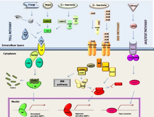
The best studied pathogen killing mechanism in Drosophila is the synthesis of antimicrobial peptides (AMP). AMP synthesis is activated by bacteria and fungi and is regulated by two intracellular signal transduction pathways (TOLL and IMD). A third pathway (JAK/STAT) is triggered in response to viruses. The recognition of pathogen associated molecular patterns in microorganisms (PAMPs) by pattern recognition receptors (PRRs) (Medzihtov & Janeway, 2002) activates a specific signal transduction pathway that results in the translocation of a cytoplasmatic NF-kB transcription factor to the nucleus, where it starts synthesis of AMPs and other immune molecules. These pathways are represented in Figure I.3.2.
Apart from AMP production, other effector mechanisms are known to be essential for immune responses in Drosophila, as phagocytosis, encapsulation, coagulation and melanization of microorganism, which have been observed upon immune challenge. However, their activation and regulation was not yet disclosed.
Additionally, molecules like the iron up-taking molecule transferrin (Yoshiga et al., 1999) and the inducible nitric oxide synthase NOS (Foley & Farrell, 2003; Nappi et al., 2004) have been implicated in microorganism killing, although a link to regulation by one of the signal transduction pathways is still missing.
The fly’s immune response is a complex and intricate net of signalling pathways that lead to the successive activation of several reactions that work together in order to restrain the microbial infection, and to heal the wounded tissues and damaged cells of the fly.
Figure I.3.2 – Immune signalling transduction pathways in Drosophila immunity. Adapted from Lemaitre & Hoffmann, 2007.
Toll pathway (left) is activated by fungi, yeast and gram-positive bacteria. Recognition involves PRR’s like Persephone (Psh), GNBP3 and GNBP1/PGRP-SA/PGRP-SD. Mutants for this pathway have a compromised survival for fungal and Gram-positive bacterial infections (Hoffmann & Reichhart, 2002). Recognition activates a Spaetzle activating enzyme (SPE) that converts pro-Spaetzle into Spaetzle (SPZ). This binds to the receptor Toll (Lemaitre et al., 1996; Imler & Hoffmann, 2001), triggering an intracellular cascade, involving Myd88, Tube and Pelle. This results in degradation of the inhibitor Cactus and release of DIF that translocates into the nucleus, starting the transcription of AMP’s like Drosomycin. Toll pathway is also associated with melanization (Ligoxygakis et al., 2002), along with hemolymph components, like serine proteases and their negative regulators, serpins. Loss-of-function mutants for serpin 43Ac have constitutive cleavage of Spz and drosomycin expression (Levashina et al., 1999; Ligoxygakis et al., 2002). Serpins seem to protect against microbial proteinases, and to regulate endogenous proteinases, preventing over activation of hemolymph coagulation, proteolytic cytokine and prophenoloxidase (Kanost, 1999).
IMD pathway is activated by Gram-negative bacteria. Bacterial peptidoglycan (PGN) is recognized by the peptidoglycan recognition protein LE (PGRP-LE). Monomeric PGN’s activate a dimeric receptor composed by PGRP-LCx and PGRP-LCa, while polymeric PGN’s activate a dimeric receptor composed by PGRP-LCx. Mutants for this pathway have a compromised survival for bacterial infection (Hoffmann & Reichhart, 2002). Recognition activates the IMD protein that in turn activates dFADD and Dredd, which cleaves the inhibitory domain of the Relish, causing it to translocate into the nucleus and start the transcription of AMP’s such as diptericin and attacin. IMD may also activate dIAP2, dTAB2 and dTAK1. The later can activate IIKγ and IKKβ leading to Relish activation, or trigger the JNK pathway. IMD is also involved in apoptosis, through the activation of the caspase-like protein DREDD (Georgel et al., 2001).
JAK/STAT pathway is triggered by virus, as mutants for this pathway are resistant to bacterial and fungal expression, but susceptible to Drosophila Virus C (Dostert et al., 2005). Recognition leads Unpaired-3 (UPD3) to bind to the receptor Domeless (Dome), activating JAK, and leading STAT to translocate into the nucleus and trigger the transcription of genes such as tep2 and turandot. Overexpression of this pathway leads to the formation of melanotic pseudotumors (Hanratty & Dearolf, 1993; Harrison et al., 1995; Luo et al., 1997). Agaisse et al.(2003) also suggested this pathway to be involved in the response to tissue damage.
I.4 – The mosquito immune response to malaria
The mosquito immune system is believed to be responsible, at least in part for the parasite losses occurring along its sporogonic development. Thus, its complete understanding will provide a powerful tool to block malaria transmission.
The completion sequencing of Anopheles gambiae genome (Holt et al., 2002) allowed a comparative analysis between the genomes of An. gambiae and D. melanogaster and the description of correspondent protein families in the mosquito. This comparison revealed that about half of the genes are orthologs 1:1 and identified 242 genes that potentially code for components of the immune system (Zdobnov et al., 2002).
The comparative analysis of 18 gene families from the fly and the mosquito revealed a 2 fold deficit in 1:1 orthologs in immune-related genes, when compared to the whole genome. In contrast these genes present specific gene expansions in the mosquito, when compared to the fly. Components of the immune system or gene families that include these components have evolved faster than the rest of the genome. Gene families coding for recognition, signal modulation and effector mechanisms are poor in ortholog genes reflecting species specific expansions and possibly a strong selective pressure (Christophides et al., 2002). On the contrary, genes belonging to intracellular immune signaling pathways are highly conserved, even more than the genome as a whole, probably due to the high range of functions in which they are involved, such as development. Although comparative genetics is of vital importance to highlight immune-related genes, it is insufficient to point out which of these genes are indeed involved in the response to Plasmodium.
Several approaches have been pursued in order to understand parasite-vector interactions and to attempt to block malaria transmission in the mosquito. These included transcription profile studies of infected and non-infected mosquitoes, and the establishment of mosquito strains refractory to malaria infection.
I.4.1 – Immune gene expression studies
Work in mosquito immune responses has focused mainly in differential expression of immune-related genes upon infection. Different transcriptional profiling approaches included:
• Reverse Transcriptase Polymerase Chain Reaction (RT-PCR) for specific immune-related genes. Dimopoulos et al., 1997 found some immune related genes to be up-regulated, both locally (midgut) and systemically, upon midgut invasion by ookinetes. These genes were also found to be up-regulated in later steps of parasite development (Dimopoulos et al., 1998).
• Subtractive libraries, enriched for genes up-regulated after bacterial challenge (Oduol et al., 2000), genes expressed in midguts after ookinete invasion and early stages of oocyst development (Abraham et al., 2004), and genes expressed in midguts during late stages of oocyst development (Srinivasan et
al., 2004).
• EST libraries constructed of immunocompetent cell lines (Dimopoulos et al., 2000), blood fed and non-blood fed mosquitoes (Ribeiro, 2003).
• Microarrays (derived from the immunocompetent cell line EST library), used to analyze genome responses to injury, bacterial challenge and malaria infection (Christophides et al., 2002; Dimopoulos et al., 2002). Microarray studies contributed to the identification of several genes with altered expression during midgut invasion (Christophides et al., 2002; Dimopoulos et al., 2002; Kumar et al., 2003; Vlachou et al., 2005). Vlachou et al. (2005) found that 7% of the assessed mosquito genes are differentially regulated at this point in infection, including genes belonging to functional classes such as cytoskeleton remodelling, apoptosis, immunity, redox metabolism, cell adhesion and extracellular matrix maintenance. Microarrays are now a widely used technique and a powerful tool to study mosquito responses to the parasite, and have been used to target differential expressed genes in the whole genome or in specific subsets of genes (immunity, stress, and others).