Correspondence to: Jelena T. Todić, Franša D Eperea 18, 34 000 Kragujevac, Serbia. Fax: +381 34 301 476, E-mail: [email protected]
O R I G I N A L A R T I C L E UDC: 577.3::616.31
DOI: 10.2298/VSP150327165T
Effects of bruxism on the maximum bite force
Uticaj bruksizma na maksimalnu zagrižajnu silu
Jelena T. Todić, Ankica Mitić, Dragoslav Lazić, Radivoje Radosavljević, Miloš Staletović
Department of Dentistry, Faculty of Medicine, University of Priština/Kosovska Mitrovica, Kosovska Mitrovica, Serbia
Abstract
Background/Aim. Bruxism is a parafunctional activity of the masticatory system, which is characterized by clenching or grinding of teeth. The purpose of this study was to determine whether the presence of bruxism has impact on maximum bite force, with particular reference to the potential impact of gen-der on bite force values. Methods. This study included two groups of subjects: without and with bruxism. The presence of bruxism in the subjects was registered using a specific clinical questionnaire on bruxism and physical examination. The sub-jects from both groups were submitted to the procedure of measuring the maximum bite pressure and occlusal contact area using a single-sheet pressure-sensitive films (Fuji Prescale MS and HS Film). Maximal bite force was obtained by multiplying maximal bite pressure and occlusal contact area va-lues. Results. The average values of maximal bite force were significantly higher in the subjects with bruxism compared to those without bruxism (p < 0.001). Occlusal contact area was significantly higher in the subjects suffering from bruxism (p < 0.001), while the maximal bite pressure values did not show a significant difference between the studied groups (p > 0.01). Maximal bite force was significantly higher in the males compa-red to the females in all segments of the research. Conclusion. The presence of bruxism influences the increase in the maximum bite force as shown in this study. Gender is a signifi-cant determinant of bite force. Registration of maximum bite force can be used in diagnosing and analysing pathophysiological events during bruxism.
Key words:
bruxism; bite force; dental occlusion; sex; male; female.
Apstrakt
Uvod/Cilj. Bruksizam je parafunkcionalna aktivnost mastika-tornog sistema, koja se karakteriše stezanjem ili struganjem zubima. Cilj ove studije bio je da se utvrdi da li prisustvo bruk-sizma ima uticaja na maksimalnu zagrižajnu silu, sa posebnim osvrtom na potencijalni uticaj pola na vrednosti zagrižajne sile.
Metode. Ova studija je obuhvatila dve grupe ispitanika: ispi-tanike sa bruksizmom i bez bruksizma. Prisustvo bruksizma kod ispitanika je registrovano upotrebom specifičnog kliničkog upit-nika za bruksizam i kliničkim pregledom. Ispitanici obe grupe bili su podvrgnuti postupku merenja maksimalnog zagrižajnog priti-ska i okluzalne kontaktne površine upotrebom jednoslojnih fil-mova osetjivih na pritisak (Fuji Prescale MS i HS Film). Maksi-malna zagrižajna sila dobijena je množenjem vrednosti maksi-malnog zagrižajnog pritiska i okluzalne kontaktne površine.
Rezultati. Prosečne vrednosti maksimalne zagrižajne sile bile su značajno veće kod ispitanika sa bruksizmom nego kod ispitanika bez bruksizma (p < 0,001). Okluzalna kontaktna površina bila je značajno veća kod ispitanika koji pate od bruksizma (p < 0,001), dok vrednosti maksimalnog zagrižajnog pritiska nisu pokazale značajnu razliku između ispitivanih grupa (p < 0,01). Maksimalna zagrižajna sila bila je veća kod muških ispitanika nego kod žen-skih ispitanika, u svim segmentima istraživanja. Zaključak. Pris-ustvo bruksizma uticalo je na povećanje maksimalne zagrižajne sile u ovoj studiji. Pol je bio značajna determinata zagrižajne sile. Registracija maksimalne zagrižajne sile može se koristiti za dijag-nozu i analizu patofizioloških događaja tokom bruksizma.
Ključne reči:
bruksizam; zagrižajna sila; zubi, okluzija; pol; muškarci; žene.
Introduction
Bruxism is a parafunctional activity of the masticatory system, which is characterized by clenching or grinding of the te-eth and/or by bracing or thrusting of the mandible 1. It may hap-pen while awake (awake bruxism) or while sleeping (sleep bruxism). Bruxism during daytime is commonly a
to-oth interference in dental occlusion, psychosocial influences, such as stress or anxiety 3, and central or pathophysiological causes involving brain neurotransmitters or basal ganglia 4. Manfredini et al. 5 indicate that occlusal factors do not seem to have any significant role in the development of bruxism. Dep-ression, increased level of hostility 6 and stress sensitivity 7 dis-tinguish a “bruxer” from a healthy individual. However, fac-tors like smoking, alcohol, drugs, diseases, and trauma may al-so be involved in the bruxism etiology 8.
Factors that may indicate the presence of bruxism in-clude physical symptoms and changes in hard and soft oral tissues. The physical symptoms of bruxism may include: he-adache, facial myalgia (muscle pain) and temporomandibular joint (TMJ) discomfort. The most common oral symptoms include: abnormal tooth wear (attrition on occlusal or incisal surfaces), fracture of the teeth and excessive tooth mobility.
In “bruxers”, the distribution of muscular force to the te-eth and surrounding tissues may result in tooth wear and oro-facial pain, as well as hyperactivity and hypertrophy of the masticatory muscles, especially the masseter muscle. In view of the fact that muscles are the main bite force generators, the changes in their function may be reflected in the maximum bi-te force (MBF) value. MBF is a result of the masticatory mus-cle activity, which is regulated by the central nervous system receptors and orofacial structures (muscle spindles, proprio-ceptors, mechanoreceptors). Previous studies report that MBF may be influenced by gender, craniofacial morphology, perio-dontal sensitivity, dental occlusal status and signs and symptoms of temporomandibular disorders 9–11.
Reports of certain studies on the effects of bruxism on MBF appear to be contradictory. Helkimo and Ingervall 12 fo-und that individuals with clenching and grinding habits had higher bite force only on the incisors, but not on the molars. On the other hand, Gibbs et al. 13 found higher bite force valu-es on the posterior region for subjects with bruxism than for the control group. Lyons and Baxendale 14 suggested that the jaw-closing muscles of subjects with bruxism might have be-nefited from a "training effect" as a result of all this activity, resulting in muscles that are stronger and possibly more resis-tant to fatigue. Cosme et al. 15 believe that bruxism does not af-fect MBF, while some of the authors find that MBF is increa-sed in 54.5% of the subjects suffering from bruxism 16. Accor-ding to Nunes 17, for some patients pain plays a modulator role in parafunctional activity, decreasing the electromyographic activity of masticatory muscles and MBF.
There seems to be no clear correlation between the MBF and bruxism. In view of the aforementioned, the main purpose of this study was to determine whether bruxism has impact on MBF, assessing the potential gender impact on the MBF values.
Methods
This trial was conducted ensuring the full adherence to the principles of the “Good Clinical Practice (GCP)” which means that the trial included only participants who had given their full informed consent to participate in writing, with a prior access to the full information about the aims and scope
of the trial. This trial was conducted with the approval of the Ethics Committee at the Faculty of Medicine, University of Priština/Kosovska Mitrovica.
The trial was conducted on the subjects selected among the students of the Faculty of Medicine in Kosovska Mitrovica and the patients who visited the Prosthodontics Clinic, Dentistry De-partment, Faculty of Medicine in Kosovska Mitrovica.
The presence/absence of bruxism in subjects were re-gistered using a specific clinical questionnaire on bruxism by Molina et al. 18 and specific physical examinations.
The Molina questionnaire included the following questions: 1) Do you wake up in the morning or during the night to find yourself grinding or clenching? 2) Do you feel fatigue or masticatory muscle pain on awakening? 3) Do you wake up in the morning or during the night with the jaws lo-cked? 4) Do you feel discomfort on the teeth on awakening? 5) Do you have recent history of chronic dislocation of per-manent or temporary restorations? 6) Do you have recent history of noises associated with nocturnal teeth grinding as reported by a third person?
Physical examination included observation of attrition on occlusal or incisal surfaces, detectable scars and buccal mucosa changes, changes on the lateral border of the tongue (tongue indentations) and verification of masticatory muscle hypertrophy by means of digital palpation in maximum in-tercuspation.
Signs and symptoms of temporomandibular disorders (TMD) were recorded by Helkimos clinical functional analysis 19. This analysis includes the case history (questionnaire relating to the signs and symptoms of TMD), clinical functional analysis of the orofacial system and occlu-sal analysis.
Group formation
The following exclusion criteria were applied for all parti-cipants: more than two missing posterior teeth (excluding third molars); previous orthodontic or prosthodontic treatment; the presence of active phase of periodontal disease; signs and symptoms of TMD or spontaneous orofacial pain; the presence of malocclusion (anterior open bite, unilateral cross bite, class II and III malocclusion according to Angle).
Further criteria for inclusion subjects in the study im-plied: the intact dental arch (third molars not taken into ac-count); the presence of no more than three fillings; Class I neutro-occlusion according to Angle's classification; age between 18 to 23 years.
The subjects included in the study, in terms of the regis-tered presence/absence of bruxism, were divided into two groups: the study and the control group. The study group consisted of 41 patients with bruxism, while the control gro-up consisted of 48 subjects without bruxism (18–23 years of age).
Registration of maximum bite force
Fig. 1 – Registration of maximum bite pressure (MBP): a) registration procedure; b) registered occlusal contacts on a prescale film.
Fig. 2 – Scale for reading color intensity of the registered occlusal contact.
Fig. 3 – Graph for determining values of bite pressure. MBF value in both the control and the experimental (study)
group. MBP was registered by means of a single sheet pres-sure-sensitive sheet (Fuji Prescale, Tokyo), type: MS and HS. MS pressure-sensitive sheet registered pressure within the range of 10–50 megapascal (Mpa), while HS sheet regis-tered the pressure of 50–130 MPa. Fuji Prescale Film technology and its principle of operation is based on indica-ting applied pressure differences as red color density variati-ons. This feature is enabled by particle size control (PCS) technology based on microcapsule layers designed to res-pond to different pressures relieving color whose intensity is proportional to the pressure applied.
The MBP registration procedure was conducted in both the study and the control group. The subjects were comfortably seated with the head erect and torso in upright position. Drying provided a relatively dry environment in bi-ting surfaces for placing a horseshoe-shaped pressure
sensi-tive sheet in-between. The subjects were instructed to bite stronger in maximum intercuspation and maintain the bite force the following 10 s (Figure 1 a and b).
The registration procedure was conducted by means of MS and HS pressure sensitive sheet in all the patients, with a 2-minute break between the two registration protocols, to allow for the masticatory muscles to relax. The films applied were further on scanned using a Canon device generating 300 dpi A4 scans. Visual comparison of the occlusal contact color and color intensity scale (0.1 to 1.5) was used for the purpose of defining color density (intensity) for each occlu-sal contact registered (Figure 2).
Based on the color density, reading of the bite pressure values was carried out for each occlusal contact (Figure 3). The graph shows two curves (A and B).
scans. Multiplying the values of MBP and OCA, gave the bi-te force for each occlusal contact observed:
F (N) = P (МPа) × А (mm2)
The sum of all occlusal forces acting in the contact po-ints registered in one patient gave MBF per patient.
Σ Fn = F1 + F2 + F3 + ... Fn
For the purpose of primary data analysis, methods of descriptive statistics were used, which included measures of central tendency (mean and median), measures of variability (standard deviation) and relative numbers. The influence of bruxism on the MBF value was determined by the Student’s t-test and the Mann-Whitney Test (Rank Sum Test). Statisti-cal hypotheses were tested at the level of statistiStatisti-cal signifi-cance of 0.01 and 0.001. For statistical data analysis, a PASW Statistics was used.
Concerning the MBP analysis, values expressed per unit area (MBP/mm2) were used in order to simplify the analysis. Similarly, OCA (mm2) was analyzed as the sum of the values of each OCA registered in one patient ( К = ΣА). However, in calculating the MBF, the values of MBP and contact surface values per occlusal contact were used.
Results
Distribution of participants in the study in relation to bruxism and gender is given in Table 1. The first segment of the analysis was conducted in order to test the impact of gender on MBF, which further determined the method of da-ta processing. Thus, comparative analysis of average MBP/mm2, OCA and MBF values was conducted between the males and females within the control group – patients without bruxism (Table 2). In the male subjects without bruxism, the values of MBF, OCA and MBP/mm2 were
significantly higher than in the female subjects (t = -2975, DF = 54, p < 0.01 for MBF; t = -6.825, DF = 54, p < 0.001 for OCA; t = -6.944, DF = 54, p < 0.001 for MBP/mm2). Since significant effects of gender on MBP/mm2, OCA and MBF were found, there was the need to test the values of these parameters comparing separately the male and female participants of both groups (the study group and controls). It was the only way to determine the actual impact of bruxism on the MBF.
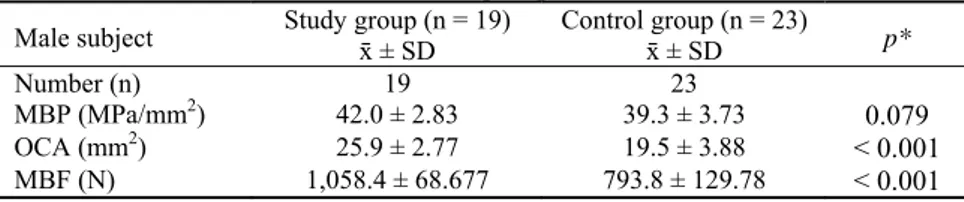
In the female subjects with bruxism, the values of MBF and OCA were significantly higher than in the females without bruxism (t = -6.5, DF = 46, p < 0.001 and t = -6786, DF = 46, p < 0.001, respectively). However, the MBP/mm2 values did not show any statistically significant difference between the female subjects with and without bruxism (Mann-Whitney test, U = 178.0; p = 0,247) (Table 3).Comparative analysis between the males of both groups showed a statistically significant difference in average values of MBF and OCA (t = 5.440, DF = 27, p < 0.001 and t = -4.288, DF=27, p < 0.001, respectively). However, in male subjects with bruxism, the MBP/mm2 values did not show statistically significant difference compared to the males without bruxism (Table 4).
Disscusion
MBF is often analyzed as an indicator of functional sta-tus of the masticatory system. Bruxism is one of the parafun-ctional activities accompanied by rapid contractions of the masseter muscle and development of forces excessively bur-dening structures of the masticatory system. Harmful effects of bruxism can be seen in non-physiological tooth wear, masticatory muscle hyperactivity and potential development of orofacial system dysfunction. The hypothesis that bruxism is capable to change the bite force by muscle strengthening is still unproven. If the bite force was truly affected by bruxism, its measurement could be an important feature in the diagnosis of such a habit.
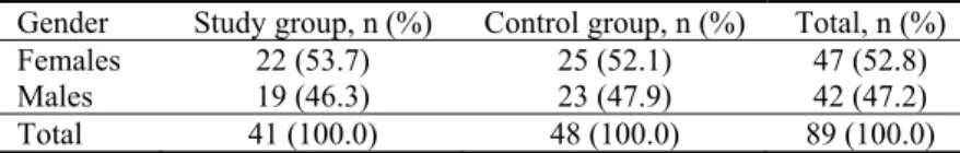
Table 1 Distribution of the subjects in relation to bruxism and gender Gender Study group, n (%) Control group, n (%) Total, n (%) Females 22 (53.7) 25 (52.1) 47 (52.8) Males 19 (46.3) 23 (47.9) 42 (47.2) Total 41 (100.0) 48 (100.0) 89 (100.0) Study group – subjects with bruxism;
Control group – subjects without bruxism.
Table 2 Comparative analysis of maximum bite pressure (MBP), occlusal contact area (OCA) and
maximum bite force (MBF) between the female and male subjects of the control group (without bruxism)
Parameter Females (n = 25) ґ ± SD
Males (n = 23)
ґ ± SD p*
Number (n) 25 23
MBP (MPa/mm2) 36.9 ± 2.50 39.3 ± 3.73 < 0.01
OCA (mm2) 12.1 ± 3.92 19.5 ± 3.88 < 0.001
MBF (N) 522.60 ± 147.99 793.80 ± 129.78 < 0.001
Table 3 Comparative analysis of maximum bite pressure (MBP), occlusal contact area (OCA)
and maximum bite force (MBF) between female subjects of both groups
Female subject Study group (n = 22) ґ ± SD
Control group (n = 25)
ґ ± SD p*
Number 22 25
MBP (MPa/mm2) 37.5 ± 3.62 36.9 ± 2.50 0.247
OCA (mm2) 20.5 ± 3.54 12.1 ± 3.92 < 0.001
MBF (N) 811.8 ± 27.60 522.6 ± 25.01 < 0.001
*p – statistical significance at the level < 0.001 for Mann-Whitney U-test and < 0.001 for Student's t-test;
Study group – subjects with bruxism; Control group – subjects without bruxism.
Table 4 Comparative analysis of maximum bite pressure (MBP), occlusal contact
area (OCA) and maximum bite force (MBF) between the male subjects of the studied groups
Male subject Study group (n = 19) ґ ± SD Control group (n = 23) ґ ± SD p*
Number (n) 19 23
MBP (MPa/mm2) 42.0 ± 2.83 39.3 ± 3.73 0.079
OCA (mm2) 25.9 ± 2.77 19.5 ± 3.88 < 0.001
MBF (N) 1,058.4 ± 68.677 793.8 ± 129.78 < 0.001 *p statistical significance (Student’s t-test);
Study group – subjects with bruxism; Control group – subjects without bruxism.
Our study showed that the average values of MBP/mm2, OCA and MBF were significantly higher in males compared to females. Some of studies support the results obtained accordingly 20. Pereira-Cenci et al. 21 and Bonakdarchian et al. 22 believe that greater muscle potential of masticatory mu-scles in males can be attributed to anatomical gender diffe-rences. Bakke 23 points out that masseter muscles of males are type II muscle fibers, which are larger in diameter com-pared to those in females. Pizolato et al. 24 suggest that hor-monal differences between sexes affect the structure of mus-cle fibers. Estimating contribution of masseter, temporal mu-scle, and anterior angle of digastric muscle to bite force, Ra-adsheer et al. 25 demonstrated that masseter thickness significantly correlates with the magnitude of MBF. However, up to 18 years of age, gender does not affect the MBF. Following a post-pubertal period, MBF tends to incre-ase significantly and to a greater extent in men than in women, becoming thus gender-related 26. According to Olt-hoff et al. 27, bite force and the number of teeth in occlusion are determining factors in masticatory performance, whereas occlusal contacts determine 10–20% of MBF variation. Fer-rario et al. 20 emphasize that dental size is larger in males, making thus the occlusal surfaces greater as well as those of the periodontal ligament, which in turn results in higher level of bite force. They stated that average value of the MBF in healthy female subjects was 522.6 ± 25.01 N, and that in men it amounted to 811.8 ± 27.6 N. These findings are con-sistent with the results of our study. However, it is noteworthy that the MBF values obtained by different studies are difficult to compare. MBF value varies depending on the type of measuring instrument applied 27, the position of the
measuring instrument within the dental arch, and the number of teeth included 28. Therefore, the literature offers MBF values ranging from 388 N to 1,109 N 29, 30.
Based on the results of this study it was found that MBF was significantly higher in participants with bruxism compared to those without it, taking into account the gender difference. The findings of our study are consistent with the findings of the study conducted by Killiaridis et al. 31. Some authors like Gibbs et al. 13 for instance, find that MBF in persons with bruxism was six times the one in those without it. However, Cosme et al. 15 did not find a significant difference between persons with bruxism and those without it, taking into account the gender dif-ference between the subjects. Similar results were reported by some other authors, as well 32. However, in these studies bite force was measured using a compressive transducer at the first molar region. Tortopidis et al. 33 addressed the issue of measu-ring instrument reliability and found that the variability in MBF values was highest when using a gnathodynamometer. The use of these measurement systems does not take into account OCA, which among other things can affect the results.
OCA, the number of occlusal contacts and the number of te-eth present are significant determining factors for MBF. Inc-reased levels of teeth clenching lead to greater intimacy between occlusal contacts in maxillary and mandibular den-tal arches. For example, with increasing teeth clen-ching/grinding levels from 30% to 100%, the occlusal con-tact areas are doubled. As our study shows, MBP/mm2 was not significantly different between persons with bruxism and those without it, whereas OCA was significantly higher in persons with bruxism; therefore, the MBF was also higher.
Using a measuring system based on the prescale pressu-re sensitive sheet, Miyaura et al. 36 found that the bite poten-tial closely correlated to the number of teeth present. Alkan et al. 37 monitored MBF values in persons with bruxism befo-re and after stabilization splint tbefo-reatment. They found that the occlusal contact area and bite force decline in patients using a splint for three months. Similar results were demonstrated by Kurita et al. 38 and Karakis et al. 39 In light of these data it is possible to comment on the muscle activity in relation to changes in the OCA and bite force.
The gold standard diagnostic method for bruxism is the use of polysomnographic recordings in a specialized sleep laboratory 40. For the purposes of our study, the questionnaire
and physical examination of the patients was used in the dia-gnosis of the patients with bruxism. Some studies compared clinical outcomes with the results of polysomnography to di-agnose bruxism and found that the clinical criteria had a reliability of 83% in patients with bruxism and 81% in asymptomatic control subjects 41. However, Baba et al. 42 did not find any associations between tooth wear status and on-going bruxism. Therefore, an insufficient reliability of clini-cal methods in the diagnosis of bruxism may somewhat af-fect the results of this study.
Conclusion
Bruxism influences the increase of MBF. It also affects the increase in OCA, but not MBP. Therefore, registration of MBF can be used in the diagnosis and analysis of pathophysiological events during bruxism.
Gender is a significant determinant of bite force, which is why gender difference must be taken into account during analysis of MBF. These results may be considered as an ini-tiative calling for further research for the sake of complete clarification of bruxism and its impact on the stomatognathic system.
R E F E R E N C E S
1. Lobbezoo F, Ahlberg J, Glaros AG, Kato T, Koyano K, Lavigne GJ, et al. Bruxism defined and graded: An international consensus. J Oral Rehabil 2013; 40(1): 2−4.
2. Huynh N, Lavigne GJ, Okura K, Yao D, Adachi K. Sleep bruxism. In: Pasquale M, Sudhansu C, editors. Sleep Disorder. Part II. Netherlands, Amsterdam: Elsevier; 2011.p. 901−11.
3. Silverman S, Eversole LR, Trulove EL. Essentials of Oral Medicine. Hamilton, London: BC Decker Inc; 2001.
4. Bader G, Lavigne G. Sleep bruxism; An overview of an
oromandibular sleep movement disorder. Sleep Med Rev 2000; 4(1): 27−43.
5. Manfredini D, Landi N, Tognini F, Montagnani G, Bosco M. Occlusal features are not a reliable predictor of bruxism. Minerva Stomatol 2004; 53(5): 231−9.
6. Manfredini D, Landi N, Romagnoli M, Bosco M. Psychic and occlusal factors in bruxers. Aust Dent J 2004; 49(2): 84−9. 7. Molina OF, dos Santos J Jr. Hostility in TMD/bruxism patients
and controls: A clinical comparison study and preliminary results. Cranio 2002; 20(4): 282−8.
8. Lobbezoo F, Naeije M. Etiology of bruxism: Morphological, pathophysiological and psychological factors. Ned TijdschrTandheelkd 2000; 107(7): 275−80. (Dutch)
9. Castelo PM, Bonjardim LR, Pereira LJ, Gavião MB. Facial dimensions, bite force and masticatory muscle thickness in preschool children with functional posterior cross-bite. Braz Oral Res 2008; 22(1): 48−54.
10.Takeuchi N, Yamamoto T. Correlation between periodontal status and biting force in patients with chronic periodontitis during the maintenance phase of therapy. J Clin Periodontol 2008; 35(3): 215−20.
11.Lasilla V, Holmlund I, Koivumaa KK. Bite force and its correlations in different denture types. Acta Odontol Scand 1985; 43(3): 127−32.
12.Helkimo E, Ingervall B. Bite force and functional state of the masticatory system in young men. Swed Dent J 1978; 2(5): 167−75.
13.Gibbs CH, Mahan PE, Mauderli A, Lundeen HC, Walsh EK. Limits of human bite strength. J Prosthet Dent 1986; 56(2): 226−9.
14.Lyons MF, Baxendale RH. A preliminary electromyographic study of bite force and jaw-closing muscle fatigue in human subjects with advanced tooth wear. J Oral Rehabil 1990; 17(4): 311−8.
15.Cosme DC, Baldisserotto SM, Canabarrosde A, Shinkai RS. Bruxism and voluntary maximal bite force in young dentate adults. Int J Prosthodont 2005; 18(4): 328−32.
16.Nishigawa K, Bando E, Nakano M. Quantitative study of bite force during sleep associated bruxism. J Oral Rehabil 2001; 28(5): 485−91.
17.Nunes LM. Association between bruxism and
temporomandibular dysfunction. Bauru: School of Dentistry, University of Sao Paulo; 2003. (Brazilian)
18.Molina OF, Junior S, Nelson SJ, Nowlin T. A clinical study of specific signs and symptoms of CMD in bruxers classified by the degree of severity. Cranio 1999; 17(4): 268−79.
19.Helkimo M. Studies of function and dysfunction of the
masticatory system. Index for anamnestic and clinical dysfunction and occlusal state. Sweden Dent 1974; 67(2): 101−21.
20.Ferrario VF, Sforza C, Serrao G, Dellavia C, Tartagilia GM. Single tooth bite forces in healthy young adults. J Oral Rehabil 2004; 31(1): 18−22.
21.Pereira-Cenci T, Pereira LJ, Cenci MS, Bonachela WC, del Belcury AA. Maximal bite force and its association with temporomandibular disorders. Braz Dent J 2007; 18(1): 65−8. 22.Bonakdarchian M, Askari N, Askari M. Effect of face form on
maximal molar bite force with natural dentition. Arch Oral Biol 2009; 54(3): 201−4.
23.Bakke M. Bite force and occlusion. Semin Orthod 2006; 12(2): 120−6.
temporomandibular disorders and bruxism. Braz Oral Res 2007; 21(3): 278−83.
25.Raadsheer MC, van Eijden TM, van Ginkel FC, Prahl-Andersen B. Contribution of jaw muscle size and craniofacial morphology to human bite force magnitude. J Dent Res 1999; 78(1): 31−42.
26.Braun S, Freudenthaler JW, Hönigle K. A study of maximum bite force during growth and development. Angle Orthod 1996; 66(4): 261−4.
27.Olthoff LW, Van Der Glas HW, Van Der Blit A. Influence of occlusal vertical dimension on the masticatory performance during chewing with maxillary splints. J Oral Rehabil 2007; 34(8): 560−5.
28.Van Der Bilt A, Tekamp A, Van der Glas H, Abbink J. Bite force and electromyograpy during maximum unilateral and bilateral clenching. Eur J Oral Sci 2008; 116(3): 217−22.
29.Kogawa EM, Calderon PS, Lauris JR, Araujo CR, Conti PC. Evaluation of maximal bite force in temporomandibular disorders patients. J Oral Rehabil 2006; 33(8): 559−65. 30.Shinogaya T, Bakke M, Thomsen CE, Vilmann A, Matsumoto M.
Bite force and occlusal load in healthy young subjects: A methodological study. Eur J Prosthodont Restor Dent 2000; 8(1): 11−5.
31.Killiaridis S, Johansson A, Haraldson T, Omar R, Carlsson GE. Craniofacial morphology, occlusal traits, and bite force in persons with advanced occlusal tooth wear. Am J Orthod Dentofac Orthop 1995; 107(3): 286−91.
32.Calderon Pdos S, Kogawa EM, Lauris JR, Conti PC. The influence of gender and bruxism on the human maximum bite force. J Appl Oral Sci 2006; 14(6): 448−53.
33.Tortopidis D, Lyons MF, Baxendale RH, Gilmour WH. The variability of bite force measurement between sessions, in different positions within the dental arch. J Oral Rehabil 1998; 25(9): 681−6.
34.Hatch JP, Shinkai RS, Sakai S, Rugh JD, Paunovich ED. Determinants of masticatory performance in dentate adults. Arch Oral Biol 2001; 46(7): 641−8.
35.Hidaka O, Iwasaki M, Saito M, Morimoto T. Influence of clenching intensity on bite force balance, occlusal contact area, and average bite pressure. J Dent Res 1999; 78(7): 1336−44. 36.Miyaura K, Morita M, Matsuka Y, Yamashita A, Watanabe T.
Rehabilitation of biting abilities in patients with different types of dental prostheses. J Oral Rehabil 2000; 27(12): 1073−6. 37.Alkan A, Bulut E, Arici S, Sato S. Evaluation of Treatments in
Patients with Nocturnal Bruxism on Bite Force and Occlusal Contact Area. A preliminary report. Eur J Dent 2008; 2: 276−82.
38.Kurita H, Ikeda K, Kurashina K. Evaluation of the effect of a stabilization splint on occlusal force in patients with masticatory muscle disorders. J Oral Rehabil 2000; 27(1): 79−82.
39.Karakis D, Dogan A, Bek B. Evaluation of the effect of two different occlusal splints on maximum occlusal force in patients with sleep bruxism: A pilot study. J Adv Prosthodont 2014; 6(2): 103−8.
40.Tosun T, Karabuda C, Cuhadaroglu C. Evaluation of sleep bruxism by polysomnographic analysis in patients with dental implants. Int J Oral Maxillofac Implants 2003; 18(2): 286−92. 41.Lavigne GJ, Rompre PH, Montplasir JY. Sleep bruxism: Validity
of clinical research diagnostic criteria in a controlled polysomnographic study. J Den Res 1996; 75(1): 546−52. 42.Baba K, Haketa T, Clark GT, Ohyama T. Does tooth wear status
predict ongoing sleep bruxism in 30-year-old Japanese subject. Int J Prosthodont 2004; 17(1): 39−44.