U
NIVERSIDADE DE
L
ISBOA
F
ACULDADE DE
C
IÊNCIAS
D
EPARTAMENTO DE
B
IOLOGIA
A
NIMAL
mRNA Metabolism: Nonsense Mediated mRNA Decay
as a Tool for Gene Therapy and the Role of Human
DIS3L2 in Transcript Degradation
Mestrado em Biologia Humana e Ambiente
Gerson Leonel Asper Amaral
Dissertação orientada por:
Doutora Luísa Romão
Professora Doutora Deodália Dias
III
U
NIVERSIDADE DE
L
ISBOA
F
ACULDADE DE
C
IÊNCIAS
D
EPARTAMENTO DE
B
IOLOGIA
A
NIMAL
mRNA Metabolism: Nonsense Mediated mRNA Decay
as a Tool for Gene Therapy and the Role of Human
DIS3L2 in Transcript Degradation
Mestrado em Biologia Humana e Ambiente
Gerson Leonel Asper Amaral
Dissertação orientada por:
Doutora Luísa Romão (Instituto Nacional de Saúde Dr. Ricardo Jorge)
Professora Doutora Deodália Dias (Departamento de Biologia Animal,
Faculdade de Ciências da Universidade de Lisboa)
V “It is finished.” – Jesus Christ (The Bible, John 19:30)
VII
ACKNOWLEDGEMENTS
_______________________________________________________________________
This dissertation is the result of the very hard work, patience and resources from a lot of people. They were instrumental in the accomplishment of this project, be it through their knowledge, plain lab work, their friendship, guidance, support or sheer trust. I am sincerely thankful that all of you made part of my life at least for this year, because without you this would never see the light of day and would remain in the darkness of night. Clichéd poetry aside, honestly, thank you all.
I want to start by thanking my main advisor, Dr. Luísa Romão, for accepting me into her brilliant lab and trusting me and my work. Thank you for sharing your vast knowledge with me, helping me, guiding me, being patient and calling my attention to my mistakes, all this without ever stopping from being pleasant! I feel so honoured and thankful.
I thank my internal advisor, Dr. Deodália Dias, for being a fantastic human being and the best guide I could ever hope for. Thank you so much for your time, savvy advice, professionalism and remarkable support and understanding. You were much more than an advisor and I was truly blessed. I’m deeply grateful.
I also want to thank Dr. João Lavinha and Dr. Glória Dias for allowing me to carry out this study at Departamento de Genética Humana from Instituto Nacional de Saúde Dr. Ricardo Jorge.
I thank Juliane Menezes and Rafaela Lacerda for their close guidance, expertise and insight into all of my problems in the lab. You were always so willing to help me and so patient, without you I couldn’t have done this. To say I learned a lot from you is a harsh understatement. You were the keys. I thank Paulo for his help in these last few months, our chats and your experience have guided me in my work and without them I’d probably be in trouble. I also thank my other lab mates, Nuno and Cláudia. Nuno for his companionship and help throughout this; Cláudia for her sympathy and for her very wise advice. It’s all of you together that make our lab the best one. Thank you for this experience.
I want to thank all the members of the Oncobiologia laboratory for your patience with us! Whenever I needed something for my work, be it materials or advice, you were always there, and with a friendly smile on your faces even though we were kind of ‘bankrupting’ you. You’re also the best.
I thank all of my friends; you are the family I chose. But especially Sandra, Filomena and Joana Neno, you were wonderful. Among the three of you, thank you for the silly funny moments that helped more than you think, the rides and all the fantastic support you gave
VIII me, especially in the stressful final phase! Also, I thank Sílvia, Ricardo and the girls next floor in the lab, Mariana and Joana, with whom I lost my breath laughing and lived many unexpected episodes several times. What a year! There are others, who didn’t directly contribute to this work, but unfortunately I can’t mention you all, you know who you are. I’d also like to thank a very special friend, Sofia Gouveia, who taught me so much but above all offered me, without asking anything in return, her unwavering support, kindness and deep understanding, in a not so easy period of my life; I’m so fortunate for having you. Lastly, I thank my mother, my aunt and my maternal grandparents. My aunt for her unconditional support; no matter what I do, even if you don’t understand it, I can always count on you to support me. My mother and grandparents for their sacrifices and patience. I thank all of you for your never-ending trust in me.
Mother, you taught me by example how to work hard every day, not to give up, and fight for my future. These and other lessons have pushed me through this process in the hope that I’m worthy enough of your sacrifices.
As Mother Teresa once said, kind words are short and easy to speak, but their echoes are truly endless.
Thank you all for believing in me. I hope I can make you proud.
IX
INDEX
_____________________________________________________
ACKNOWLEDGEMENTS ... VII
FIGURES ... XI
ABBREVIATIONS ... XII
ABSTRACT ... XV
RESUMO ... XVII
1.
INTRODUCTION ... 1
1.1. Gene Expression in Eukaryotes ...1
1.2. mRNA Translation ...1
1.2.1. Translation Initiation ...2
1.2.2. Translation Elongation ...4
1.2.3. Translation Termination...5
1.3. Nonsense Mediated mRNA Decay ...6
1.3.1. PTC Recognition and NMD Eliciting...6
1.3.2. mRNA Degradation via NMD ...8
1.3.3. NMD Targets... 10
1.3.4. NMD and Human Disease ... 10
1.3.5. β-thalassemia as a Model ... 11
1.3.6. Nonsense Suppression Therapy... 11
1.4. Nonstop mRNA Decay ... 13
1.5. Eukaryotic Exosome ... 14
1.6. Human Dis3L2 ... 15
2.
AIMS ... 17
2.1. Chapter I... 17
2.2. Chapter II ... 17
3.
MATERIALS AND METHODS ... 19
3.1. Plasmid Constructs ... 19
3.2. Cell Culture and Plasmid Transfections ... 19
3.3. Knockdown Experiments... 19
3.4. RNA isolation ... 20
X
3.6. SDS-PAGE and Western Blotting ... 20
3.7. SOEing PCR ... 21
3.8. Digestions and Ligation Reactions... 21
3.9. Statistical Analysis ... 22
CHAPTER I ... 23
4.
RESULTS & DISCUSSION ... 25
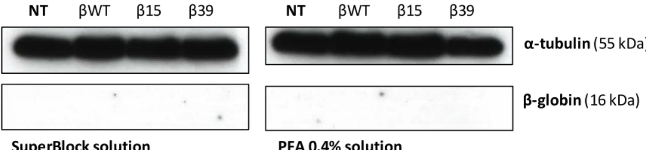
4.1. Human β-globin protein wasn’t detectable by western blot analysis ... 25
4.2. SOEing PCR was not successful enough as an approach to clone a FLAG-tag into the C-terminal region of Human β-globin... 26
5.
FINAL CONSIDERATIONS & FUTURE DIRECTIONS... 31
CHAPTER II ... 33
4.
RESULTS & DISCUSSION ... 35
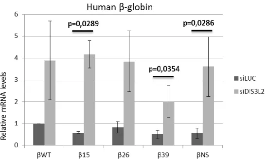
4.1. DIS3L2 knockdown correlates with increased levels of β-globin mRNAs independently from their decay mechanism... 35
4.2. hDIS3L2 knockdown correlates with increased HFE mRNA levels ... 36
5.
FINAL CONSIDERATIONS & FUTURE DIRECTIONS... 38
6.
REFERENCES ... 39
ANNEXES... 47
ANNEX I ... 48
XI
FIGURES
_______________________________________________________________________
Figure 1 – Canonical model of cap-dependent translation initiation. ... 3
Figure 2 – Translation Elongation ... 5
Figure 3 – Translation Termination. ... 6
Figure 4 - Simplified model of mammalian EJC-dependent NMD eliciting. ... 8
Figure 5 – Simplified decay pathway of human NMD ... 9
Figure 6 – Suppression of translation termination. ... 12
Figure 7 – Eukaryotic exosome structures. ... 15
Figure 8 – Model for Dis3 domains’ structure. ... 15
Figure 9 – mRNA degradation pathways in the cytoplasm of S. pombe ... 16
Figure 10 – Western blot analysis of cell lysates... 25
Figure 11 – Ilustrations for SOEing PCR 1, 2 and 3 ... 27
Figure 12 – Model for SOEing 4 PCR and Ligation to pTRE vectors ... 28
Figure 13 – Gel Electrophoresis of the SOEing PCR reactions products. ... 29
Figure 14 – Gel electrophoresis of the colony PCR products ... 30
Figure 15 – RT-qPCR analysis of Dis3L2 knockdown experiments performed for the
Human β-globin gene ... 35
Figure 16 – RT-qPCR analysis of Dis3L2 or UPF1 knockdown experiments performed for
the HFE gene ... 36
XII
ABBREVIATIONS
_____________________________________________________
A: Adenine A-site: aminoacyl-site Bp: base pairsCCR4: Chemokine (C-C Motif) receptor 4 cDNA: complementary deoxyribonucleic acid C-terminal: carboxyl-terminal
CD: (any) codon
CFTR: Cystic Fibrosis Transmembrane Conductance Regulator CSD: cold shock domain
DCP: decaping protein DEAD: Asp-Glu-Ala-Asp motif DECID: decay inducing complex
Dis3L: DIS3-Like Exosome 3'-5' Exoribonucleases DMEM: Dulbecco's modified Eagle medium DNA: deoxyribonucleic acid
eEF: eukaryotic translation elongation factors eIF: eukaryotic initiation factors
EJC: exon junction complex
eRF: eukaryotic translation release factor E-site: exit-site
FBS: foetal bovine serum G: guanine
GDP: guanosine diphosphate GTP: guanosine triphosphate HBB: Haemoglobin beta chain
HeLa: human cervical cancer cell line HFE: human hemochromatosis protein MAGOH: mago-nashi homolog
Met: metionin
Met-tRNAiMet: methionine-loaded initiator transfer ribonucleic acid
XIII miRNA: micro ribonucleic acid
mRNA: messenger ribonucleic acid
mRNP: messenger ribonucleoprotein particle NGD: no-go mRNA decay
NMD: nonsense-mediated mRNA decay NOT: negative regulator of transcription NSD: nonstop mRNA decay
nt: (any) nucleotide
N-terminal: amino-terminus ORF: open reading frame PABP: poly(A)-binding protein
PABPC1: cytoplasmic poly(A)-binding protein 1 PAGE: polyacrylamide gel electrophoresis PAN: poly(A) nuclease
P-bodies: Processing bodies PBS: phosphate buffered saline PCR: polymerase chain reaction PIC: pre-initiation complex PIN: PilT N terminus PLB: passive lysis buffer
PNPase: Polynucleotide phosphorylase Poly(A): poly-adenilate
PP2A: protein phosphatase 2A
Pre-mRNA: messenger ribonucleic acid precursor P-site: peptidyl-site
PTC: premature translation termination codon PVDF: polyvinylidene difluoride
RNA: ribonucleic acid RNB: Ribonuclease domain RNase: ribonuclease
Rrp: Ribosomal RNA processing protein RT: reverse transcription
XIV SDS: sodium dodecyl sulphate
sec: seconds
siRNA: small interfering ribonucleic acid
SMG: suppressor of morphological defects on genitalia SURF: SMG1-UPF1-eRFs complex
T: thymine
TBS: tris-buffered saline tRNA: transfer ribonucleic acid U: uracil
uORF: upstream open reading frame UPF: up-frameshift protein
UTR: untranslated region WT: wild type
XRN1: 5’-3’ exoribonuclease 1
β15: human β‐globin transcript with a PTC at codon 15 β26: human β‐globin transcript with a PTC at codon 26 β39: human β‐globin transcript with a PTC at codon 39
βNS: human β‐globin transcript without an in-frame stop codon βWT: normal/wild type human β‐globin transcript
XV
ABSTRACT
_______________________________________________________________________
Eukaryotic cells have developed elaborate mechanisms of mRNA quality control that secure gene expression fidelity through the detection and degradation of abnormal transcripts. NMD (nonsense-mediated mRNA decay), which detects and degrades transcripts containing premature translation termination codons (PTCs), and NSD (nonstop mRNA decay), that detects and degrades transcripts without in-frame stop codons, are just two examples. Nonsense-mediated mRNA decay (NMD) in particular, is a conserved surveillance system in all eukaryotic cells and is also the most extensively studied. PTC-containing mRNAs could, without NMD, give rise to C-terminally truncated proteins toxic for the cell. The physiological importance of NMD is further manifested by the fact that about one third of genetic disease-associated mutations generate PTCs. Recently, some studies have shown that aminoglycosides, low molecular weight compounds, and non -aminoglycosides can suppress PTCs in cystic fibrosis, Duchenne’s muscular dystrophy others, as a novel therapeutic approach, suppression therapy, which uses these compounds to induce recoding of a PTC into a sense codon. It is unclear whether β-thalassaemia would also be responsive to suppression therapy. Some recent studies show positive results for the compound PTC124 in suppressing nonsense mutations in the CFTR gene and others; also preliminary results obtained in our lab have shown that the aminoglycoside G418 can suppress a nonsense mutation at codon 39 of the human β -globin mRNA, although at low levels in cultured erythroid cells. As a first part of this work, we decided to investigate if suppression therapy can restore enough β-globin protein to correct the disease manifestations of β-thalassaemia. We intended to test whether G418 and/or PTC124 were able to induce efficient levels of suppression in a dose -dependent manner in HeLa cells transfected with plasmids containing the human β-globin wild type gene (βWT) or the other variants carrying a nonsense mutation at codon 15 (β15) or 39 (β39). However, we weren’t successful in this approach due to difficulties in gene cloning. The next step for RNAs targeted by NMD or NSD, as well as normal transcripts, which don’t accumulate indefinitely, is degradation. Generally the exosome complex, a multi-subunit ribonuclease complex, is responsible for the 3’-5’ degradation of every type of RNA in the cell, with its main catalytic component being either Dis3 or Dis3L1 in humans. However, another ribonuclease has been identified: DIS3L2. This protein is thought to be a
XVI cytoplasmic exosome-independent 3’-5’ ribonuclease, with special affinity for urydilated transcripts. Nonetheless, not much else is known for certain about its activity, especially in humans, including if it is coupled to NMD or NSD mRNA degradation. As a consequence, we intended to evaluate hDIS3L2’s possible involvement in mRNA degradation pathways, by performing knockdown of hDIS3L2, with siRNAs (small-interfering RNAs) in HeLa cells transfected with plasmids containing the human β-globin wild type gene (βWT), the variants carrying a nonsense mutation at codon 15 (β15), 26 (β 26) or 39 (β39), and also a variant lacking an in-frame stop codon (nonstop) (βNS). We then evaluated the human β-globin mRNA levels, as well as HFE’s, which is an NMD natural target. Our results show that human DIS3L2 is involved in NMD, NSD and possibly normal transcript degradation (mRNA turnover).
Key-words: Nonsense-mediated mRNA decay (NMD); Nonstop mRNA Decay (NSD); G418; PTC124; hDIS3L2.
XVII
RESUMO
_______________________________________________________________________
A expressão génica nos eucariotas envolve uma série de passos interligados e acoplados entre si, tendo a molécula de RNA (ribonucleic acid) como mensageiro entre os grandes passos. Resumidamente, a mensagem codificada pelas bases nucleotídicas do ácido desoxirribonucleico (DNA) (deoxyribonucleic acid) é transferida para uma molécula de RNA (transcrição), que, após processamento no núcleo, é transferida para o citoplasma onde é lida e transformada numa cadeia polipeptídica (tradução). Por vezes, contudo, podem ocorrer erros, em qualquer uma das fases da expressão, erros esses que podem resultar em mRNAs aberrantes que, se forem traduzidos, podem dar origem a proteínas truncadas com possíveis efeito deletérios. Para contornar este problema, as células eucarióticas desenvolveram mecanismos de controlo de qualidade do mRNA de modo a assegurarem a fidelidade da expressão génica através da detecção e degradação de transcritos aberrantes. O decaimento do mRNA mediado por mutações nonsense (NMD;
Nonsense-mediated mRNA decay) é o mais conhecido, detecta e degrada transcritos que contêm
codões de terminação prematuros (CTPs). O decaimento nonstop (NSD; Nonstop mRNA
decay) detecta e degrada transcritos que não possuem codões de terminação em fase na
grelha de leitura, mas existem outros, como o NGD (No-go decay) que evoluiu para lidar com os transcritos que possuem uma qualquer mutação que impeça a normal elongação da tradução. O NMD em particular, é um mecanismo de vigilância conservado em todas as células eucarióticas e é também o mais estudado. Os mRNAs que contêm CTPs poderiam dar origem, sem o NMD, a proteínas truncadas na extremidade C-terminal tóxicas para a célula, que podem adquirir um ganho de função prejudicial ou um efeito dominante -negativo. A importância fisiológica do NMD é ainda adicionalmente demonstrada p elo facto de que cerca de um terço das doenças genéticas associadas a doenças gerarem CTPs. Recentemente, alguns estudos têm vindo a demonstrar que compostos de baixo peso molecular, aminoglicósidos e não-aminoglicósidos podem suprimir CTPs em contexto de fibrose cística, distrofia muscular de Duchenne e difeciência em carnitina palmitoltransferase 1A como uma nova abordagem terapêutica, a terapia de supressão, a qual usa estes compostos para induzir a recodificação de um codão nonsense num codão
sense. Uma doença também associada a mutações nonsense é a talassémia. A
XVIII os fenótipos mais agressivos requer inevitavelmente transfusões regulares sanguíneas, com os riscos que isso acarreta como a acumulação excessiva de ferro no organismo. Uma cura que se possa chamar definitiva ainda não existe e, portanto, qualquer nova abordagem terapêutica constitui uma mais valia. O NMD é um modelador do fenótipo da β-talassémia, podendo contribuir para o melhoramento das manifestações da doença. Em relação à terapia de supressão, permanece ainda por esclarecer se a β-talassémia responderia também à mesma. Alguns estudos recentes mostram resultados positivos para o composto PTC124 (ou Ataluren) na supressão de mutações nonsense no gene da CFTR (associadas à Fibrose Cística) bem como noutros genes, associados à Distorifia Muscular de Duchenne, Síndrome de Usher e Defeciência em Carnitina Palmitoltransferase 1C; para além disso, resultados obtidos previamente pelo nosso laboratório demonstraram que o aminoglicósido G418 pode suprimir uma mutação nonsense no codão 39 do mRNA do gene da β-globina humana, embora a baixos níveis em células eritróides em cultura. Tendo em conta esta informação, como uma primeira parte deste trabalho, decidimos investigar se a terapia de supressão pode restaurar β-globina suficiente para conseguir corrigir as manifestações da β-talassémia. Propusémo-nos a testar se o G418 e/ou o PTC124 seriam capazes de induzir níveis suficientes de supressão, duma maneira dependente da dose, em células HeLa transfectadas com plasmídeos que contêm o gene da β-globina humana, variante selvagem, wild type (βWT), ou as outras variantes contendo uma mutação nonsense no codão 15 (β15) ou 39 (β39). Contudo, não fomos bem sucedidos nesta primeira parte do projecto devido a dificuldades intransponíveis associadas à clonagem de genes.
O próximo passo para os RNAs sinalizados pelas maquinarias do NMD ou do NSD, bem como os RNAs normais, que não se acumulam indefinidamente, é a degradação. A degradação é, na verdade, uma parte essencial do metabolismo do mRNA, constituindo até um mecanismo pelo qual a expressão génica é regulada. Na degradação do mRNA, o exossoma é um componente essencial. O exossoma eucariótico é um complexo ribonucleolítico multi-subunidade, e é responsável pela degradação na direcção 3’-5’ de todos os tipos de RNA na célula, entre outras coisas. As proteínas humanas Dis3 ou Dis3L1 são os seus elementos catalíticos. Contudo, outra ribonuclease foi também identificada, a Dis3L2, da qual pouco ainda se sabe, especialmente em humanos. Pensa-se que esta proteína seja uma ribonuclease citoplasmática na direcção 3’-5’, independente do exossoma, que parece ter especial afinidade para transcritos uridilados. Tendo em conta
XIX as lacunas no conhecimento acerca da Dis3L2 humana (hDis3L2) e da importância dos mecanismos de vigilância do mRNA e da sua degradação, decidimos avaliar a possibilidade da Dis3L2 humana estar efectivamente envolvida nas vias de degradação do mRNA NMD e/ou NSD. Para esse efeito, foram efectuadas experiências de silenciamento do gene da Dis3L2 humana recorrendo à tecnologia de RNA de interferência, mais especificamente siRNAs (small interfering RNAs) em células HeLa transfectadas com plasmídeos contendo a variante selvagem do gene da β-globina humana (βWT), com as variantes contendo uma mutação nonsense no codão 15 (β15), 26 (β26) ou 39 (β39), e também a variante que não contém um codão de terminação na grelha de leitura, nonstop, (βNS). Seguidamente avaliámos os níveis celulares de mRNA da β-globina humana, bem como os do HFE, que é um alvo natural do NMD. Os nossos resultados sugerem que a hDis3L2 está envolvida no NMD, NSD e possivelmente também na degradação de transcritos normais e consequentemente no turnover do RNA.
Palavras-chave: Decaimento do mRNA mediado por mutações nonsense (NMD); Decaimento do mRNA nonstop (NSD); G418; PTC124; hDIS3L2.
1
1. INTRODUCTION
_______________________________________________________________________
1.1. Gene Expression in Eukaryotes
The central dogma of molecular biology states that protein synthesis starts with transcription of a DNA template into a complementary messenger RNA molecule (mRNA) in the nucleus of the eukaryotic cell, which is then transported to the cytoplasm where it will be translated into protein by the ribosomes1. This seemingly straightforward mechanism involves, in reality, a series of interconnected steps and a complex coupled network of machineries that guarantees the efficiency and specificity of gene expression, with messenger RNAs being the key intermediaries throughout the whole process2,3. Briefly, the DNA is transcribed into a precursor mRNA (pre-mRNA) molecule by the ribosome, to which a 5’cap and a 3’poly(A) tail will be added mainly for protection against degradation. Also this pre-mRNA molecule will be subject to splicing, which consists in removing introns and joining together the exons. By the end of this stage we have a mature mRNA molecule. This mRNA is transported to the cytoplasm where it will finally be translated into protein and eventually degraded3,4.
However, sometimes errors occur, at any step of gene expression, and aberrant mRNAs are produced4. If translated, these result in aberrant proteins that can be detrimental to the cell5. To deal with this problem, evolution came up with mRNA surveillance mechanisms. These mechanisms target and degrade aberrant transcripts before they can be translated thus avoiding the possible deleterious effects of aberrant proteins. There are three main mRNA surveillance pathways, although they are not the only ones, which are: Nonsense-Mediated Decay (NMD), Nonstop Decay (NSD) and No-Go Decay (NGD); they target and degrade, respectively, transcripts containing a premature termination codon (PTC), mRNAs lacking a termination codon and mRNAs containing any mutation that stalls the ribosome and impedes normal translation elongation6. Of these three pathways, NMD and NSD are the ones relevant for this study.
1.2. mRNA Translation
One of the most important steps in mRNA life and gene expression is the translation of messenger RNA into protein. Like virtually all the steps in gene expression, mRNA
2 translation is a tightly regulated process, aiding the fine tuning of eukaryotic gene regulation7. In order to better understand the translation-dependent mechanisms of NMD and NSD, as well as gene therapy itself, it is useful to get familiar with the basic concepts of mRNA translation.
Translation is conceptually divided into three stages (initiation, elongation and termination) where the codon sequence in the mRNA directs the synthesis of a polypeptide chain, with ribosomes, which are two-subunit ribonucleoproteins, being the key players in the reaction8,9,10. The three stages are explained bellow.
1.2.1. Translation Initiation
In the canonical, cap-dependent, mechanism of translation, the interaction of the ternary complex with the 40S ribosomal subunit marks the beginning. This complex is formed by the eukaryotic initiation factor eIF2 bound to both the methionyl-initiator tRNA (Met-tRNAiMet) and GTP11. When the ternary complex, along with eIF1, 1A, 3 and 5, binds to the 40S ribosomal subunit, the 43S pre-initiation complex (PIC) is formed7,11,12,13. This complex, according to Kozak’s scanning model has the task of scanning the 5’UTR (5’ untranslated region) until it finds the AUG initiation codon, where translation starts14. But before that happens, another complex must be present: eIF4F. The eukaryotic initiation factor 4F recognizes the 5’-cap proximal region of mRNA and its functions are illustrated by each of its three comprising elements: the ATP-dependent DEAD-box RNA-helicase eIF4A, which resolves secondary structures in the 5’UTR, making the scanning process possible; eIF4E which is the cap-binding element; and eIF4G that acts as a scaffold protein for the interaction of the other two and also interacts with the poly(A)-binding protein (PABP) and by doing so, circularizes the mRNP (messenger ribonucleoprotein) molecule, because PABP is bound to the 3´-poly(A)-tail15,16,17.
When both complexes, eIF4F and PIC, are present and bound to their respective targets, scanning, by PIC, of the 5’UTR in the 5’-3’ direction takes place until it finds an AUG codon in an optimum context, which is the start codon. When this happens, complementary base pairing takes place between the start codon, which by now is at the peptidyl-tRNA (P) site of the 40S subunit, and the anticodon present in the Met-tRNAiMet. Then, eIF5 activates GTPases, which hydrolyze the ternary complexes’ GTP bound to the eIF2 and this triggers the displacement of the initiation factors, eventually leading to the joining of the 60S
3 ribosomal subunit, catalyzed by eIF5B. The 80S eukaryotic ribosome is formed and the translation initiation phase has ended18,19,20. The entire process is pictured in figure 1.
Figure 1 – Canonical model of cap-dependent translation initiation. A: The ternary
Complex (TC) binds to the 40S ribosomal subunit, along with eIF1, 1A, 3 and 5 forming the 43S pre-initiation complex (PIC). B: Also, eIF4F recognizes the 5’-cap proximal region of mRNA and interacts with PABP via the eIF4G element. C: When both eIF4F and PIC are bound to the mRNP, PIC scans the 5’UTR in the 5’-3’ direction until it finds the AUG initiation codon. D: The start codon is recognized, complementary base pairing takes place between the nucleotide elements of the Met-tRNAiMet and the
mRNP. Then, eIF5 activates GTPases, which hydrolyze the ternary complexes’ GTP bound to the eIF2, triggering the displacement of the initiation factors. E: Joining of the 60S ribosomal subunit to the 40S takes place, catalyzed by eIF5B. The 80S eukaryotic ribosome is formed and the translation initiati on phase is complete. Adapted from Sonenberg & Hinnebusch (2009).
A B
C
D
4 1.2.2. Translation Elongation
This stage consists of the sequential decoding of the mRNA nucleotide sequence, in the 5’ -3’ direction, into a polypeptide chain21,22; the order of the mRNA codons specifies the order in which amino acids are added23.
By the end of initiation, we are left with the initiator aminoacyl-tRNA located at the P-site of the ribosome, base-paired with the start codon, and the adjacent A-site empty and ready to receive a cognate aminoacyl-tRNA, thereby continuing the decoding process of mRNA into protein10,11,18,24. In this sequential process, the eukaryotic elongation factor eEF1A has the task of bringing the aminoacyl-tRNAs to the ribosome A-site: if codon and anticodon are complementary, then eEF1A activates GTP hydrolysis, which in turn induces eEF1A release, thereby permitting the entry of aminoacyl-tRNA to the A site24,. Then, the two tRNAs present at the moment at the ribosome are translocated, thanks to the aid of the GTP-dependent eEF2: the deacylated tRNA is translocated from the P to the E-site (E from “exit”) and the peptidyl-tRNA from the A to the P-site (figure 2) and the ribosome moves 3 nucleotides in the 5’-3’ direction23. Also eEF1A interacts with eEF1B, which promotes the exchange of GDP for GTP, recycling the GTP-bound eEF1A23. After translocation, the A-site is once again empty and ready for the next aminoacyl-tRNA. This process goes on until the ribosome reaches a stop codon.
5 Figure 2 – Translation Elongation. Starting at the top: the elongation factor eEF1A brings the aminoacyl-tRNA to the ribosomal A-site, where complementary base pairing occurs. Then eEF1A activates GTP hydrolysis, inducing eEF1A release, which allows the entry of the aminoacyl -tRNA to the A-site. GDP is recycled to eEF1A-GTP by the exchange factor eEF1B. After the tRNA accommodation into the A-site, peptide bond formation occurs. Then, the two tRNAs present at the ribosome are translocated, by binding of the GTP-dependent eEF2. The ribosome is now ready for the next cycle of elongation with release of the deacylated tRNA from the E-site and binding of the appropriate eEF1A-GTP-aminoacyl-tRNA to the A-site. In this figure GTP is depicted as a green circle and GDP as a red one.
In Dever & Green (2012).
1.2.3. Translation Termination
Translation termination takes place when the ribosome recognizes a stop codon, making the whole translation process come to an end and the nascent protein is released25. It all starts when a stop codon, either UAG, UGA or UAA, is present at the A -site. Because there is no cognate aminoacyl-tRNA for a stop codon, the ribosome stalls and eRF1 recognizes the stop codon instead and also forms a complex with the GTPase eRF3, which causes GTP hydrolysis. This hydrolysis, in turn, induces a conformational change in eRF1 that allows it to move closer to the P-site and promote cleavage of the peptidyl-tRNA ester bond, finally leading to the release of the newly synthesized polypeptide chain23,26,27 and both ribosome subunits dissociate, ready to be recycled23,24,28.
6 Figure 3 – Translation Termination. From left to right, when a stop codon, UAG, UGA or UAA, is present at the A-site, the ribosome stalls and eRF1, complexed with the GTPase eRF3, recognizes the stop codon, which causes GTP hydrolysis. This hydrolysis induces a conformational change in eRF1 that allows it to move closer to the P-site and promote cleavage of the peptidyl -tRNA ester bond, leading to the release of the newly synthesized polypeptide chain. Adapted from Keelint et al. (2014).
1.3. Nonsense Mediated mRNA Decay
NMD is a post-translational surveillance mechanism that is evolutionary conserved in eukaryotic cells. It is also the most studied quality control mechanism in eukaryotes6. Briefly, NMD function is to identify and eliminate mRNAs that contain premature translation termination codons (PTCs), also known as nonsense codons, thus preventing the synthesis and accumulation of C-terminally truncated proteins that can have a dominant-negative or gain-of-function effect potentially deleterious for the cell4,26,29,30,31. It is important to note, however, that in mammals there are other quality control mechanisms, such as NSD (nonstop mRNA decay), which we will also approach in this work, and NGD (no-go mRNA decay) 8,6. In order to understand the type of gene therapy that we tried to achieve in this work, which was one where we used the knowledge of the NMD mechanism to our advantage, one must first comprehend NMD itself.
1.3.1. PTC Recognition and NMD Eliciting
Generally, PTCs can arise from nonsense or frameshift mutations in the DNA sequence, transcription errors, anomalous splicing events, among others31,32,33. When these PTCs are
7 located at more than 50-55 (or 50-54 depending on the references; for convenience purposes, we will refer to this boundary as 50-55) nucleotides (nts) upstream the last exon-exon junction, they are indeed recognized as what they are, aberrant stop codons, and are thus able to elicit NMD, but if they are located downstream of this boundary, NMD is not elicited4,27,32. This happens as such because, when pre-mRNA splicing occurs in the nucleus of the eukaryotic cell, multiprotein complexes appropriately called exon junction complexes (EJCs) are deposited approximately 20-24 nts upstream of each exon-exon junction. During the pioneer round of translation, the 80S ribosome displaces the EJCs assembled in the open reading frame (ORF), but, if the transcript has a PTC positioned at ≥ 50-55 nts upstream the last exon-exon junction, the ribosome will reach a stop codon (which is, obviously, premature in this case) before it actually can be in a close enough position to displace the next EJC (figure 4) and therefore, the EJC will not be removed as it normally would34,35. The EJC will trigger recruitment the NMD factors UPF3 and UPF2, which may be attributable to the MAGOH/Y14 heterodimer (a component of the EJC)31,36. It has been shown, however, that AUG-proximal PTCs fail to elicit NMD, in spite of fulfilling the rule of being positioned at ≥ 50-55 nts upstream the last exon-exon junction; this implies that the EJC-dependent model is not the only mechanism by which NMD is triggered32,37. How is NMD elicited then? Work previously done in our lab, as well as other studies, have suggested that the mechanism behind this is a competitive relationship between PABP and UPF1 for binding eRF3. When PABP is in close proximity to a PTC (which happens when the PTC is AUG-proximal; this is made possible by the circular conformation of the mRNA), it interacts with the release factor eRF3 and promotes translation termination; however when the PTC is not physically close enough to PABP, then it won’t interact with eRF3, instead, the NMD factor UPF1 will, and this in turn will trigger NMD38,39,40.
8 Figure 4 - Simplified model of mammalian EJC-dependent NMD eliciting. On the left, a PTC is located more than 50-55 nts upstream the last exon-exon junction, therefore it will elicit NMD and hence, degradation of the transcript will occur. On the right, there’s a PTC located within the 50-55 nts boundary and as such it won’t be recognized by the NMD machinery and, as a consequence, that transcript won’t be rapidly degraded and will result in a truncated protein. In the figure, “Ter” represents the canonical termination codon. In Khajavi et al. (2006).
1.3.2. mRNA Degradation via NMD
Following PTC recognition, the next step for the NMD machinery is the degradation of the faulty mRNA molecule. However, unlike the previous step, mRNA degradation via NMD is not a very well understood process to date; still, some studies did manage to provide insights and models.
It is known that, in NMD, phosphorylated UPF1 has better affinity for RNA and marks the mRNA for degradation27,41. UPF1 phosphorylation occurs as follows: once UPF1 has been recruited to the terminating ribosome, as it has already been mentioned, it will interact with eRF3 and form the SURF complex (SMG1-UPF1-eRF1-eRF3)42. The SURF complex, via UPF1, binds to UPF2 and UPF3, the former, in turn, binds to the EJC; all these elements together constitute the DECID complex (Decay-Inducing Complex). Finally, the DECID complex promotes UPF1 phosphorylation by SMG143,44,45,46. This phosphorylated UPF1 recruits either SMG6, which has endonucleolytic activity, or the SMG7/SMG5 heterodimer46.
If SMG6 is the one recruited and interacts with phosphorylated UPF1, endonucleolytic cleavage of the transcript in the vicinity of the PTC occurs by the action of SMG6 itself. This cleavage produces two cleavage products: one 3’ product, which contains the EJC and NMD components, and another 5’ product that includes the PTC. The 5’ cleavage product undergoes 3’-5’ degradation, as it seems, by the exosome, while the 3’ product is first stripped off of its proteins by the action of UPF1 (because when it binds UPF2, as is the
9 case in the DECID complex, UPF1’s helicase activity is enabled) and then is cleaved in the 5’-3’ direction by XRN1 following decaping47,48,49.
If, however, SMG7/SMG5 heterodimer is recruited, then its interaction with the phosphorylated UPF1 induces deadenylation, carried out by the PAN2/PAN3 and C CR4-NOT complex; also, decaping by the action of DCP1/DCP2 takes place. This allows the transcript to be degraded in the 3’-5’ direction by the exosome and in the 5’-3’ direction by the exonuclease XRN132,50,51,52.
Additionally, SMG7 and SMG6 recruit protein phosphatase 2a (PP2A), which induces UPF1 dephosphorylation and dissociation, thereby allowing recycling for the next round of NMD53. The mRNA degradation via NMD pathway is simplified bellow on figure 5.
Figure 5 – Simplified decay pathway of human NMD. UPF1 binds to the stalled ribosome through interaction with eRF3 and is subsequently phosphorylated. Phosphorylated UPF1 binds either SMG6 or the SMG5/SMG7 heterodimer. Binding of the endonuclease SMG6 results in RNA cleavage in the vicinity of the PTC, producing decay intermediates that will be rapidly degraded in both 5’-3’ (light blue PacMan) and 3’-5’ directions (violet PacMan) by exonucleases. On the other hand, binding of SMG5/SMG7 results in the recruitment of deadenylases (purple PacMan) and decaping enzymes (dark PacMan). After being both decapped and deadenylated, the RNA is degraded by exonucleases. In Mühlemann & Lykke-Andersen (2010).
10 1.3.3. NMD Targets
During recent years, it has been increasingly clear that NMD isn’t simply an mRNA quality control mechanism targeting nonsense mRNAs, as wide variety of physiological transcripts that don’t contain any PTC whatsoever are regulated by it54,55. In fact, studies have revealed that NMD participates in the control of steady-state levels of 3-10% of the transcriptome56. So the question arises: what renders these non-faulty mRNAs NMD-sensitive? Some characteristics possessed by these NMD-sensitive transcripts include upstream ORFs (uORFs), long 3’UTRs or signals for programmed frameshift; mRNAs encoding proteins that contain selenocysteine, where the UGA codon is interpreted either as a stop codon or a PTC depending on selenium concentration; bicistronic mRNAs, among others33,57,58. Many of these aforementioned features make the stop codons of these mRNAs, called natural NMD targets, to be in a position where they are recognized as de
facto PTCs by the NMD machinery, in spite of them being canonical ones. NMD natural
targets perform a variety of functions in the cell, ranging from regulation of alternative splicing to stress response and cell-cycle progression59,60. One of those natural NMD targets, which is important in this work, is HFE mRNA, whose gene, when mutated, might be involved in hereditary hemochromatosis, a disease of iron metabolism where an excessive intestinal iron absorption is present, leading to organ damage59.
1.3.4. NMD and Human Disease
One can easily infer the clinical relevance of NMD from the fact that approximately one third of all human genetic disorders are attributable to PTCs, many of which are NMD targets29,61. There are several disease phenotypes that result from nonsense or frameshift mutations that are indeed modulated by NMD29,34. Examples of disease clinical phenotypes modulated by NMD include cystic fibrosis, Duchenne muscular dystrophy and β-thalassemia, among others.
In diseases where NMD is a clinical manifestation modulator, the surveillance mechanism impedes the nonsense mRNA to be translated into truncated proteins. These truncated proteins can have lost their function, have acquired a dominant-negative effect or gain of function, but they can also be still functional or residually functional and in those cases, it’s obvious that NMD, instead of protecting the cell against the deleterious effect of proteins resulting from faulty mRNAs, is in fact having a detrimental effect instead. So, NMD can either be beneficial or detrimental to the clinical manifestation of a given disease26,30.
11 Cystic fibrosis constitutes an example where NMD worsens the clinical outcome of a disease. Some truncated proteins encoded by the mutated CFTR gene partially retain normal function, however, NMD targets and degrades their PTC-containing mRNAs, thereby downregulating those proteins, which of course aggravates the clinical manifestations of the disease26,62.
In β-thalassemia, on the other hand, NMD has the opposite effect; it has a well-documented beneficial role. PTC-containing β-globin mRNAs are targeted and degraded by NMD and this prevents the synthesis of C-terminally truncated proteins toxic for the cell26,63.
1.3.5. β-thalassemia as a Model
The β-thalassemias comprise a heterogeneous group of inherited human anaemias, and are among the most common genetic diseases worldwide arising most frequently as a result of nonsense or frameshift mutations in the β-globin gene. It generally has a pattern of autosomal recessive inheritance, except when nonsense mutations occur on the last exon, being dominant in that case. The disease is characterized by reduced or absent β-globin chains, which causes low levels of haemoβ-globin and reduced production of red blood cells64,65.
Several nonsense mutations have been characterized for the β-globin gene, of with CD15, CD26 and CD39 are of particular importance for this study. In CD15-thalassemia the PTC, being on codon 15, is near the 3’-end of the transcript and this proximity to PABP will inhibit NMD, making this transcript produce a C-terminally truncated protein. In CD39-thalassemia, however, there is no AUG-proximal effect and this PTC will elicit NMD66,67. The profusion of this disease in the world, the small size of the β-globin gene and the wide range of nonsense mutations that it can harbour, “reacting” in different ways to NMD, are all reasons that turn this genetic disorder a good model for studying this surveillance mechanism64,66.
1.3.6. Nonsense Suppression Therapy
In the most aggressive manifestation of β-thalassemia (Thalassemia Major) and in some phenotypes of Thalassemia Intermedia, patients require regular red blood cell transfusions and appropriate chelation therapy to counterbalance the iron overload often present due to those necessary transfusions. Bone marrow transplantation remains the only cure, but it doesn’t constitute by any means a true solid hope for patients, given the compatibility
12 issues and other risk factors like the extent of iron accumulation present at the time of transplantation. Given the fact that NMD is a modulator of the clinical manifestation of β-thalassemia, it constitutes a promising and needed therapeutic strategy for this disease and many others caused by PTC-generating mutations that don’t have effective treatments22,33,64,68. At the current point in time, these therapies have already been analysed in clinical trials for Cystic Fibrosis and Duchenne’s muscular dystrophy69.
In nonsense suppression therapy, one takes advantage of the fact that some truncated proteins arising from PTC-containing mRNAs are still functional but, however, they do elicit NMD and are therefore down-regulated. If this NMD eliciting could be hampered, then the protein would still be produced and hence at least phenotype amelioration of the disease would take place33,70. The way this is done is through the PTC “readthrough” strategy, which is the process that recodes a stop codon, in this case premature, into a sense codon, thereby allowing translation elongation to continue until the canonical stop codon is reached and a full-length protein is produced, which would otherwise be impossible70. Mechanistically, suppression therapy works by facilitating the ability of near-cognate aminoacyl tRNAs (tRNAs that are complementary for two of the three codon nucleotides) to be incorporated into the ribosomal A-site (figure 6), thereby effectively increasing the number of times a PTC will be read as a sense codon and a full -length protein is produced, restoring enough normal protein function in the cell70.
Figure 6 – Suppression of translation termination. From left to right, a near-cognate aminoacyl-tRNA complementary base pairs (at two of the three bases , of course) with the premature termination codon with the help of eEF1A. The aminoacid carried by this near-cognate tRNA is added to the growing polypeptide chain, translocation occurs and premature translation termination is aborted, taking place normal translation elongation instead, until the canonical stop codon is eventually reached. Adapted from Keeling & Bedwell (2011).
13 The ribosomal readthrough is accomplished by the use of small molecular weight compounds, namely aminoglycoside antibiotics such us gentamicin and geneticin (G418), and also non-aminoglycoside compounds such as ataluren (PTC124). This drugs bind to the ribosomal decoding centre, decreasing the accuracy of complementary base pairing between codon and anticodon and, in this way, facilitates near-cognate aminoacyl-tRNA incorporation. Aminoglycosides have antibacterial activity, may cause cell toxicity and, at an organismal level, nephrotoxicity; PTC124, a non-aminoglycoside, on the other hand, is free from the previously mention disadvantages, however its effectiveness is controversial, with one study71 reporting that there is no evidence for PTC inducing readthrough, however, the majority of studies point to successful PTC readthrough in mammalian cells and it is currently undergoing clinical trials for several diseases33,70,72,73,74,75,76.
Suppression therapy efficiency, besides being different for different compounds and depending on each individual, also depends on the PTC nature, the ranking order for aminoglycoside readthrough efficiency being UGA≥UAG>UAA; and PTC context: in this regard, the nucleotide immediately downstream of the PTC is the most important one, being the ranking order of readthrough efficiency according to the it, C>U>A>G, but up to 6 nucleotides downstream and 2 nucleotides upstream of the PTC can have an effect on readthrough efficiency77,70,69.
1.4. Nonstop mRNA Decay
Nonstop decay is by far much less well understood than NMD and this lack of knowledge is even more apparent for human cells; a great amount of what is known for the process relates to yeasts. Nonstop decay has evolved as a surveillance mechanism that targets and degrades transcripts that lack an in-frame stop codon. These transcripts don’t allow normal translation termination to take place and instead the ribosome reads the ORF and into the 3’UTR, where it eventually stalls, as translation of poly(A) results in positively charged poly-lysine that interacts with the very negative exit channel of the ribosome ; as little as 6 incorporated lysine residues are enough to incite ribosome stalling8,78,79,80. Nonstop mRNAs can arise from nonstop or frameshift mutations in the DNA that disrupt the stop codon, instances when transcription aborts or even from cryptic poly(A) sites, which cause premature polyadenylation80,81,82. In particular, when cryptic poly(A) sites are present, upstream of the stop codon, these mRNAs can give rise to truncated proteins that, in similarity with NMD, can acquire deleterious dominant negative or gain of function
14 effects79,80,83. It is estimated that nonstop mRNAs can arise from premature polyadenylation 5-10% of the time83,84. However, there are many questions regarding the initial recognition of NSD-targeted ribosome complexes on a mechanistic level8.
In yeasts, nonstop mRNA degradation probably starts when the Ski7 GTPase recognizes the stalled ribosome through its C-terminal domain, promoting transcript degradation by the exosome83,85. Ski7 belongs to a family of proteins that also includes the eukaryotic release factor 3 (eRF3) and the eukaryotic elongation factor eEF1α, however, Ski7 is different in two ways: it doesn’t act as an heterodimer and may not be dependent on GTP, which is an integral part of the specificity of both eRF3 and eEF1α86,87,85. Nonstop mRNA degradation, as it has already been previously mentioned, is accomplished by the 3’ -5’ exonucleolytic action of the exosome, without the need for prior deadenylation82.
1.5. Eukaryotic Exosome
The exosome, as well as its cytoplasmic factors are conserved in all eukaryotes88,89. It is a 3’-5’ multi-subunit ribonuclease complex of approximately 400 kDa, present in nuclear, cytoplasmic and nucleolar forms, whose main function is degrading almost every type of RNA in the cell, thereby guaranteeing the following: the degradation of RNA processing by-products, elimination of aberrant transcripts in various mRNA surveillance pathways (including NMD and NSD), removal of incorrectly spliced mRNAs and it also prevents defective rRNA incorporation into the ribosome structure. Furthermore the exosome plays a role in RNA molecules maturation in the nucleus, such as ribosomal RNAs, for instance90,91,92,93.
The eukaryotic exosome has a conserved structure constituted by a 9-subunit core, in similarity with bacterial PNPase and the archaeal exosome, which is made up of a hexameric ring and a “cap” consisting of three RNA-binding subunits (figure 7). Depending on the subcellular localization, to this 9-subunit core can bind Rrp44 (Ribosomal RNA Processing Protein) also known as Dis3, Rrp6, or both Rrp44 (Dis3) and Rrp6; it is possible that the exosome co-factors influence how a “binding path” is chosen (Rrp6 or Dis3) by competing for interaction with the exosome core or by influencing activities of both subunits94,95.
15 Figure 7 – Eukaryotic exosome structures. On the left, we can see the cytoplasmic 10-subunit exosome, at the centre it is pictured the canonical nuclear 11-10-subunit exosome and on the right, we have the nucleolar 10-subunit exosome. The 9-subunit core is present in all three forms. In the figure, the red circles represent phosphorolytic and hydrolytic exoribonuclease active sites and the pale yellow circles represent endoribonuclease active sites. Adapted from Januszyk & Lima (2014).
Dis3 is the largest subunit of the exosome (approximately 110 kDa) and the responsible for the major exoribonucleolytic activity of the complex. Dis3 is indispensable for cellular viability and is predominantly present in the nucleus, but also in the cytoplasm . Its structure consists of: an N-terminal pilus-forming N-terminus domain (PIN) with three aspartic acid residues; the PIN domain mediates the majority of the contacts with the N -terminal of the Rrp41 subunit of the exosome core, so its main function is really to bind Dis3 to the core; two cold shock domains (CSD1 and CSD2) responsible for RNA-binding; central ribonuclease domain (RNB), which contains an active site for exoribonuclease activity; and a C-terminal S1 domain also responsible for RNA binding (figure 8). Dis3 binds both to Rrp41 and Rrp4594,95.
Figure 8 – Model for Dis3 domains’ structure. On the left one can see the N-terminal exosome-binding domain, PIN, followed by the RNA-binding CDS domains. Next are the catalytic RNB site and the C-terminal RNA-binding S1 domain. In Tomecki et al. (2010).
1.6. Human Dis3L2
Recent studies indicate that human DisL2 (DIS3 Like 3'-5' Exoribonuclease 2) protein is a cytoplasmic processive 3’-5’ ribonuclease that contributes to RNA homeostasis and whose activity is independent to that of the exosome. Regarding its structure, it is known that it lacks a PIN domain, hence it not binding to the exosome. Furthermore, Dis3L2 doesn’t
co-16 precipitate or co-localize with the exosome components. It also has an extended RNA-binding CSD1 domain, which can explain Dis3L2’s stronger activity for structured substrates with short two nucleotide overhangs or blunt-ended double stranded RNA when compared to hDis396,97. Dis3L2 main targets constitute poly-urydilated transcripts (figure 9), being inhibited by poly(A) sequences, and it has recently been linked to apoptosis-triggered global RNA degradation, which involves 3’-urydilated intermediaries96,98. Transcript urydilation can trigger decaping and 5’-3’ degradation and protect 3’ ends from exoribonucleases; however, it can also promote 3’-5’ degradation. This only makes sense because of the spatial separation between 5’ -3’ degradation and decaping, as well as Dis3L2 localization on docked structures to processing bodies (P-bodies), which is where 5’-3’ degradation takes place in the eukaryotic cell; even more, under stress conditions, Dis3L2 completely co-localizes with P-bodies, which can be a way of accelerating the degradation of urydilated RNAs96,97. Recently, human Dis3L2’s role in mRNA metabolism has been highlighted and it has been seen that it may well be essential for cell growth regulation and division, as its depletion causes mitotic abnormalities due to deregulation of mitotic control proteins, being associated with Perlman syndrome and susceptibility to Wilms tumor99; furthermore, it has a role in target directed miRNA degradation100. Weather Dis3L2 is coupled to NMD or NSD is still unclear.
Figure 9 – mRNA degradation pathways in the cytoplasm of S. pombe. From left to right, mRNAs can de degraded in the 5’-3’ direction by Xrn1 exonuclease, in the P-bodies; in the 3’-5’ direction by Dis3L2 that can be localized on P-bodies or not; and again in the 3’-5’ direction by the exosome in the cytoplasm. What applies to S. pombe may also apply to humans. In Malecki et al. (2013).
17
2. AIMS
_______________________________________________________________________
The aims for this work are divided in two parts, corresponding to the two chapters of this dissertation.
2.1. Chapter I
β-thalassemia is one of the most common genetic diseases worldwide and its treatment can be bothersome and inconvenient, with no risk-free and easy to attain cure64,65,68. Furthermore, β-thalassemia happens mainly as a result of nonsense mRNAs committed to degradation by NMD; with NMD being a clinical modulator of the manifestations of this disorder, it constitutes a hope for new treatments26,63,101. Under that light, if one can use suppression therapy to induce PTC readthrough on the faulty mRNAs that trigger this disease, one would, in theory, be able to induce synthesis of full-length protein that could maintain normal physiological function. Already preliminary results in our lab have shown that G418 can suppress a PTC at codon 39 of the human β-globin mRNA, although at low levels in cultured erythroid cells; also, some recent studies have shown high readthrough efficiencies for a non-aminoglycoside compound, PTC12474,75,76.
The aim for chapter I of this work was to test weather suppression therapy can restore enough β-globin to correct the manifestations of the disease and, under that light, we decided to test weather β15 (nonsense mutation CD15) or β39 (nonsense CD39) mutated versions of the HBB gene allow their suppression by either G418 or PTC124 to occur, in a dose-dependent manner, using for this effect HeLa cells transiently transfected with plasmids containing either the wild-type (βWT), β15 or β39 versions of the human β-globin gene.
2.2. Chapter II
DIS3L2 is a novel 3’-5’ exonuclease independent of the exosome of which little is yet known, especially in humans96,97. Currently it is known that this protein has a preference for poly-urydilated transcripts, but either if this protein is linked to the degradation coupled to surveillance mechanisms or just to global urydilated mRNA is yet to be known96,97,98,99. Also, recently, mutations in the germline DIS3L2 gene have been linked to mitotic abnormalities and human diseases99. Given the fact that surveillance mechanisms are vital to the cell and are involved in many human diseases, and taking into account the
18 possible important role in human disease and the current gap in the knowledge regarding hDis3L2, it would be of interest investigate if this protein is involved in the degradation coupled to surveillance mechanisms, augmenting the overall knowledge of mRNA metabolism.
The aim for the chapter II of this work was to investigate if hDis3L2 is i nvolved in the NMD and/or NSD mRNA degradation, as well as normal transcript degradation. To this effect, we utilized a knockdown approach using siRNAs specific to hDis3L2 in HeLa cells transiently transfected with plasmids containing either the βWT, β15¸ β26, β39 or βNS (a variant which lacks a stop codon) mutated version of the human β-globin gene.
19
3. MATERIALS AND METHODS
_______________________________________________________________________
3.1. Plasmid Constructs
The plasmids containing βWT (wild type version of the β-globin gene), β15 (with nonsense mutation at codon 15) [CD 15 (TGG→TGA)], β26 (with nonsense mutation at codon 26) [CD 26 (GAG→TAG)], β39 (with nonsense mutation at codon 39) [CD 39 (CAG→TAG)], or βNS (without the canonical stop codon) of the human β-globin gene were obtained as previously described in Silva et al. (2006). All variants were created within the 428-bp NcoI-BamHI fragment of the β-globin gene template by overlap-extension PCR. Competent
Escherichia coli were transformed with the plasmid DNA, and transformants were selected
on luria-bertani (LB) agar/ampicillin plates. The corresponding plasmid DNAs were purified from overnight cultures of single colonies with the NZYMini prep kit (NZYTech, Portugal) following the manufacturer’s instructions. Confirmation of the correct cloned sequences containing the relevant mutation was carried out by automatic sequencing.
3.2. Cell Culture and Plasmid Transfections
HeLa cells were grown in Dulbecco’s modified Eagle’s medium (DMEM 1x + GlutaMAXTM-I; Gibco® by Life Technologies™, USA) supplemented with 10% (v/v) foetal bovine serum (FBS; Gibco® by Life Technologies™, USA), incubated at 37°C in a humidified atmosphere of 5% CO2.
Transient transfections were performed using Lipofectamine 2000 Transfection Reagent (Invitrogen® by Life Technologies™, USA), following the manufacturer’s instructions, in 35-mm plates containing HeLa cells plated 24h prior to transfection, using 200 ng, of plasmid DNA of each variant (βWT, β15 or β39). Cells were lysed, 24h after transfection, via solubilisation in Passive Lysis Buffer (PLB; Promega, USA) for posterior protein analysis.
3.3. Knockdown Experiments
HeLa cells were plated into P-60mm cell plates to a concentration of approximately 100x103 cells/mL and 24h later, the cells were transfected with the siRNAs to a final concentration of 20nM using Lipofectamine 2000 Transfection Reagent (Invitrogen® by Life Technologies™, USA), following the manufacturer’s instructions. Approximately 36h later, a second siRNA transfection was performed in the same way as the first one and, at the same time, the cells were transiently transfected with 400ng of the plasmid containing the respective variant (βWT, β15, β26, β39 and βNS) using Lipofectamine 2000
20 Transfection Reagent (Invitrogen® by Life Technologies™, USA), following the manufacturer’s instructions. Cells were lysed, 24h after this last transfection, by solubilisation in RA1 Buffer (NZYTech, Portugal) for posterior RNA isolation.
3.4. RNA isolation
Total RNA from cultured HeLa cells was isolated using the RNA extraction kit NucleoSpin RNA II (Macherey-Nagel, Germany) according to the manufacturer’s instructions. RNA samples were treated with RNase-free DNase I (Ambion® by Life Technologies™, USA).
3.5. Reverse transcription-coupled quantitative PCR (RT-qPCR)
cDNA synthesis was carried out using 2 μg of total RNA and Reverse Transcriptase (NZYTech, Portugal), according to the manufacturer’s instructions. Real -Time quantitative PCR (RT-qPCR) was performed in ABI Prism 7000 Sequence De tection System, using SybrGreen Master Mix (Applied Biosystems® by Life Technologies™, USA). Primers specific for the gene of interest, β-globin (primer forward 5’-CTCAAGGGCACCTTTGCCAC-3’ and primer reverse 5’-CAGCACACAGACCAGCACGT-3’) and for the control, luciferase reporter gene were used. Quantification was performed using the relative standard curve method (ΔΔCt, Applied Biosystems® by Life Technologies™, USA). The following cycling parameters were used for the cDNA synthesis: 10 min at 25°C, then 50°C for 40 min and 5 min at 85°C. Technical triplicates from each experiment were assessed in all cases.
3.6. SDS-PAGE and Western Blotting
Cells lysates were denatured for 10 minutes at 95°C. Five μl of SDS sample buffer 5x [Bromophenol blue (0.25%), DTT (dithiothreitol; 0.5 M), Glycerol (50%), SDS (sodium dodecyl sulfate; 10%), Tris-Cl (0.25 M, pH 6.8)] was added to 20 μl of purified lysates and these were loaded into a 12% polyacrylamide gel and resolved for 1 hour. Afterwards, they were transferred to a PVDF membrane (Bio-Rad, USA) at 30V for 30minutes. The membranes were then either fixated with a 0,4% PFA (v/v) solution in PBS 1x (Phosphate Buffer Saline) for 30min at room temperature and blocked in TBS 5% (w/v) non-fat dry milk for 1 hour, or immediately blocked in SuperBlockTM Blocking Buffer (ThermoFisher Scientific, USA). Membranes were probed using mouse anti-α-tubulin antibody (loading control; Roche, Switzerland) at 1:10 000 dilution and mouse monoclonal anti -β-globin (Santa Cruz Biotechnology, USA) at 1:200 overnight. After incubation with the primary antibody, membranes were washed 3 times in TBS 1x. Detection was carried out by
21 incubating the membranes for 1 hour with the secondary antibodies, peroxidise -conjugated anti-mouse IgG (Bio-Rad, USA), anti-rabbit IgG (Bio-Rad, USA) antibodies, followed by enhanced chemiluminescence reaction.
3.7. SOEing PCR
SOEing (Gene Splicing by Overlap Extension) PCR: four SOEing PCRs were required in order
to extract the FLAG sequence
(GACTACAAGGATGACGATGACAAAGACTACAAGGATGACGATGACAAA) from pTRE+mTOR plasmid to pTRE2pur+(β-globin variant). The first three SOEings were carried out using the following conditions: 5 μL Pfu Buffer 10x, 5 μL DMSO 100%, 1 μL MgSO4, 5μL dNTPs (2mM), 0,85μL BSA, 1μL Pfu enzyme, 2μL of each primer (forward and reverse) and 1μL template DNA and water to the final volume of 50 μL. The cycling program was: 95oC during 4min30sec.; 40 cycles of 95oC 1min, 54oC 30sec., 72oC 1min.; and 72oC 10min. The final SOEing4 was carried out using a final reaction volume of 100 μL, seeing that 15 μL of that volume was the template DNA (4 μL of SOEing1 PCR reaction, 8 μL of SOEing2 and 3 μL of SOEing 3, respecting equimolar concentrations). The sequence of these primers is listed in Annex I.
3.8. Digestions and Ligation Reactions
Following purification form the agarose gel using the NZYGelpure kit (NZY-Tech, Portugal), following the manufacturers’ instructions, 6,5μL of SOEing4 PCR product reaction was submitted to an A-tailing reaction following the manufacturer’s instructions in NZY-A PCR cloning kit (NZYTech Potugal). After A-tailing, ligation was performed using T4 DNA Ligase and a proportion of 1 part vector to 3 parts fragment in a 10μL final volume at 4oC overweekend following the manufacturer’s instructions in the NZY-A PCR cloning kit. Following that, digestion of the ligation product was performed with the enzymes BstXI (10 U/µL) (Thermo Scientific) and BspLU11I (10 U/µL) for 2 hours at 37oC. We used the volume corresponding to 2μg of the ligation product, 1μL of each enzyme, 5μL of Buffer3 (100mM NaCl; 50mM Tris-HCl; 10mM MgCl2; 100μg/ml BSA) in a final volume of 50μL.
22
3.9. Statistical Analysis
Results are expressed as mean ± standard deviation of 3 experiments in which the mRNA levels expressed from β15, β26, β39 and βNS-containing plasmids are normalized to the mRNA levels of the βWT transfected with siRNA for Luciferase, which are arbitrarily set to 1. Student’s t test was used for estimation of statistical significance (unpaired, two-tailed). Significance for statistical analysis was defined as a p< 0.05.
23