UNIVERZITA PAVLA JOZEFA ŠAFÁRIKA V KOŠICIACH
MEDICAL FACULTY
Pregnancy-related liver disorders
JOANA PIMENTA FALCÃO MARQUES
DIPLOMA THESIS
UNIVERZITA PAVLA JOZEFA ŠAFÁRIKA V KOŠICIACH
MEDICAL FACULTY
1
stDepartment of Internal Medicine
Pregnancy-related liver disorders
JOANA PIMENTA FALCÃO MARQUES
DIPLOMA THESIS
Thesis supervisor: MUDr. Martin Janičko, PhD.
Analytical sheet
Author Joana Pimenta Falcão Marques
Thesis title Pregnancy-related liver disorders
Language English
Type of thesis Diploma thesis
Number of pages 80
Academic degree Magister
University Pavol Józef Šafárik University
Faculty Medical Faculty
Department 1st Department of Internal Medicine
Study branch General Medicine
City Košice
Thesis supervisor MUDr. Martin Janičko, PhD. Date of submission June 2017
Date of defense September 2017
Key words Pregnancy, Liver disease, hyperemesis gravidarum, intrahepatic cholestasis of pregnancy, HELLP syndrome - hemolysis, elevated liver enzymes, low platelets, preeclampsia, acute fatty liver of pregnancy.
Thesis title in Slovak language
Ochorenia pečene súvisiace s tehotenstvom.
Key words in Slovak language
Tehotnosť, ochorenia pečene, hyperemesis gravidarum, intrahepatálna cholestáza tehotných, HELLP syndróm - hemolýza, elevované pečeňové testy a nízke trombocyty, preeklampsia, akútne stukovatenie pečene tehotných.
Abstrakt
Ochorenia pečene sa vyskytujú približne v 3% tehotenstiev. Tieto ochorenia sa dajú zaradiť k dvom typom porúch, a to k ochoreniam v priamom súvise s tehotenstvom a nesúvisiacich s tehotenstvom. Ochorenia pečene v priamej súvislosti s tehotenstvom sú najčastejšími príčinami dysfunkcie pečene vyskytujúcej sa počas tehotenstva a sú spojené s morbiditou a mortalitou plodu ako aj matky. Ochorenia pečene, ktoré sa vyskytujú iba počas tehotenstva, sú viazané na špecifické trimestre a zahŕňajú: hyperemesis gravidarum, intrahepatálnu tehotenskú cholestázu, preeklampsiu, HELLP syndróm a akútnu tehotenskú steatózu pečene.
Rýchla diferenciálna diagnostika špecifickej tehotenskej hepatopatie je potrebná u žien, ktoré majú už v anamnéze poruchu funkcie pečene. Základným kameňom je včasné rozpoznanie ochorenia, a pri ťažkom priebehu určitých typov ochorení je jediným riešením urgentný pôrod dieťaťa. Nedávne pokroky v diagnostike a liečbe znížili morbiditu a mortalitu týchto ochorení, napriek tomu je stále potrebných viac výskumov a klinických skúšok v tejto oblasti.
Táto diplomová práca má za cieľ poskytnúť kompletný prehľad a revíziu mechanizmov výskytu týchto ochorení a zdôrazňuje najdôležitejšie aspekty diagnostiky a manažmentu pacienta v klinickej praxi.
Abstract
Liver disorders occur in approximately 3% of all pregnancies. They fall into pregnancy-related and pregnancy-unrelated disorders. Pregnancy-related liver disorders are the most frequent causes of liver dysfunction occurring during pregnancy and are associated with both fetal and maternal morbidities and mortality. Liver disorders unique to pregnancy are most often trimester-specific and include: hyperemesis gravidarum, intrahepatic cholestasis of pregnancy, preeclampsia, HELLP syndrome and acute fatty liver of pregnancy.
Differential diagnosis between liver diseases presenting during pregnancy can be challenging in most of the cases. The cornerstone is early diagnosis and at the severe end of the disease spectrum, promptly delivery. Recent advances have improved morbidity and mortality rates of these diseases, nevertheless more research and clinical trials are required.
This diploma thesis aims to carry out a complete review and update underlying mechanisms of their occurrence and highlighting most relevant to clinical practice to
Declaration on honor
I hereby certify that this diploma thesis is entirely the result of my own work and I have faithfully and properly cited all sources used in the thesis.
In Košice on 21st June 2017
...
Acknowledgement
I would like to express my gratitude to my supervisor MUDr. Martin Janičko for introducing me to the topic, for the useful comments and remarks and support on the way. I would like to thank my loved ones, who have supported me throughout entire process, both by keeping me harmonious and helping me putting pieces together. I will be grateful forever for your love.
L i s t o f S y m b o l s a n d A b b r e v i a t i o n s ... 12 L i s t o f F i g u r e s ... 15 L i s t o f T a b l e s ... 16 I n t r o d u c t i o n ... 19 1. P r e g n a n c y - i n d u c e d p h y s i o l o g i c c h a n g e s ... 21
1.1.Hepatobiliary changes in pregnancy
21
2. Approach to liver diseases occurring during pregnancy
... 24 3. P r e g n a n c y - r e l a t e d l i v e r d i s o r d e r s ... 28 3.1.Hyperemesis gravidarum 28 3.1.1.Definition 28
3.1.2.Incidence and risk factors 28
29 3.1.5.Diagnosis 29 3.1.6.Complications 32 3.1.6.1. M a t e r n a l c o m p l i c a t i o n s 32 3.1.6.2. F e t a l a n d p e r i n a t a l c o m p l i c a t i o n s 33 3.1.7.Treatment 33
3.2.Intrahepatic cholestasis of pregnancy
34 3.2.1.Definition 34 3.2.2.Epidemiology 34 3.2.3.Risk factors 35 3.2.4.Etiopathogenesis 35 3.2.5.Clinical presentation 36 3.2.6.Complications 37 3.2.7.Diagnosis 38 3.2.8.Treatment 40
40 3.3.1.Preeclampsia 41 3.3.1.1. D e f i n i t i o n 41 3.3.1.2. E p i d e m i o l o g y 42 3.3.1.3. R i s k f a c t o r s 42 3.3.1.4. E t i o p a t h o g e n e s i s 43 3.3.1.5. C l i n i c a l p r e s e n t a t i o n 48 3.3.1.6. C o m p l i c a t i o n s 49 3.3.1.7. D i a g n o s i s 50 3.3.1.8. M a n a g e m e n t a n d t r e a t m e n t 51 3.3.2.HELLP Syndrome 54 3.3.2.1. D e f i n i t i o n 54
54 3.3.2.3. R i s k f a c t o r s 54 3.3.2.4. E t i o p a t h o g e n e s i s 54 3.3.2.5. C l i n i c a l p r e s e n t a t i o n 56 3.3.2.6. C o m p l i c a t i o n s 57 3.3.2.7. D i a g n o s i s 57 3.3.2.8. M a n a g e m e n t a n d t r e a t m e n t 60
3.3.3.Hepatic hematoma, rupture and infarction 61
3.4.Acute fatty liver of pregnancy
63
3.4.1.Definition 63
3.4.2.Epidemiology 63
List of Symbols and Abbreviations
kg kilogram, SI mass unit g gram, 10-3 kilograms
mg milligram, 10-6 kilograms
L Liter, SI volume unit
dl deciliter, 10-1 liters
ml milliliter, 10-3 liters
µL microliter, 10−6 liters
mol mole, SI amount of substance unit mmol millimole, 10-3 moles
µmol micromole, 10−6 moles
mmHg millimeter of mercury
AFLP Acute Fatty Liver of Pregnancy
AHP Asymptomatic Hypercholanemia
ALP Alkaline Phosphatase ALT Alanine Transaminase
APPT Activated Partial Thromboplastin Time AST Aspartate Transaminase
AT1-AA Angiotensin II Type-1 Receptor Autoantibody
BA Bile Acids
BP Blood Pressure
BUN Blood Urea Nitrogen
CA Cholic Acid
CBC Complete Blood Count
CD Cluster of Differentiation CDCA Chenodeoxycholic acid CRP C-Reactive Protein
DCA Deoxycholic Acid
DIC Disseminated Intravascular Coagulation FA Fatty Acids
FAO Fatty Acid Oxidation
FAOD Fatty Acid β-Oxidation Disorder GFR Glomerular Filtration Rate
GGT Gamma-Glutamyl Transferase
GSTA Glutathione S-Transferase Alpha
Hb Hemoglobin
hCG Human Chorionic Gonadotropin
HELLP Hemolysis, Elevated Liver Enzymes, Low Platelets HG Hyperemesis Gravidarum
HUS Hemolytic-Uremic Syndrome
ICP Intrahepatic Cholestasis of Pregnancy ICU Intensive Care Unit
IL Interleukin
INR International Normalized Ratio ISSHP Study of Hypertension in Pregnancy IU International Units
LCA Lithocholic Acid
LCHAD Long-Chain 3-Hydroxyacyl CoA Dehydrogenase LFTs Liver Function Tests
LHD Lactate Dehydrogenase
MHC Major Histocompatibility Complex MMP Matrix Metalloproteinase
MRI Magnetic Resonance Imaging MTP Mitochondrial Trifunctional Protein
NICE National Institute for Health and Care Excellence
NK Natural Killer
NVP Nausea and Vomiting in Pregnancy
PCT Phosphocholine Transferase
PE Preeclampsia
PlGF Placental Growth Factor
PT Prothrombin Time
PUQE Pregnancy-Unique Quantification of Emesis
RBC Red Blood Cells
sFlt-1 Soluble fms-like tyrosine kinase-1 STOX1 Storkhead box 1 gene
SVR Systemic Vascular Resistance
TG Triglycerides
TNF- α Tumor Necrosis Factor Alpha tPA Tissue Plasminogen Activator
TTP Thrombotic Thrombocytopenic Purpura UDCA Ursodeoxycholic Acid
USG Ultrasonography
VEGF Vascular Endothelial Growth Factor
WBC White Blood Cells
List of Figures
Figure 1. Workup abnormal liver tests in pregnant woman ... 20
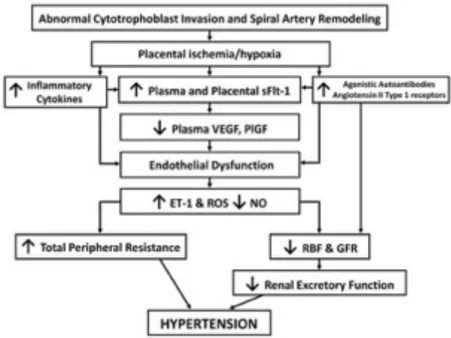
Figure 2. Pathophysiological events in preeclampsia ... 40
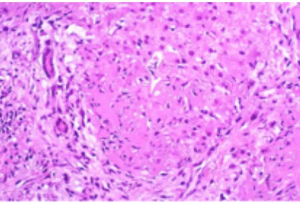
Figure 3. Necrosis of periportal hepatocytes with abundant fibrin deposition ... 44
Figure 4. Peripheral blood smear from a patient with a microangiopathic hemolytic anemia with marked red cell fragmentation ... 53
Figure 5. Clinical course of hemolysis, elevated liver enzymes, and low platelets syndrome (HELLP) syndrome ... 55
Figure 6. Abdominal CTs of pregnant women with (a) HELLP syndrome and (b) antiphospholipid syndrome ... 57
Figure 7. Acute fatty liver of pregnancy ... 63
List of Tables
Table 1. Liver diseases during pregnancy ... 15
Table 2. Typical reference ranges for liver enzymes by pregnancy and trimester ... 18
Table 3. Patterns of abnormal liver function tests (LFTs) ... 21
Table 4. Typical presentation of pregnancy-related liver disorders ... 22
Table 5. Differential diagnosis in sustained nausea and vomiting in pregnancy ... 25
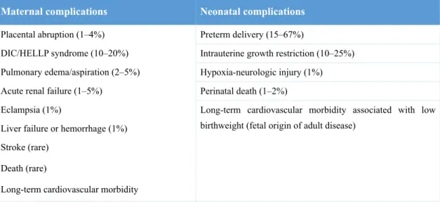
Table 6. Maternal and fetal complications in severe preeclampsia ... 43
Table 7. International Society for the Study of Hypertension in Pregnancy (ISSHP) definition of preeclampsia ... 44
Table 8. Reported frequency of signs and symptoms of HELLP syndrome ... 50
Table 9. Tennessee Classification and Mississippi classification for HELLP syndrome ... 53
Table 10. Comparison of frequency of signs, symptoms, and laboratory findings in TTP, HUS, HELLP and AFLP ... 54
Table 11. Summary of laboratory findings in acute fatty liver of pregnancy ... 60
Introduction
Pregnancy is a dynamic process in which physiologic adaptations occur in the mother in response to the demands. These physiologic events can result in changes in liver biochemical profile which are normal in pregnancy. However, up to 3% of all pregnancies are complicated by liver disorders with different clinical outcomes ranging from self-limiting to rapidly fatal (1,2).
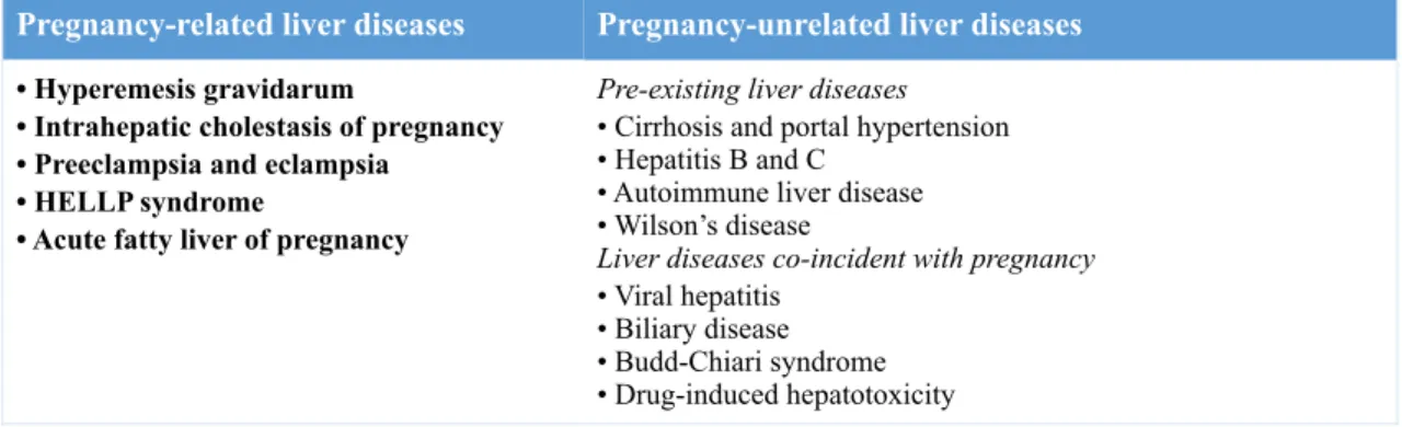
Liver disorders during pregnancy can be related or unrelated to pregnancy. Unrelated disorders can be further differentiated into preexisting disorders, that might worsen during pregnancy and those presenting -de novo (3) (see table 1).
Disorders specific to pregnancy include hyperemesis gravidarum (HG), intrahepatic cholestasis of pregnancy (ICP), preeclampsia (PE), hemolysis, elevated liver enzymes and low platelets (HELLP) syndrome and acute fatty liver of pregnancy (AFLP). Liver dysfunction unrelated to pregnancy can occur any time, contrarily to liver disorders specific to pregnancy, which display characteristic trimester-specific clustering of occurrence. Moreover, they have been shown to be the most common reasons for abnormalities in liver test results during pregnancy and can carry a high mortality rate for both mother and baby. A rapid evaluation of pregnant women to distinguish between those is essential to facilitate appropriate management, which at the severe end of the spectrum require urgent delivery (4).
This diploma thesis will be focusing on pregnancy-related liver disorders by summarizing possible causes, underlying mechanisms of their occurrence and a methodological approach to their diagnosis and treatment.
Table 1. Liver diseases during pregnancy
Pregnancy-related liver diseases Pregnancy-unrelated liver diseases
• Hyperemesis gravidarum
• Intrahepatic cholestasis of pregnancy • Preeclampsia and eclampsia
• HELLP syndrome
• Acute fatty liver of pregnancy
Pre-existing liver diseases
• Cirrhosis and portal hypertension • Hepatitis B and C
• Autoimmune liver disease • Wilson’s disease
Liver diseases co-incident with pregnancy
• Viral hepatitis • Biliary disease • Budd-Chiari syndrome • Drug-induced hepatotoxicity
1. Pregnancy-induced physiologic changes
During pregnancy, significant anatomical and physiological changes occur in order to accommodate to the developing fetus. Cardiovascular changes are characterized by an increased maternal heart rate, cardiac output (CO), together with a marked fall in systemic vascular resistance (SVR) (5). Arterial blood pressure (BP) remains unaffected or demonstrates some tendency toward lower diastolic pressure. Plasma volume increases progressively throughout pregnancy, peaking in the second trimester (50%). Expansion in plasma volume is greater than the increase in red blood cell (RBC) mass, which results in fall of hemoglobin (Hb) concentration, hematocrit and RBC count. However, mean corpuscular volume and mean corpuscular hemoglobin concentration do not change. Platelet count falls progressively but remains in normal reference values. Gestational thrombocytopenia (100 000 to 150 000 cells/µL), occurs in 5 to 10% of pregnant women, mostly in late gestation, with no apparent risk to either mother or fetus (6,7).
Regional blood flow to kidney, lung, skin and uterus are increased in normal pregnancy. Renal blood flow changes, result in an increased glomerular filtration rate (GFR) (30 to 50%), with consequent decrease in serum blood urea nitrogen (BUN) and creatinine levels, together with increased renal protein excretion. Rising estrogen levels stimulate renin-angiotensin-aldosterone system, increasing tubular reabsorption of sodium. In contrast, distal tubular reabsorption of glucose decreases, hence glycosuria maybe a normal finding (7).
Physiologically, water retention is greater in pregnancy which is partly mediated by fall in plasma osmolality. This, together with increased venous pressure bellow uterine level, as a consequence of partial inferior vena cava obstruction, favors edema, which becomes worse at late pregnancy.
Lastly, pregnancy causes an increase in iron, folate and vitamin B12 requirements (6).
1.1.Hepatobiliary changes in pregnancy
Physical examination of liver is compromised during pregnancy, since it is shifted upwards and posteriorly, especially during the third trimester, yet it does not change in size. Increased plasma volume does not influence hepatic blood flow as SVR is lower and the liver receives a lower percentage of the CO (28%) (8,9).
Finding which might suggest liver disease such as spider nevi and palmar erythema are often normal during pregnancy. These are attributed to the increased circulating hormonal levels of estrogens. Moreover, increased levels of progesterone in pregnant women, have been associated with dilation of gallbladder and biliary duct system by its action on smooth muscle (by cholecystokinin inhibition). In addition, during pregnancy hepatic cholesterol synthesis increases, however bile acids (BA) levels remain within normal limits during pregnancy. Even so, several studies have documented increased BA levels in late pregnancy, compared to first trimester values. Overall, overproduction of selected BA (especially chenodeoxycholic acid) and decreased concentration of biliary water, reduce the ability of bile to solubilize cholesterol. The aforementioned alterations result in an increased lithogenecity of bile, stasis and higher prevalence of cholelithiasis among pregnant women (8).
Furthermore, hormonal changes according gestational age, can also affect metabolism by altering expression of cytochrome P450 system. Expression of hepatic CYP1A2 is decreases while CYP2D6 and CYP3A4 increases (10).
Coagulation pathway and its changes during pregnancy are complex and out of the scope of this diploma thesis. In general, physiological changes in pregnant women result in a hypercoagulable state. Procoagulant activity is enhanced by elevation of fibrinogen, factors V, VII, VIII, IX, X, XII and von Willebrand factor. Antithrombin levels are decreased. In contrast, activated partial thromboplastin time (APTT) and prothrombin time (PT) remain unchanged (4,9). Although fibrinolysis has been suggested to be augmented during pregnancy, recent studies have produced conflicting results, suggesting reduced fibrinolytic activity. Fibrinolytic process is normally mediated by tissue plasminogen activator (tPA) that converts plasminogen into plasmin, ensuring fibrin degradation. Interestingly, tPA activity has been shown to be decreased in pregnant women along with raised levels of plasminogen activator inhibitors 1 and 2. These events are countered by increased levels of plasminogen and decreased level of another plasmin inhibitor (α2 antiplasmin), ensuring hemostatic balance during normal pregnancy. Overall, body is adapting to minimize blood losses in delivery, however, these same changes increase mother’s risk of thrombotic events from the first trimester until 12 weeks postpartum (6).
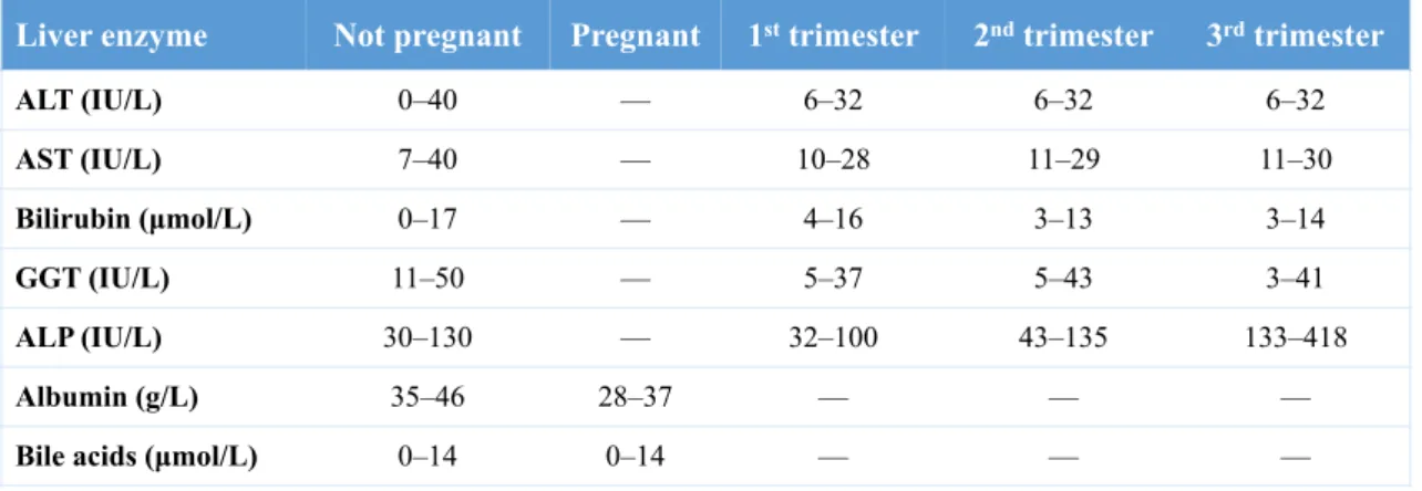
Table 2. Typical reference ranges for liver enzymes by pregnancy and trimester (11)
ALT, alanine transaminase; AST, aspartate transaminase; GGT, gamma-glutamyl transferase; ALP, alkaline phosphatase.
Physiological changes occur in liver function tests (LFTs) during pregnancy (see table 2). Important liver parameters such as alanine transaminase (ALT), aspartate aminotransferase (AST), gamma-glutamyl transferase (GGT) remain unchanged. However, superior limit is slight altered due to hemodilution effect. Expansion in plasma volume also affects serum albumin and bilirubin concentrations. Serum alkaline phosphatase (ALP) levels are higher and peak in the third trimester when noticeable placental ALP and fetal bone isoenzyme are produced. This makes it has a poor diagnostic tool in pregnancy. To a lesser extent, 5-nucleotidase activity also increases in the second and third trimesters.
Lastly, lactate dehydrogenase (LDH) levels are unchanged throughout gestation (6,8).
Liver enzyme Not pregnant Pregnant 1st trimester 2nd trimester 3rd trimester
ALT (IU/L) 0–40 — 6–32 6–32 6–32 AST (IU/L) 7–40 — 10–28 11–29 11–30 Bilirubin (µmol/L) 0–17 — 4–16 3–13 3–14 GGT (IU/L) 11–50 — 5–37 5–43 3–41 ALP (IU/L) 30–130 — 32–100 43–135 133–418 Albumin (g/L) 35–46 28–37 — — —
Bile acids (µmol/L) 0–14 0–14 — — —
2. Approach to liver diseases occurring during pregnancy
Abnormal liver studies can be an early indicator of serious pathology. Differentiating liver disorders related to pregnancy from those unrelated is essential for management of these diseases and to achieve good outcomes. Standard workup in a patient presenting with abnormal liver enzymes, as with any non-pregnant individual, comprises several steps that one must follow. A complete history (including past LFTs), physical exam and standard serological workup should be performed as indicated by clinical presentation. LFTs can change significantly during normal pregnancy, making challenging interpretation of it. They might fall under 3 categories:
i. Tests of the liver’s capacity to transport organic anions and to metabolize drugs- serum bilirubin, urine bilirubin and urobilinogen;
ii. Tests that detect injury to hepatocytes (serum enzyme tests) – aminotransferases, ALP, GGT and 5-nucleotidase; and
iii. Tests of the liver’s biosynthetic capacity- serum proteins, albumin, prealbumin, PT, α-fetoprotein, serum ceruloplasmin, procollagen III peptide and α1 antitrypsin (12).
Thus, approach to a pregnant woman with abnormal LFTs, can be simplified by following questions:
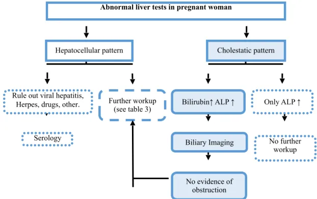
1) What is the pattern of abnormal LFTs? (see figure 1) (see table 3)
Three main patterns of LFTs results are recognize: hepatocellular, cholestatic and mixed. Raised aminotransferases levels out of proportion to ALP suggests hepatocellular injury, with inflammation or necrosis of hepatocytes. Levels of ALT are more specific as AST can be found ubiquitously throughout the body (8). Slight AST or ALT elevations (within 1.5 times the upper limits of normal) do not necessarily indicate liver disease, however these women should be followed up. Additional, mild to moderate increase in transaminases might suggest specific disorders like HG, AFLP, ICP whereas marked increase is often seen in severe PE, HELLP syndrome and liver rupture. Concerning pregnancy-unrelated liver disorders, cholelithiasis and alcoholic hepatitis usually present with mild elevation, in contrast with drug-induced hepatotoxicity, and viral hepatitis, that can result into extensive damage to hepatocytes with striking raised levels of transaminases (13). One should rule out hepatitis, by appropriate serological tests.
Cholestatic pattern suggests obstruction of biliary flow, that results in hepatobiliary disturbances, with high levels of ALP out of proportion to aminotransferases. Accessing to 5-nucleotidase, GGT, and bilirubin levels, which are increased in cholestatic pattern, can be helpful to confirm pathological ALP levels in pregnant women. Raised serum BA are present in almost any disease of the hepatobiliary system, yet serum BA are a sensitive but nonspecific marker of hepatic and biliary disease, when presenting alone (8). Severe biliary obstruction can lead to high amounts of serum conjugated bilirubin, resulting in a dark urine and pale stools (9). Imagining of biliary system may be helpful in cases of suggesting obstruction. Ultrasonography (USG) remains the safest imaging. If further detailed imaging is needed, magnetic resonance imaging (MRI) without contrast can be safely done (2).
Mixed pattern is usually presenting in cases of expanding masses, punching into hepatic structures, resulting in a concomitant elevation of both transaminases and cholestasis parameters.
Figure 1. Workup abnormal liver tests in pregnant woman. Adapted (5)
ALP, alkaline phosphatase; ↑, increased; ↓ decreased.
*25
Hepatocellular pattern Cholestatic pattern
Bilirubin↑ ALP ↑ Rule out viral hepatitis,
Herpes, drugs, other.
Abnormal liver tests in pregnant woman
Further workup
(see table 3) Only ALP ↑
No further workup Biliary Imaging No evidence of obstruction Serology
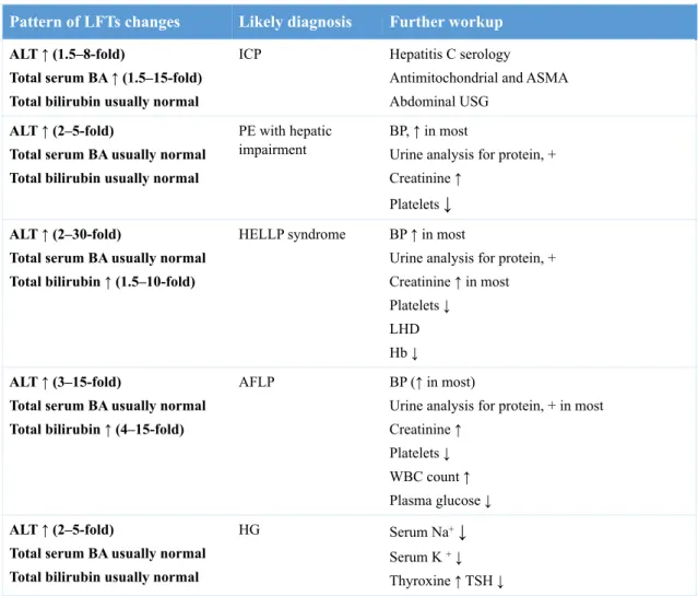
Table 3. Patterns of abnormal liver function tests (LFTs) (11).
ALT, alanine aminotransferase; BA, bile acids; ICP, intrahepatic cholestasis of pregnancy; PE, preeclampsia; HELLP, hemolysis, elevated liver enzymes and low platelets; AFLP, acute fatty liver of pregnancy; HG, hyperemesis gravidarum; ASMA, anti-smooth muscle antibodies; USG, ultrasonography; BP, blood pressure; LDH, lactate dehydrogenase; Hb, hemoglobin; WBC, white blood cells; TSH, thyroid stimulating hormone; ↑, increased; ↓ decreased; + positive.
2) In which trimester is pregnant women presenting abnormal LFTs?
Gestational age can be a clue to identify liver diseases in pregnancy, as pregnancy-related disorders present in a specific trimester in most of the cases. Contrastingly, pregnancy-unrelated liver disorders can occur throughout the gestation (4).
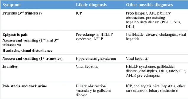
3) Does patient present additional signs? (see table 4)
Laboratory findings must be interpreted according presenting symptoms. Liver involvement usually presents with fatigue, jaundice, pruritus, abdominal pain, nausea and vomiting. As patient presents with signs of liver dysfunction, one might search for constellations of signs typical for each disease, together with past/present history and severity of complains. During physical examination measurement of BP, weight, presence of jaundice and edema, might help to decide further investigations (11).
Pattern of LFTs changes Likely diagnosis Further workup
ALT ↑ (1.5–8-fold)
Total serum BA ↑ (1.5–15-fold) Total bilirubin usually normal
ICP Hepatitis C serology
Antimitochondrial and ASMA Abdominal USG
ALT ↑ (2–5-fold)
Total serum BA usually normal Total bilirubin usually normal
PE with hepatic impairment
BP, ↑ in most
Urine analysis for protein, + Creatinine ↑
Platelets ↓
ALT ↑ (2–30-fold)
Total serum BA usually normal Total bilirubin ↑ (1.5–10-fold)
HELLP syndrome BP ↑ in most
Urine analysis for protein, + Creatinine ↑ in most Platelets ↓
LHD Hb ↓
ALT ↑ (3–15-fold)
Total serum BA usually normal Total bilirubin ↑ (4–15-fold)
AFLP BP (↑ in most)
Urine analysis for protein, + in most Creatinine ↑
Platelets ↓ WBC count ↑ Plasma glucose ↓
ALT ↑ (2–5-fold)
Total serum BA usually normal Total bilirubin usually normal
HG Serum Na+↓
Serum K + ↓
Table 4. Typical presentation of pregnancy-related liver disorders (11)
ICP, intrahepatic cholestasis of pregnancy; AFLP, acute fatty liver of pregnancy; PBC, primary biliary cirrhosis; PSC, primary sclerosing cholangitis; DILI, drug induced liver injury; HELLP, hemolysis, elevated liver enzymes, and low platelets.
Symptom Likely diagnosis Other possible diagnoses
Pruritus (3rd trimester) ICP Preeclampsia, AFLP, biliary obstruction, pre-existing
hepatobiliary disease (PBC, PSC), DILI
Epigastric pain
Nausea and vomiting (2nd and 3rd
trimesters)
Headache, visual disturbance
Pre-eclampsia, HELLP
syndrome, AFLP Gallbladder disease, cholangitis, viral hepatitis
Nausea and vomiting (1st trimester) Hyperemesis gravidarum Viral hepatitis
Jaundice Viral hepatitis HELLP syndrome, gallbladder disease, cholangitis, DILI, rarely ICP, AFLP, pre-eclampsia
Pale stools and dark urine Biliary obstruction secondary to gallstone disease
ICP, cholangitis, viral hepatitis, other rare causes of biliary obstruction
3. Pregnancy-related liver disorders
3.1.Hyperemesis gravidarum
3.1.1.Definition
Nausea and vomiting in pregnancy (NVP) is a common symptom among pregnant women. About 50 to 80% (14) of pregnant women experience these symptoms in a mild and self-limiting way and improved without intervention in the second trimester. However, some women develop a severe form of these symptoms. This condition, known as HG, is characterized by intractable vomiting leading to dehydration, electrolyte abnormalities and significant weight loss (at least 5% of body weight) requiring hospitalization. HG is mostly self-limited and presents often during the first trimester (typically 4-10 weeks).
HG is not a liver disease by itself, but leads to liver dysfunction in 50% of cases (4).
3.1.2.Incidence and risk factors
HG is a condition present in 0.8 to 3.6% of all pregnancies and the most common cause of pregnancy-related hospitalization in the first half of gestation (15). Asian women seem to be more affected then Caucasian women (16). It’s a multifactorial condition and has been associated with many risks factors (17). These include: younger age (<20), multiple pregnancy, nulliparity, obesity, metabolic disturbances, trophoblastic and psychological disorders (18). Specific diseases that have been showed to increase risk of HG include hyperthyroid disorders, gastrointestinal problems, pre-existing diabetes and asthma. Maternal smoking and age (>30) have been associated with decreased risk (19).
3.1.3.Etiopathogenesis
Etiology is not yet fully understood, but it is believed to be multifactorial. The most relevant etiological factors are hormonal, immunological and genetic (20).
High levels of human chorionic gonadotropin (hCG), have been shown to correlate with the severity of HG, which stimulate the secretory function of the gastrointestinal tract and thyroid function (3). Transient hyperthyroidism may occur in 60% of gestations owing to the thyroid-stimulating activity of hCG (4). Other hormonal factors
Available data indicate that HG is associated with a hyperactivity of the immune system (21). Increased levels of interleukin 6 (IL-6) (22), tumor necrosis factor alpha (TNF-α) (23), immunoglobulins G and M, and complement proteins C3 and C4 have been shown in patients with HG. Furthermore, lymphocytes count, natural killer (NK) cells and extrathymic T-cells are higher than normal (24).
Recent studies also point for the potential role of Helicobacter pylori (H. pylori) in HG. H. pylori was found, histologically, in gastric mucosa of 95% of pregnant women with HG. Further studies identified H. pylori genome in saliva of 61.8% HG patients compared with 21.6% of pregnant women without symptoms (18). Although these associations have not determined if H. pylori infection is a cause or not, administration of antibiotics in these women improved their symptoms (25).
3.1.4.Clinical presentation
The clinical symptoms are mostly unspecific and uncharacteristic (20). The main symptom is intractable vomiting that results in loss of volume, weight loss, and electrolytes abnormalities.
HG typically starts in the first trimester , peaks in approximately by the ninth week and resolves by the twentieth week in 90% of women (26). However, it is not uncommon for HG to continue into the late second and third trimesters.
Frequent findings include metabolic acidosis, caused by a poor nutritional intake or metabolic alkalosis by the loss of hydrochloric acid and hypokalemia (2). Metabolic ketoacidosis and ketonemia may present with a “fruity smell on the breath” and ketonuria (18). Pyrexia, gastric pain, headache or neurologic signs point to other causes, although the latter finding may, in rare cases, result from severe and prolonged NVP.
Laboratory findings include raised BUN and creatinine, and decreased phosphate and magnesium. These disorders resolve with the cessation of vomiting.
Hepatic involvement occurs in 50 up to 60% of cases. The most common alteration is raised transaminases levels, with higher values of ALT when compared to AST (2). Clinical jaundice rarely occurs, but if does, bilirubin levels rarely exceed 100 µmol/L (27). The persistence of these alterations should raise alternative diagnoses (2).
3.1.5.Diagnosis
A c c o r d i n g t h e r e c e n t g u i d e l i n e s f r o m t h e R o y a l C o l l e g e o f Obstetricians and Gynecologists “NVP should only be diagnosed when onset is in the
first trimester of pregnancy and other causes of nausea and vomiting have been excluded”. HG should be diagnosed when there is protracted NVP with the triad of more than 5% pregnancy weight loss, dehydration and electrolyte imbalance (26).
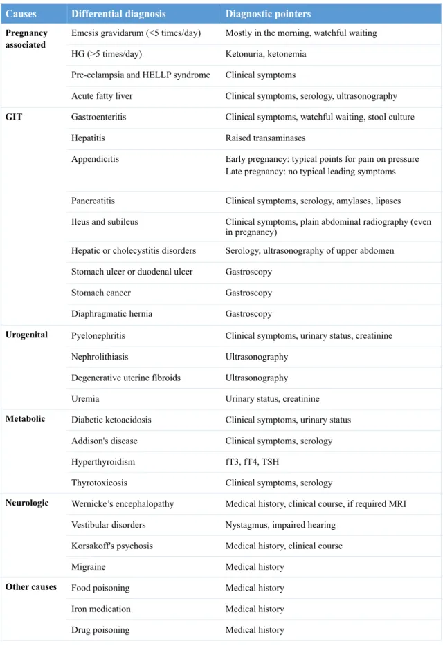
HG diagnosis is mainly by exclusion, from other conditions associated with emesis such as: other pregnancy associated conditions (emesis gravidarum, PE, HELLP syndrome and AFLP), gastrointestinal, urogenital, metabolic and neurologic causes (see table 5), and based on clinical symptomatology.
Table 5. Differential diagnosis in sustained nausea and vomiting in pregnancy (28).
HG, hyperemesis gravidarum; HELLP, hemolysis, elevated liver enzymes and low platelets; GIT, gastrointestinal; MRI, magnetic resonance imaging; fT3, free triiodothyronine; fT4, free thyroxine; TSH, thyroid stimulating hormone.
History and complete examination together with investigations are essential for differential diagnosis. USG (ultrasonography) scan should be scheduled to confirm viability and gestational age and to exclude multiple pregnancy or trophoblastic
Causes Differential diagnosis Diagnostic pointers
Pregnancy associated
Emesis gravidarum (<5 times/day) Mostly in the morning, watchful waiting HG (>5 times/day) Ketonuria, ketonemia
Pre-eclampsia and HELLP syndrome Clinical symptoms
Acute fatty liver Clinical symptoms, serology, ultrasonography
GIT Gastroenteritis Clinical symptoms, watchful waiting, stool culture
Hepatitis Raised transaminases
Appendicitis Early pregnancy: typical points for pain on pressure Late pregnancy: no typical leading symptoms Pancreatitis Clinical symptoms, serology, amylases, lipases Ileus and subileus Clinical symptoms, plain abdominal radiography (even
in pregnancy)
Hepatic or cholecystitis disorders Serology, ultrasonography of upper abdomen Stomach ulcer or duodenal ulcer Gastroscopy
Stomach cancer Gastroscopy
Diaphragmatic hernia Gastroscopy
Urogenital Pyelonephritis Clinical symptoms, urinary status, creatinine
Nephrolithiasis Ultrasonography
Degenerative uterine fibroids Ultrasonography
Uremia Urinary status, creatinine
Metabolic Diabetic ketoacidosis Clinical symptoms, urinary status Addison's disease Clinical symptoms, serology
Hyperthyroidism fT3, fT4, TSH
Thyrotoxicosis Clinical symptoms, serology
Neurologic Wernicke’s encephalopathy Medical history, clinical course, if required MRI Vestibular disorders Nystagmus, impaired hearing
Korsakoff's psychosis Medical history, clinical course
Migraine Medical history
Other causes Food poisoning Medical history
Iron medication Medical history
Drug poisoning Medical history
disorder (29). Complete blood count (CBC), serum electrolytes, urinalysis and hepatic, renal and thyroid parameters should be performed. Liver biopsy is not recommended.
To classify the severity of NVP two validated and objective scores have been used: Pregnancy-Unique Quantification of Emesis (PUQE) score and Rhodes Index. The PUQE score is based on shorter disease-specific questionnaire and can be used to determine whether the NVP is mild, moderate or severe (see Annex A) and to track progress with treatment (26). Rhodes Index was originally validated to measure emesis in patient undergoing chemotherapy, but has subsequently been used for NVP (30).
3.1.6.Complications
3.1.6.1. Maternal complications
Pregnant women suffering from HG are at risk for complications related to excessive vomiting and dehydration with its subsequent consequences of malnutrition, electrolytes abnormalities and metabolic disturbances (29).
Nowadays, in its extreme forms HG may cause severe maternal malnutrition. End-organ damage may follow it manifesting as oliguria and abnormal liver tests, however permanent liver damage and associated death is rare (17).
Hematemesis, esophageal rupture, Mallory–Weiss tears and pneumothorax, were described in severe cases due to prolonged vomiting (31,32).
The mean nutrients dietary intake of most nutrients falls below 50% in women with HG that ultimately may lead to vitamin deficiencies and low protein status. Suboptimal biochemical status of vitamin A, B1 (thiamine), B2 (riboflavin), B6 (pyridoxine), B12 (cyanocobalamin) and retinol-binding protein have been shown in 60% of HG patients (33). Wernicke’s encephalopathy is rare but can be caused by thiamine deficiency and precipitated by carbohydrate-rich food and glucose infusions (34).
Profound hyponatremia may result in personality changes, muscle cramps and weakness, ataxia, drowsiness and lately convulsions (15). Furthermore, central pontine myelinolysis, characterized by damage of corticospinal and corticobulbar pathways, was associated with rapid correction of plasma sodium levels and reported to co-exist with Wernicke’s encephalopathy in HG patients (33).
Hypokalemia, when profound, together with hypovolemia may result in rare consequences, such as rhabdomyolysis, pancreatitis, and acute renal failure with resolution of symptoms after electrolyte and hydration repletion (35).
The combination of pregnancy, dehydration and further immobility in HG women increases the risk of thromboembolic events (33).
HG has been reported at increased risk for cognitive, behavioral and emotional dysfunction in pregnancy. Moreover, authors described an increased risk for depression, anxiety and mental health difficulties (17).
3.1.6.2. Fetal and perinatal complications
HG has been reported to be associated with an increased risk for adverse pregnancy outcomes such as low birth weight, preterm birth and small-for-gestational age infants (36-38) . However, not all studies found adverse outcomes in HG (39,40). A systematic review identified no association with Apgar scores, congenital anomalies, or perinatal death (39).
Pregnancy outcome in HG patients are determine mainly by maternal characteristics but other factors associated with HG, such as insufficient weight gain during pregnancy, maternal stress responses during pregnancy, or Helicobacter pylori (H. Pylori) infection involving the placenta, could adverse birth outcomes associated with HG (17).
Risk of psychological and behavioral problems in adulthood is increased in two studies concerning the neurodevelopment (15). Moreover, a recent study showed that insulin sensitivity in children of hyperemetic mothers is decreased (41). Others found that early pregnancy weight loss was associated with increased BP in offspring (42). Lastly, vitamin K deficiency due to nutritional maternal deficiencies was purposed as risk factor for fetal intracranial hemorrhage owing its potential on fetal blood coagulability (43).
3.1.7.Treatment
HG is often a self-limited disorder and its treatment is essentially supportive and should be adapted to the severity of the clinical picture. The main objectives are to control nausea and vomiting, correction of fluids and electrolyte imbalances and, if needed, thromboprophylaxis (3,29).
Mild cases can be controlled only with nutritional counselling and some changes in lifestyle (18). Pregnant women should be encouraged to avoid stimuli that cause nausea. Meals should be light and frequent, high in carbohydrates and low in fat content (1). When correction of dehydration is necessary, intravenous fluid therapy is performed.
Treatment of NVP with vitamin B6 or vitamin B6 plus doxylamine is recommended as first-line therapy, when available. Second-line therapy includes anti-emetic drugs such as antihistamines, namely promethazine and prochlorperazine. Metoclopramide, a dopamine antagonist is also safe and approved, as well as ondansetron and corticosteroids for refractory cases (3,44).
Supplementation with folates and thiamine should be weighted, especially in cases of vomiting with longer evolution time (29,34,44). Since in HG the administration of glucose enhances the risk of Wernicke encephalopathy, this should be accompanied by administration of thiamine (34) .
In severe cases, nasogastric tube feeding and/or total parenteral nutrition may be required (14).
Currently, alternative options such as ginger root and acupuncture are available for treatment, and results seem favorable (29).
3.2.Intrahepatic cholestasis of pregnancy
3.2.1.Definition
ICP is the most common pregnancy-specific liver disease. It presents in most of the cases with the triad of pruritus, typically of the palms and soles, abnormal LFTs, and raised serum BA levels (45). Biochemical abnormalities and symptoms usually resolves after delivery but recurrences in later pregnancies and with the use of combined hormonal contraceptives were often described (27). It classically presents in the third trimester but it can present as early as in 7 weeks of gestation (2). It has been associated with increased rates of adverse fetal and pregnancy outcomes.
3.2.2.Epidemiology
Sweden where there was a higher incidence of cholelithiasis and hepatitis C virus in ICP women (27). The incidence in Europe and USA is lower (about 1%), and rarely reported in African countries (3). Higher incidence occurs in multiple pregnancies (20 to 22%) and after in vitro fertilization (2.7%) (45).
3.2.3.Risk factors
Several risk factors are listed in the literature. Those include history of previous cholestasis during oral contraceptive use, family history of cholestasis, multiparity, multiple pregnancy, advanced maternal age, personal and/or family history of cholelithiasis, and hepatitis B virus infection (2,45).
Risk of recurrence in future pregnancies has been shown to range from 40 to 60% (46).
3.2.4.Etiopathogenesis
ICP has a multifactorial etiology to which genetic, hormonal and environmental factors contribute (47).
Genetic defects of canalicular transporters have been identified for the development of ICP, which may be further influenced by reproductive hormones (specially progesterone and estrogen). The etiology of fetal complications is likely to be related to the destructive effects of toxic BA, which accumulate in the fetal compartment.
Hormonal effects are based on following four circumstances: Firstly, ICP usually starts in the last trimester (highest maternal estrogen and progesterone concentrations) (48). Secondly, the incidence is higher in multiple pregnancies (49,50), in pregnancies following in vitro fertilization (2.7% vs 0.7% in normal conception) (49). Thirdly, studies have shown ICP in pregnant women given oral progesterone to prevent preterm labor; and finally, ICP resolves promptly after delivery and recurs in half of the patients during subsequent pregnancies (3).
Rodent studies have shown that estrogen has a key role in the development of cholestasis by causing reduced expression of hepatic biliary transport proteins and through internalization of the bile acid transporter bile salt export pump (45). Moreover, sulphated progesterone metabolites can act as partial agonists of farnesoid X receptor, thereby reducing the function of the main hepatic BA receptor and subsequent impairment of hepatic BA homeostasis (51).
The genetic etiology of ICP is largely unknown. However, several studies have identified genetic variations in genes encoding biliary transport proteins and BA receptor (primarily farnesoid X receptor). Positive familial history was reported in UK in 14% of pregnant women with ICP. Some studies have suggested an autosomal-dominant, sex-limited pattern (50). Overall, ten different mutations have been identified (2). About 15% of cases have genetic mutations in ABCB11, ABCB4 (3) and in a smaller amount of cases in ATP8B1 (45). Mutations in these genes cause recessively inherited progressive familial intrahepatic cholestasis, which is a rare, early-onset condition associated with intrahepatic cholestasis in infancy or early childhood and resulting in death (52). ABCB4 encodes the multiresistant protein 3 (MRP3), a phosphatidylcholine flippase that transports phatidylcholine to the inner leaflet of the canicular membrane (45). Mutations in this gene may affect BA transport and lead to a rise in BA concentrations. These have been linked to a severe form of ICP (52).
Various single nucleotide polymorphisms in ABCB11 (encodes bile salt excretory protein) and ABCB4 genes are associated with ICP. ATP8B1, which encodes phosphatidylserine flippase 1, is also a candidate gene for ICP but small number of cases were identified (45).
Some of the exogenous factors implicated in the genesis of ICP are selenium deficits, seasonality and hepatitis C virus infection. Low serum selenium levels were reported in ICP pregnant women, and it is known that their dietary intake is low in Chile and Finland, countries where the incidence of this syndrome is higher (48). Interestingly the incidence peaks in winter months in these countries, when is known that selenium levels are lower. Selenium deficiency may cause defective bile formation or secretion since it is a cofactor for several oxidative hepatic enzymes (45). Not to mention that vitamin D levels are likely to be lower in the winter months, and deficiency of this vitamin has been reported in women with ICP (53).
3.2.5.Clinical presentation
Pruritus is a cardinal symptom of ICP, being present in 95% of the cases (54). It usually arises in the third trimester, after 25 weeks (55), and starts in the palms and soles. It often becomes worsen and generalized (27). The pathophysiology of the pruritus is still unknown but it has been suggested that results from bile salts deposition on nerve endings (56). The pruritus is most severe at night and can cause insomnia
must be excluded such as atopic dermatitis, allergic or drug reaction, prurigo of pregnancy and atopic eruption of pregnancy (45). Skin excoriations can be find flowing scratching (54).
Jaundice affects about 10 to 15% of pregnant women with ICH and manifests two to four weeks after the onset of pruritus (45,55). It is usually mild, associated with bilirubin levels not higher than 6 mg/dL (102.6 µmol/L) and keeps stable throughout gestation. Rarely, severe cases can present with pale stools and dark urine (27).
Pascual et al., describes the phenomena of asymptomatic hypercholanemia (AHP) of pregnancy present in 10% of pregnant population (57). It consists in elevated serum BA in pregnancy in the absence of symptoms and other biochemical markers of ICP. Furthermore, Castaño et al., described AHP in 40% of pregnant women in Argentina with normal pregnancy outcomes (58).
In severe cases ICP can cause steatorrhea with decreased absorption of fat-soluble vitamins and weight loss (59). This can further result in vitamin K insufficiency and prolonged PT (60).
There is usually spontaneous resolution of pruritus within the first 48 hours postpartum and analytical changes resolve within the next two to eight weeks (3).
3.2.6.Complications
ICP is usually associated with a good maternal prognosis. Malabsorption of vitamin K may lead to an increase in PT, and if not corrected it can lead to life-threatening postpartum hemorrhages, however it is a rare event. In women affected by ICP, cholestasis may recur with the use of oral contraceptives and/or with hormone replacement therapy (45).
ICP is associated with several adverse outcomes with considerable perinatal morbidity and mortality.
There are no concrete explanations for the occurrence of fetal complications, but these seem to be related to the raised maternal serum BA. BA induce contraction of the placental chorionic veins and increase the sensitivity of the myometrium to the action of oxytocin (27).
Several studies have shown that the risk of fetal complications increases 1 to 2% for each increase of one µmol/L of serum BA. The risk is significant higher when serum BA values exceed 40 µmol/L (61,62).
The main complication is prematurity. Incidence varies between 19 and 60%. Intrauterine death (0.4 to 4.1%) may also occur, as well as fetal distress (22 to 33%), meconium staining of the amniotic fluid (approximately 15%) (27) and fetal bradycardia (˃14%) (55).
The rate of abortion and malformations is not increased in this disease (27).
3.2.7.Diagnosis
Laboratory findings, clinical presentation, and exclusion of other causes, make the diagnosis of ICP.
Liver tests, including direct bilirubin, PT, transaminases, GGT and serum BA (45) should be performed. The study of these pregnant women should also include the serology for the exclusion of hepatitis A, B, C and cytomegalovirus virus infection, as well as evaluation of renal function and CBC with platelets. Abdominal USG allows visualization of hepatic morphology and main bile ducts (63). USG usually reveals no dilatation of the intrahepatic nor extrahepatic bile ducts, but fasting and ejection volumes of the gallbladder are greater than in normal pregnant women. Thirteen percent of women with ICP are found to have gallstones in USG examination (27).
Generally, liver biopsy is not required for the diagnosis of ICP to be established. When performed, it reveals centrilobular cholestasis without associated inflammation and deposition of bile agglomerates in hepatocytes and canaliculi (48).
The differential diagnosis includes other forms of hepatic diseases. In patients with very high serum levels of aminotransferases, hepatitis A, B, C, severe PE, the HELLP syndrome, acute fatty liver of pregnancy, and drug toxicities should be excluded (3).
Increased serum BA levels have been used widely to diagnose ICP. Classification of ICP according the levels of BA seems to correlate with severity of fetal outcomes (64). A BA levels between 10-14 µmol/L (6 µmol/L if fasted woman) and 40 µmol/L are classified as mild, whereas >40 µmol/L is described as severe ICP. The primary BA cholic acid (CA) and chenodeoxycholic acid (CDCA) are the end products of hepatic cholesterol metabolism. These are conjugated with taurine or glycine before transport across canicular membrane. Within terminal ileum and colon, conjugated BA undergo bacterial modifications to form the secondary BA – deoxycholic acid (DCA) and lithocholic acid (LCA). In normal pregnancy, it is accepted and increase of levels of BA up to 1.5 µmol/L. More accurate parameter is CA/CDCA ratio which is reported to be
particularly tauroconjugates of cholic and chenodeoxycholic acid have been purposed as the most suitable biochemical marker for both diagnosis and monitoring of ICP, whereas CA/CDCA ratio, as being the most sensitive indicator of early diagnosis of the disease (27). Moreover, analysis of urine from women with ICP shows an increased excretion of CA and CDA, but lower values of DCA and LCA. The BA profile shows a shift from glycine to taurine conjugation and increased proportions of sulfated species. Nevertheless, a recent study in the Chinese population has shown, that unlike findings in other populations, total serum BA levels was not a reliable indicator of ICP. Instead, the authors of this study have found more specific biomarkers for this condition: MMP-2 and MMP-9 (metalloproteinases). Both markers were elevated in serum of ICP pregnant women and did correlate with the disease severity. MMPs also play a role in remodeling within the liver and have been studied as potential markers of fibrosis and cirrhosis. Elevated levels were also found in hepatitis C virus, cirrhosis and hepatocellular carcinoma (65). Prior studies in China also reported a better correlation to severity of ICP with indicators such as cholyglycine (a serum BA), serum bilirubin levels, in addition to AST and ALT levels (66,67).
In most of the cases liver transaminases are slightly elevated. This may occur before or after rise of serum BA, indicating in both scenarios hepatocellular damage. ALT seems to be the more sensitive indicator than AST (68).
The levels of serum bilirubin are elevated in the most severe form of ICP, and the level of total bilirubin is associated with preterm labour (69). The activity of GGT is increased in about 10 up to 15% but is commonly normal (48).
Glutathione S-transferase alpha (GSTA) is a phase II detoxification enzyme found within hepatocytes and in high concentrations within the liver. Therefore, plasma GSTA is expected to raise following acute liver damage. Dann et al., has identified high serum GSTA levels as a useful indicator of liver dysfunction also in ICP and might help to distinguish ICP from pruritus gravidarum (70).
In a recent study, high serum autotaxin activity was suggested as a sensitive marker and correlated with the onset of ICP-related pruritus (71).
Other routine laboratory parameters can also predict an adverse perinatal outcome in ICP. Raised mean platelet volume is associated with preterm delivery in ICP patients (69). The neutrophil-to-lymphocyte ratio is elevated in pregnancies complicated with ICP and may be a predictor of the severity of ICP (72). Finally, a recent study showed
that RBC distribution width, was associated with ICP and was suggested as a diagnostic and prognostic marker in ICP (73).
3.2.8.Treatment
Treatment of ICP is mostly symptomatic to pregnant women, and ensure, as far as possible, a good fetal prognosis.
Currently, ursodeoxycholic acid (UDCA), a hydrophilic BA, is the treatment of choice at the dose of 500 mg twice daily or 15 mg /kg/day. UCDA shows a good effect in reduction of pruritus, improvement in liver tests and allows the prolongation of pregnancy, thus reducing prematurity. It is well tolerated by woman and is not associated with adverse maternal and/or fetal reactions. In higher doses, woman may complain of gastrointestinal upset and diarrhea, but is a rare finding (27).
Cholestyramine (8 to 16 g/day) is another viable option (62). This therapeutic choice reliefs pruritus, but does not decrease BA levels and there may even be worsening of vitamin K deficiency (2).
S-adenosyl-L-methionine and dexamethasone, may also be used. However, those are far less effective when compared to UDCA.
All pregnant women diagnosed with ICP should be supplemented with vitamin K (3).
Lastly, in severe cases, induction of labour between 37 and 38 weeks of gestations may be necessary (74). The decision must be taken on a case by case basis, considering the risks of prematurity and the possible complications inherent to this pathology.
3.3.Hypertension-associated liver dysfunction of pregnancy
Hypertensive pregnancy disorders include a broad range of clinical conditions, including chronic hypertension; gestational hypertension (transient hypertension of pregnancy or chronic hypertension identified in the latter half of pregnancy; formerly known as pregnancy-induced hypertension) (75); PE -de novo or superimposed on chronic hypertension (76) . These disorders are considered to be major causes of maternal, fetal, and neonatal morbidity, and may be life-threatening for both mother and fetus (44).
HELLP syndrome used to be categorized as a separate syndrome, but current thinking considers it as a severe manifestation of PE (77). However, is still controversial so will be discussed separately from PE.
Finally, hypertensive disorders in pregnancy may be associated with serious hepatic manifestations, including infarction, hemorrhage, and rupture.
3.3.1.Preeclampsia 3.3.1.1. Definition
PE is defined by the International Society for the Study of Hypertension in Pregnancy (ISSHP) as the onset of hypertension in previously normotensive pregnant women after the 20th week of pregnancy, of at least 140/90 mmHg, combined with new onset of one or more of the following : proteinuria (greater than 300mg in 24 hours); and/or signs of maternal organ dysfunction (renal, hepatic or neurologic involvement) and/or signs of uteroplacental dysfunction (fetal growth restriction) (78),. Detailed diagnostic criteria are further reviewed in subchapter “Diagnosis”. PE must resolve completely by the 6th postpartum week (79). It is considered as multisystemic disease that affects every maternal organ, predominantly the vascular, renal, hepatic, cerebral and coagulation systems (77). If PE presents with one or more convulsions, it becomes defined as eclampsia. PE can be classified as mild or severe which influences following approach to this condition. Severe PE is associated with one or more of the following findings: severe hypertension, BP ≥160 mmHg systolic, or ≥110 mmHg diastolic, on two different occasions at least 4 hours apart; proteinuria ≥500 mg in a 24-hour urine collection (dipstick [3+] or [4+]); or symptoms of organ dysfunction such as headache,
hyperreflexia, oliguria, epigastric or right upper quadrant pain, impaired liver function, hepatomegaly, or thrombocytopenia (80).
Controversy remains as to whether fetal growth, without any other maternal feature of PE, should be considered to define it. Some authors support that this should apply, given that PE is more often itself a primary placental disorder (78).
Liver dysfunction in pregnancy complicated by PE and eclampsia ranges from 20 to 30 % (81).
3.3.1.2. Epidemiology
Hypertensive disorders affect 5 to 10% of all pregnancies. According to the World Health Organization (WHO), they are the main single cause of maternal death, accounting for approximately 16% of them in industrialized countries.
Pregnancy complicated by PE accounts for approximately 10% of all gestations and accounts for >50000 maternal deaths worldwide per year (4,82). In developed countries, the maternal mortality is practically nil, while in developing countries it can reach 15 to 20%. Fetal mortality is reported to range from 1 to 2% (1).
In 2002, incidence in Slovakia was estimated in 0.29/ 1 000 pregnancies. Average maternal age was about 23 years old, 86.7% of deliveries were cesarean and without fetal nor maternal deaths (83).
3.3.1.3. Risk factors
Complication rates are directly related to the severity and duration of elevated BP. Patients with severe hypertension in the first trimester have a greater than 50% risk of developing superimposed PE (75). All hypertensive patients should undergo increased surveillance, laboratory tests throughout pregnancy, USG scans to follow intrauterine growth, and antenatal testing (84).
Early identification of PE, and prevention when possible, it’s the main principle in an adequate management. Maternal characteristics that are associated with an increased risk of PE are:
• Past PE history (especially when severe or early-onset); • Underlying antiphospholipid antibody syndrome;
• Pre-existing medical conditions - chronic hypertension, chronic renal disease, or insulin-dependent diabetes mellitus (85);
• Multiple pregnancy (79).
Other factors less strongly associated with PE include: • Primigravidity;
• Interval between two pregnancies greater than 5 (86); • Primipaternity – changed paternity (87);
• Short duration of sexual relationship (88); • Obesity;
• African American race;
• Advanced maternal age, >40 years old; • Family history of PE (89).
The incidence of
HELLP
syndrome is
approximately
0.6%
of all
pregnancies and
is considered a
variant of severe
pree-
eclampsia.
Seventy percent
of cases occur
between weeks
27
and 37, with 20%
occurring within
48 hours of
delivery.
Features of
preeclampsia
occur in the
majority of
patients
presenting with
HELLP
syndrome. Ten
percent to 20%
of
patients with
severe
preeclampsia will
develop HELLP.
Liver
involvement in
preeclampsia is
not common,
however, if
present signifies
severe disease.
Table 1 lists the
prevalence
of liver
disorders, unique
to pregnancy
throughout the
gestalt-
tion period and
their association
with
preeclampsia
The incidence of
HELLP
syndrome is
approximately
0.6%
of all
pregnancies and
is considered a
variant of severe
pree-
clampsia.
Seventy percent
of cases occur
between weeks
27
and 37, with 20%
occurring within
48 hours of
delivery.
Features of
preeclampsia
occur in the
majority of
patients
presenting with
HELLP
syndrome. Ten
percent to 20%
of
patients with
severe
preeclampsia will
develop HELLP.
Liver
involvement in
PEpsia is not
common,
however, if
present signifies
severe disease.
Table 1 lists the
prevalence
of liver
disorders, unique
to pregnancy
throughout the
gesta-
tion period and
their association
with preeclampsi
The incidence of
HELLP
syndrome is
approximately
0.6%
of all
pregnancies and
is considered a
variant of severe
pree-
clampsia.
Seventy percent
of cases occur
between weeks
27
and 37, with 20%
occurring within
48 hours of
delivery.
Features of
preeclampsia
occur in the
majority of
patients
presenting with
HELLP
syndrome. Ten
percent to 20%
of
patients with
severe
preeclampsia will
develop HELLP.
Liver
involvement in
preeclampsia is
not common,
however, if
present signifies
severe disease.
Table 1 lists the
prevalence
of liver disorders
unique to
pregnancy
throughout the
gesta-
tion period and
their association
with preeclampsi
Significant progress has been made in developing tests to predict risk of PE. Measuring angiogenic profiles, including placental growth factor (PlGF), inflammatory markers (CRP), or newer tests involving other metabolites, are promising tools to early management of this condition (79,82).3.3.1.4. Etiopathogenesis
Mechanisms responsible for the pathogenesis of PE are not fully understood. Growing evidence point to the central role of placenta in the development of hypertension, that explains the remission of this condition after delivery (90). Thus, placental ischemia/hypoxia is thought to lead to widespread dysfunction of the maternal vascular endothelium. Angiogenic unbalance leads to generalized vasoconstriction throughout the vascular system, ultimately compromising renal regulation of arterial pressure (77). Numerous factors including genetic, behavioral, and environmental factors have been implicated in the pathogenesis of PE (91) (see figure 2).
During normal placental development, fetal cytotrophoblasts induce changes in uterine spiral arteries that leads to changes and differentiation of the endothelium. Overall, this remodeling ultimately results in a conversion of the high-resistance, small diameter vessels into a high-capacitance, low-resistance vessels to accommodate to the increased maternal circulation needed for adequate placental perfusion (77). Multiple studies have suggested that in PE somehow this process is prevented, resulting in hypoperfusion and some degree of placental hypoxia (75).
Pathogenesis of this process has been discussed and different approaches have been proposed. These include: immune-mediated, angiogenic factors unbalance, cardiovascular maladaptation and vasoconstriction, genetic predisposition, platelet activation and vascular endothelial damage or dysfunction.
Current thinking enhances the role of immunological factors (92). Variability of immune system genes that code for major histocompatibility complex (MHC) molecules and NK cell receptors (93), was recently suggested to play an important role on human placentation. Colucci et al., proposed that specific combinations of fetal MHC and maternal NK cell receptors genes in humans, correlate with the risk of PE, recurrent miscarriage and fetal growth restriction (94). Thus, interaction between maternal NK cell and trophoblastic cells seems to fail in PE, as well as macrophage-derived TNF-α (95). Evidence support of this theory is that the risk of PE is highest in a first pregnancy (77).
Other specific factors recently implicated in placentation include: 1) the Notch signaling pathway; 2) the transcription factor storkhead box 1 (STOX1); 3) components of the renin-system (96); and 4) the intracellular serpin proteinase inhibitor-9. Notch signaling pathway is thought to play an important role in vasculogenesis by modulating differentiation and function during cell-cell contact. A recent report has shown that the absence of Notch2 gene in mice is associated with reduced spiral artery diameter and placental perfusion (97). Doridot et al., reported that aberrant and mutated forms of STOX1 induced in mouse leads to a phenotype that mimics PE, with severe hypertension during gestation and rise in levels of soluble fms-like tyrosine kinase-1 (sFlt-1) (98).
While abnormal placentation leads to placental insufficiency and fetal growth restriction, the development of PE may not occur. Further systemic changes are needed