UNIVERSIDADE DE LISBOA
FACULDADE DE CIÊNCIAS
DEPARTAMENTO DE BIOLOGIA ANIMAL
Study of the effects of the culture condition in the permeation of
reconstructed human skin
Mafalda Soares Ferreira Pinto de Pádua
Mestrado em Biologia Humana e Ambiente
Dissertação orientada por:
Professor Doutor Abel Oliva
Professora Doutora Deodália Dias
Study of the effects of the culture condition in the permeation
of reconstructed human skin
Mafalda Soares Ferreira Pinto de Pádua
Masters in Human Biology and Environment
This work was held in the Biomolecular Diagnostic Laboratory from ITQB NOVA – Instituto
de Tecnologia Química e Biológica António Xavier, an institute of the Universidade NOVA
de Lisboa
The bibliographic references were written according to the criteria of Nature
Dissertation mentored by:
Abel Oliva, PhD
1Deodália Dias, PhD
21 Biomolecular Diagnostic Laboratory, Instituto de Tecnologia Química e Biológica (ITQB NOVA) from Universidade Nova de Lisboa, Av. da República, 2780-157 Oeiras, Portugal
2 Animal Biology Department, Faculdade de Ciências da Universidade de Lisboa, 1749-016 Lisboa, Portugal
A
CKNOWLEDGEMENTS
/A
GRADECIMENTOS
Ao ITQB NOVA - Instituto de Tecnologia Química e Biológica António Xavier, por me ter disponibilizado as suas instalações e equipamento para realizar o trabalho proposto.
Ao meu orientador, Professor Doutor Abel Oliva, por me ter aceite no seu grupo e me ter permitido fazer investigação numa área que tanto me interessa. Um grande obrigado por toda a paciência, acompanhamento e orientação.
À minha orientadora, Professora Doutora Deodália Dias, pela permanente disponibilidade e apoio demonstrados.
Aos meus colegas de laboratório, Ricardo, Ana Filipa, Patrícia, Vânia e Sara, pela vossa amizade, pela paciência que tiveram para me aturar e por todo o apoio e encorajamento que me deram durante esta jornada.
Ao IGC - Instituto Gulbenkian de Ciência, particularmente à Unidade de Histopatologia pelo processamento das amostras para análise histológica.
Aos meus amigos, por não terem desistido de mim apesar de eu ter estado mais ausente e por todo o apoio e força que me deram.
Por último, à minha família e especialmente aos meus pais, por todo o apoio e encorajamento que me deram durante este percurso.
S
UMÁRIO E
P
ALAVRAS
-C
HAVE
A indústria farmacêutica e a indústria cosmética estão entre as que mais investem em pesquisa e desenvolvimento, o que leva ao constante aparecimento de novos fármacos e cosméticos. Todos estes produtos, antes de serem introduzidos no mercado, necessitam de passar por uma avaliação criteriosa relativamente à sua eficácia e toxicidade, pois podem provocar efeitos nocivos no organismo humano. Anteriormente, estas avaliações eram realizadas recorrendo à experimentação animal, no entanto com o passar dos anos desenvolveu-se uma consciencialização relativamente ao uso de animais para este fim, o que levou a que restrições legais fossem criadas (em diversos países de todo o mundo) e por conseguinte a uma utilização muito limitada desta prática. Em substituição deste método começaram a desenvolver-se métodos alternativos que permitissem a reprodutibilidade, quantidade e predictibilidade das avaliações.
No que diz respeito aos produtos com aplicação tópica, não só a sua eficácia e toxicidade têm de ser testadas mas também a sua eficiência de penetração. A pele sendo a interface entre o organismo e o seu ambiente externo, constitui uma barreira de proteção contra todas as interferências exteriores e é dividida fundamentalmente em 3 camadas: a hipoderme, a derme e a epiderme, esta última a principal responsável pela função de barreira apresentada por este orgão. A epiderme é a camada mais exterior da pele e é constituida por queratinócitos os quais sofrem um processo continuo de diferenciação originando um tecido estratificado. Para que o produto aplicado topicamente penetre a pele e tenha o efeito desejado, esta camada tem de ser transposta.
Hoje em dia já existem diversos modelos in vitro de pele humana para testar estes produtos, alguns contendo apenas a epiderme, outros apenas a derme e outros ainda contendo ambas as camadas (epiderme e derme). Todos estes modelos se assemelham morfologicamente e bioquimicamente à pele in vivo, no entanto ainda não exibem uma adequada propriedade de barreira, evidenciando maiores permeablidades que a pele nativa. Esta disparidade parece estar relacionada com a camada apical da epiderme - stratum corneum - que integra células mortas e achatadas envoltas numa matriz lipídica, onde se observam variações tanto no perfil lipídico como na sua estruturação. Dado verificar-se uma maior permeabilidade nos tecidos in vitro do que nos tecidos in vivo, sempre que um produto é submetido a avaliações toxicológicas e de eficiência de penetração, extrapolações têm de ser realizadas para fases clínicas o que torna o processo pouco preciso. Surge assim a necessidade de descobrir quais os paramêtros que influenciam a formação da barreira da pele durante a cultura de equivalentes epidermicos humanos in vitro, de forma a gerar um modelo que mais se assemelhe com a epiderme humana nativa, tanto em termos de morfologia como de propriedade barreira.
Assim, este projeto pretendeu produzir equivalentes epidérmicos in vitro sob quatro condições distintas, resultantes da alteração independente de quatro fatores relativamente às condições standard de cultura, de forma a avaliar o efeito dessas alterações nos tecidos produzidos e nas suas propriedades de barreira. Para tal, queratinócitos humanos primários isolados foram cultivados em monocamada, cumprindo as regras básicas de cultura celular, e quando estes se encontraram em 4ª passagem foram semeados em insertos com filtro de policarbonato para reconstrução de epidermes in vitro, as quais foram obtidas ao fim de onze dias de interface ar-liquido. Após obtenção dos tecidos epidérmicos, foram realizadas análises histológicas de forma a avaliar as caracteristicas morfológicas e ensaios de permeabilidade a três fármacos de diferentes polaridades (cafeína, hidrocortisona e testosterona) em células de difusão de Franz para avaliar as propriedades de barreira. Os resultados das análises histológicas foram avaliados por observação microscópica enquanto que os resultados dos ensaios de permeabilidade foram avaliados
por leitura espectrofotométrica, a partir da qual se obteve a quantidade de fármaco permeada pelo tecido ao longo do ensaio, que permitiu calcular o seu fluxo.
As quatro alterações testadas, que foram implementadas durante a fase de exposição da cultura à interface ar-líquido, foram: (1) o aumento da disponibilidade de factores de autocrinos e homocrinos; (2) o aumento da disponibilidade de nutrientes via suplementação do meio de cultura com FBS; (3) a diminuição da temperatura de incubação; (4) o aumento da disponibilidade de oxigénio. A alteração relativa aos factores autocrinos e homocrinos resultou da dedução de que as células dispunham de pouco tempo em contacto com os factores de sinalização produzidos por elas. Como tal, primeiramente variou-se o periodo de renovação dos meios de modo a avaliar qual o periodo ótimo entre renovações consecutivas e posteriormente cultivou-se tecido epidérmico com um periodo entre renovações estendido de forma a aumentar a sua exposição aos factores. A alteração relativa à suplementação do meio de cultura com FBS adveio de resultados contraditórios descritos na bibliografia, em que certos estudos descrevem o FBS como sendo benéfico para o desenvolvimento da cultura, enquanto que outros descrevem-no como prejudicial. Como tal, realizou-se uma cultura em que este suplemento foi adicionado, numa concentração de 10%, ao meio de cultura de forma a avaliar os seus efeitos. A alteração da temperatura derivou da constatação de que os queratinócitos que integram a epiderme crescem naturalmente a 32 ̊C, temperatura registada à superfície da pele, contrariamente à maioria das células que crescem à temperatura fisiológica humana (37 ̊C). Como tal, inicialmente avaliou-se o impacto da redução da temperatura, para 32 ̊C, no periodo de desenvolvimento dos queratinócitos e posteriormente desenvolveu-se uma cultura sob essas mesmas condições de forma a avaliar o efeito da temperatura natural na formação da epiderme. A alteração da disponibilidade de oxigénio surgiu de um resultado proveniente de um estudo realizado anteriormente no laboratório, o qual indiciava que as células estavam sob condições limitantes de oxigénio. Como tal, aumentou-se a superfície de contacto entre o meio e a atmosfera de forma a permitir a solubilização de mais oxigénio no meio que nutre as células.
Os resultados obtidos em cada alteração foram comparados com os resultados obtidos em equivalentes epidérmicos humanos produzidos in vitro segundo o protocolo adoptado como standard. Verificou-se que à excepção da cultura suplementada com FBS, todas as alterações geraram tecidos com a propriedade de barreira melhorada, ou seja, com uma permeabilidade mais reduzida. Quando a exposição aos factores autocrinos e homocrinos foi aumentada, assim como quando a temperatura de incubação foi diminuida, foram produzidos tecidos bem diferenciados e com uma espessura média superior à apresentada pelos tecidos epidérmicos cultivados em condições standard. Consequentemente, a permeabilidade destes tecidos à cafeina e à testosterona melhorou significativamente (p < 0.05), contudo não alterou a permeabilidade à hidrocortisona (p > 0.05). Quando se aumentou a disponibilidade de oxigénio produziram-se tecidos bem diferenciados, mas com uma espessura média bastante próxima à apresentada pelos tecidos epidérmicos cultivados em condições standard. Consequentemente, a permeabilidade destes tecidos à cafeina e à hidrocortisona não mostraram alterações significativas (p > 0.05), contudo apresentaram melhorias significativas na permeabilidade à testosterona (p < 0.05). No que diz respeito ao aumento da disponibilidade de nutrientes via suplementação do meio de cultura com FBS, constatou-se que o tecido produzido era pouco diferenciado e estratificado além de pouco compacto. Isto refletiu-se na sua permeabilidade, a qual piorou significativamente relativamente à cafeina e à hidrocortisona (p < 0.05), apesar de não ter alterado significamente a permeabilidade à testosterona (p > 0.05).
Estes resultados parecem promissores para o desenvolvimento de um protocolo otimizado, porém outros testes complementares à análise histológica e aos ensaios de permeabilidade deveriam ser realizados para uma melhor compreensão do impacto das alterações nos tecidos produzidos.
Assim, este trabalho contribui para a percepção de como estes parâmetros podem ser variados para produzir um equivalente epidérmico humano in vitro mais semelhante à epiderme humana nativa. No entanto, é claro que mais estudos são necessários, pois os valores de permeabilidade ainda estão longe dos observados in vivo.
Palavras-Chave: Epiderme; equivalentes epidérmicos humanos in vitro; permeabilidade; função de
A
BSTRACT AND
K
EYWORDS
The skin is the interface between the organism and the external environment, and its outermost layer - epidermis - is the major responsible for the barrier function of this organ. As such, epidermis is a crucial obstacle to be overcome by any substance intended to penetrate the skin.
Cosmetic and pharmaceutical products for topical administration, prior to their placement in the market, are evaluated as to their safety and efficacy as well as penetration efficiency. Previously these tests were performed using animal experimentation. However due to legal demands, this use was discontinued and replaced by in vitro human skin equivalents. Although the in vitro epidermal models currently produced resemble the morphological and biochemical characteristics of the human epidermis, their barrier function is still reduced when compared to in vivo skin, evidencing higher permeability values which require extrapolation to clinical stages. Thus, it is necessary to discover the parameters that influence the skin barrier formation during epidermal tissue culture, in order to generate an in vitro model that better resembles the in vivo human epidermis.
This work aimed to produce human epidermal equivalents under four different conditions in order to evaluate the effect of these alterations on the cultured tissue barrier function. The four studied conditions were: increased availability of cellular growth factors; increased availability of nutrients via FBS supplementation; lowered incubation temperature; increased availability of oxygen. Subsequently their morphology was assessed by histological analysis and their permeability by diffusion studies in Franz diffusion cells using three model drugs with different polarities (caffeine, hydrocortisone and testosterone).
Out of the four conditions tested, the ones related to cellular growth factors, temperature and oxygen have shown to produce well stratified and differentiated tissues containing all the expected strata. Additionally, these tissues have evidenced statistically significant improvements in reducing permeability. The same was not true for FBS medium supplementation which produced tissues that were poorly consistent and differentiated without a well-defined stratification. These tissues evidenced statistically significant worsening in reducing permeability.
This work provides a preliminary insight into how these parameters can be adjusted to produce an in vitro human epidermal equivalent that better resembles the in vivo human epidermis. Although it is clear that further study is needed as permeability values are still far from those observed in vivo.
I
NDEX
A
CKNOWLEDGEMENTS/A
GRADECIMENTOS...
IIS
UMÁRIO EP
ALAVRAS-C
HAVE...
IIIA
BSTRACT ANDK
EYWORDS...
VII
NDEX...
VIIT
ABLEI
NDEX...
IXF
IGUREI
NDEX...
XN
OMENCLATURE...
XIII
NTRODUCTION... 1
1.1. The Human Skin ... 2
1.1.1. Skin structure ... 2
1.1.2. Permeation routes through skin ... 5
1.2. In vitro Human Skin Models ... 7
1.2.1. Human Epidermal models ... 8
1.2.2. Attempts to improve the in vitro epidermal models ... 9
1.3. New attempts to improve the in vitro epidermal models ... 10
1.3.1. Conditions intended to screen ... 10
1.3.2. Franz diffusion cells ... 11
M
ATERIALS ANDM
ETHODS... 14
2.1. Cell culturing and in vitro reconstruction of human epidermis ... 14
2.1.1. Preparation of solutions ... 14
2.1.2. Monolayer cell culturing ... 15
2.2. Assessment of epidermis’ permeability ... 18
2.2.1. Testosterone solubility ... 18
2.2.2. Diffusion studies ... 19
2.2.3. Samples analysis ... 19
2.2.4. Data analysis ... 20
2.3. Histological analysis of reconstructed epidermis ... 21
R
ESULTS AND DISCUSSION... 22
3.1. Testosterone Solubility ... 22
3.2. In vitro reconstruction of human epidermis and assessment of its permeability ... 23
3.3. Attempts to improve the barrier properties of in vitro epidermis ... 28
3.3.1. Availability of autocrine and homocrine factors ... 28
3.3.2. Nutrients availability ... 31 3.3.3. Temperature ... 34 3.3.4. Oxygen availability ... 38
C
ONCLUSION... 42
R
EFERENCES... 44
A
PPENDIXA ... 50
T
ABLE
I
NDEX
Table 2.1 - Variations assessed in the culture conditions. 17
Table 3.1 - Values obtained for the permeability parameters of the different model drugs in the
reconstructed human epidermis (RHE) grown under standard conditions (average ± SD). 25
Table 3.2 - Values obtained from bibliographic references for the permeability parameters of the
different model drugs in commercial models and in human cadaver skin (average ± SD). 26
Table 3.3 - Comparison of the values obtained for the permeability parameters of the different model
drugs in the reconstructed human epidermis (RHE) grown in culture medium renewed only every 72 hours and in the reconstructed human epidermis (RHE) grown under standard conditions (average ±
SD). 29
Table 3.4 - Comparison of the values obtained for the permeability parameters of the different model
drugs in the reconstructed human epidermis (RHE) grown in culture medium additionally supplemented with 10% FBS and in the reconstructed human epidermis (RHE) grown under standard conditions
(average ± SD). 32
Table 3.5 - Comparison of the values obtained for the permeability parameters of the different model
drugs in the reconstructed human epidermis (RHE) grown at 32 ̊C for 22 days and in the reconstructed human epidermis (RHE) grown under standard conditions (average ± SD). 36
Table 3.6 - Comparison of the values obtained for the permeability parameters of the different model
drugs in the reconstructed human epidermis (RHE) grown in petri dishes and in the reconstructed human epidermis (RHE) grown under standard conditions (average ± SD). 39
F
IGURE
I
NDEX
Figure 1.1 - Structure of human skin: The 3 different skin layers (epidermis, dermis and hypodermis,
from the outer to the innermost layer, respectively) and their accessory structures. 2
Figure 1.2 - Epidermal differentiation: The different strata that compose epidermis and their main
constituents. 3
Figure 1.3 - Pathways for permeation through the skin: Molecules diffuse either through the intercellular
spaces, through keratinocytes or even through skin appendages. 6
Figure 1.4 - Crucial steps for the reconstruction of epidermal equivalents. 9
Figure 1.5 - Three main types of diffusion cells: (A, C) Franz-type diffusion cell and Flow-through
diffusion cell, respectively, both mainly used for in vitro percutaneous absorption studies; (B) Side-by side diffusion cell, mainly used for evaluation of skin immersed conditions. 12
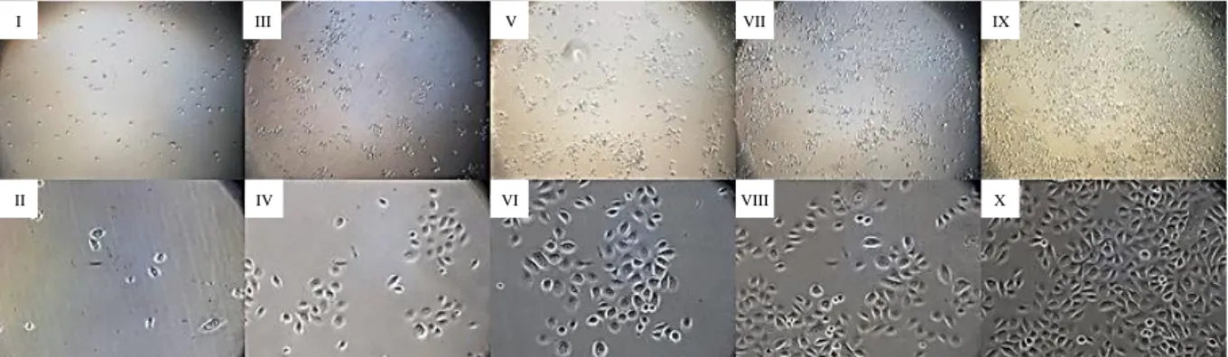
Figure 2.1 - Monitorization of culture growth: Keratinocytes covering 10% (I, II); 20% (III, IV); 40%
(V, VI); 60% (VII, VIII); 80% (IX, X) of t-flask surface. Magnifications: 40x (I, III, V, VII, IX); 200x
(II, IV, VI, VIII, X). 16
Figure 2.2 - Diffusion studies equipment: Hand-blown Franz-type diffusion cell (A) and the complete
experimental apparatus (B), in which 5 diffusion cells are dipped in a thermostatic bath maintained at a
constant temperature. 18
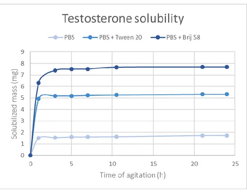
Figure 3.1 - Unsolubilized testosterone, by visual inspection, in the different solutions after 24 hours of
agitation: (A) PBS solution with a lot of testosterone non-solubilized, (B) PBS + 1% Tween®20 with some testosterone non-solubilized and (C) PBS + 1% Brij®58 with almost none testosterone
non-solubilized. 22
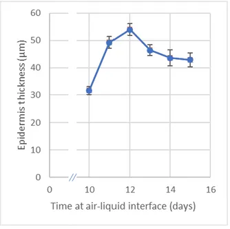
Figure 3.2 - Testosterone solubility profile over 24 hours of agitation in the different solutions. 23 Figure 3.3 - Evolution of epidermis thickness when cultivated at 37 ̊C, during the days surrounding the
standard air-liquid interface period (n = 6, average ± SD). 24
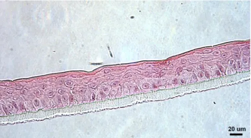
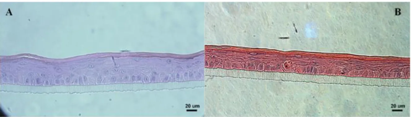
Figure 3.4 - Fully differentiated epidermis with the strata that compose epidermis pointed out (approx.
54 µm thick). Magnification 400x. 24
Figure 3.5 - Morphology of the reconstructed human epidermis grown under standard conditions, after
12 days at air-liquid interface (approx. 52 µm thick). Magnification 400x. 25
Figure 3.6 - Caffeine permeability profile over 24 hours of sampling in reconstructed human epidermis
(RHE) grown under standard conditions (n = 5, average ± SD). 26
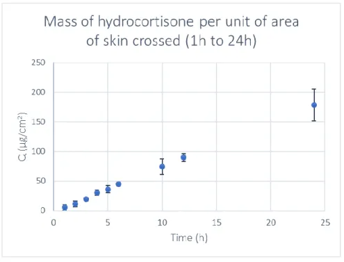
Figure 3.7 - Hydrocortisone permeability profile over 24 hours of sampling in reconstructed human
epidermis (RHE) grown under standard conditions (n = 4, average ± SD). 27
Figure 3.8 - Testosterone permeability profile over 24 hours of sampling in reconstructed human
epidermis (RHE) grown under standard conditions (n = 4, average ± SD). 27
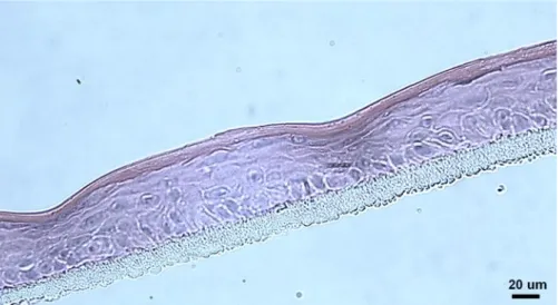
Figure 3.9 - Morphology of (A) the reconstructed human epidermis grown in culture medium renewed
medium renewed every 96 hours (approx. 41 µm thick), after 12 days at air-liquid interface.
Magnification 400x. 28
Figure 3.10 - Morphology of the reconstructed human epidermis grown in culture medium renewed
only every 72 hours, after 12 days at air-liquid interface (approx. 65 µm thick). Magnification 400x. 29
Figure 3.11 - Caffeine permeability profile over 24 hours of sampling in reconstructed human epidermis
(RHE) grown in culture medium renewed only every 72 hours. (n = 5, average ± SD). 30
Figure 3.12 - Hydrocortisone permeability profile over 24 hours of sampling in reconstructed human
epidermis (RHE) grown in culture medium renewed only every 72 hours. (n = 5, average ± SD). 30
Figure 3.13 - Testosterone permeability profile over 24 hours of sampling in reconstructed human
epidermis (RHE) grown in culture medium renewed only every 72 hours. (n = 5, average ± SD). 31
Figure 3.14 - Morphology of the reconstructed human epidermis grown in culture medium additionally
supplemented with 10% FBS, after 12 days at air-liquid interface (approx. 40 µm thick). Magnification
400x. 32
Figure 3.15 - Caffeine permeability profile over 24 hours of sampling in reconstructed human epidermis
(RHE) grown in culture medium additionally supplemented with 10% FBS. (n = 5, average ± SD). 33
Figure 3.16 - Hydrocortisone permeability profile over 24 hours of sampling in reconstructed human
epidermis (RHE) grown in culture medium additionally supplemented with 10% FBS. (n = 5, average
± SD). 33
Figure 3.17 - Testosterone permeability profile over 24 hours of sampling in reconstructed human
epidermis (RHE) grown in culture medium additionally supplemented with 10% FBS. (n = 5, average
± SD). 34
Figure 3.18 - Evolution of epidermis thickness when cultivated at 32 ̊C, starting on the 13th day of standard air-liquid interface period and ending on the 30th day of air-liquid interface (n = 6, average ±
SD). 35
Figure 3.19 - Morphology of the reconstructed human epidermis grown at 32 ̊C, after 22 days at
air-liquid interface (approx. 86 µm thick). Magnification 400x. 36
Figure 3.20 - Caffeine permeability profile over 24 hours of sampling in reconstructed human epidermis
(RHE) grown at 32 ̊C for 21 days. (n = 5, average ± SD). 37
Figure 3.21 - Hydrocortisone permeability profile over 24 hours of sampling in reconstructed human
epidermis (RHE) grown at 32 ̊C for 22 days. (n = 4, average ± SD). 37
Figure 3.22 - Testosterone permeability profile over 24 hours of sampling in reconstructed human
epidermis (RHE) grown at 32 ̊C for 23 days. (n = 5, average ± SD). 38
Figure 3.23 - Morphology of the reconstructed human epidermis grown in petri dishes, after 12 days at
air-liquid interface (approx. 56 µm thick). Magnification 400x. 39
Figure 3.24 - Caffeine permeability profile over 24 hours of sampling in reconstructed human epidermis
(RHE) grown in petri dishes. (n = 5, average ± SD). 40
Figure 3.25 - Hydrocortisone permeability profile over 24 hours of sampling in reconstructed human
epidermis (RHE) grown in petri dishes. (n = 5, average ± SD). 40
Figure 3.26 - Testosterone permeability profile over 24 hours of sampling in reconstructed human
N
OMENCLATURE
% – percent λ – Wavelength µ (unit prefix) – micro 2D – Two-Dimensional 3D – Three-Dimensional BMI – Body-Mass Index
Brij®58 – Polyoxyethylene-20-cetyl ether BSA – Bovine Serum Albumin
̊C – degree Celsius (SI Unit) c (unit prefix) – centi CaCl2 – Calcium Chloride CO2 – Carbon Dioxide Da – Dalton
DFBS – Dialysed Fetal Bovine Serum DMSO – Dimethyl sulfoxide
ECVAM – European Center for the Validation of Alternative Methods EDTA – Ethylenediaminetetraacetic acid
EU – European Union FBS – Fetal Bovine Serum FFAs – Free Fatty Acids g – force of gravity g – gram (SI Unit) h – hour
H & E – Hematoxylin and Eosin H2O – Water
HEKn – neonatal Human Epidermal Keratinocytes HKGS – Human Keratinocyte Growth Supplement HU – Histopathology Unit
IGC – Instituto Gulbenkian de Ciência
Igepal® – Octylphenoxy poly-ethyleneoxy-ethanol ITQB – Instituto de Tecnologia Química e Biológica
Jmax – Maximum Flux KCl – Potassium Chloride
KGF – Keratinocyte Growth Factor KH2PO4 – Monopotassium phosphate L – Liter
log P – Octanol-water partition coefficient m – meter (SI Unit)
M – Molar (SI Unit) m (unit prefix) – milli n (unit prefix) – nano N2 – Nitrogen
Na2HPO4 – Disodium Hydrogen Phosphate NaCl – Sodium Chloride
O2 – Oxygen
OECD – Organization for Economic Co-operation and Development OR – Oregon
PBS – Phosphate-buffered saline PG – Propylene Glycol
pH – potential of Hydrogen
Q24 – Cumulative Corrected Amount of compound crossed per unit of area of skin in 24 hours RH – Relative Humidity
RHE – Reconstructed Human Epidermis SB – Stratum Basale SC – Stratum Corneum SD – Standard Deviation SG – Stratum Granulosum SL – Stratum Lucidum SS – Stratum Spinosum
Tween®20 – Polyoxyethylenesorbitan monolaurate USA – United States of America
UV – Ultraviolet
I
NTRODUCTION
Healthcare industry is among the industries that most invest in research and development, which allows large pharmaceutical, medical and biotechnological innovations to emerge in short periods of time. Any new drug or biologic developed in this industry, whether of topical, enteral or parenteral administration, must be tested as to its toxicity and safety for the organism, prior to their placement in the market. Previously, these studies relied on animal experimentation, however over the years a consciousness regarding the use of animals began to arise and non-animal methods began to be prospected1–3. In 1959, a major step towards the awareness of animal testing was taken with the publication of The Principles of Humane Experimental Technique by William Russell and Rex Burch, which brought to the attention of the scientific community three simple principles (“3 Rs”) that should be taken into account when animal experimentation was considered. These principles encouraged researchers to
reduce the number of animals used in an experiment to the minimum necessary, prioritizing the use of
animals with lower neurological development; to refine the conditions to which animals were exposed so that the pain and distress caused on the animals were minimal; and to replace the use of animals with alternative non-animal methods whenever possible. The above-mentioned concepts gained such a recognition that were incorporated into the EU Directive 86/609 in 1986 and reinforced in several legislations all over the world3–5.
Despite Russel and Burch’s effort to raise awareness on this subject, the number of animals used in laboratory practices continued to increase, so in 2013 European Parliament and European Council banned the animal testing for toxicity evaluation of cosmetic ingredients in Europe (Directive 2003/15/EC). As in Europe, also in other countries such as Japan and United States of America (USA) legislations limiting animal testing appeared4,6.
Since legal restrictions on animal experimentation were imposed, non-animal alternative methods were taken more seriously and implemented on several basic and applied research studies. Considering that a large percentage of the animals used for scientific purposes in the European Union were used in toxicological and other safety assessments (80%, according to data from 20055), various methods have been proposed as alternatives in drug testing and toxicological screenings, which allow the reproducibility, quantity and predictability required in these evaluations. Included on these methods are the cell and tissue cultures, namely of human origin2,3,5.
Regarding topically administered drugs, in vitro engineered human skin models were recognized as the best alternative to the use of animal models. Even though ex vivo human skin samples, mainly obtained either from cadavers or biopsies, were for a while also a plausible alternative as it successfully replicated in vivo skin properties, its limited availability as well as poor reproducibility due to variation on skin properties depending on factors such as donor’s ethnicity, age, gender, skin type, body-mass index (BMI) and lifestyle, caused this approach to be discarded. Thus, increased efforts were devoted to the
development of consistent three-dimensional (3D) human skin equivalents that allowed the performance of release-rate estimates besides toxicity assessments6–9.
Although these alternative approaches are currently of great value on helping to explain and predict several in vivo behaviors, the existing models are still not able to fully portray all the biological features and processes. In order to improve these models and make better predictions of human biological responses, more commitment on the part of the scientific community must be directed towards the optimization of cell cultures, so that better and more accurate models emerge2,4,5,10.
1.1.
T
HEH
UMANS
KINThe skin is the largest organ of the human body and the first line of defense against the innumerable external aggressions that it can suffer, functioning as a protective interface between the organism and its external environment. This organ, along with the accessory structures, such as hair, glands and nails, form the integumentary system. In order to carry out its numerous functions, whether of protection, regulation, metabolic, immune and nervous, the skin presents a massive complexity, being constituted by a multiplicity of different types of cells, along with accessory structures, that interact with each other7–9,11–14.
1.1.1.
S
KIN STRUCTUREThe skin is divided fundamentally into 3 different layers: hypodermis, dermis and epidermis (Figure 1.1).
Hypodermis is the innermost layer of the integumentary system and is essentially composed by adipocytes although containing some fibroblasts and macrophages. Despite working as a connection between the skin and the muscles or bones and as a supplier of blood vessels and nerves to the other layers of the skin, its major functions are as thermal insulator, nutritional storage and shock absorber9,11,14,15.
Dermis, accounting for around 90 % of the weight of the skin, is the middle layer of the integumentary system, being delimited inferiorly by the hypodermis and superiorly by the epidermis. This layer consists of connective tissue, where collagen, elastin and glycosaminoglycans, collectively, comprise
Figure 1.1 - Structure of human skin: The 3 different skin layers (epidermis, dermis and hypodermis, from the outer to the innermost layer, respectively) and their accessory structures. (Adapted from13)
the extracellular matrix, produced by the primary cell type of dermis, fibroblasts. Its main role is to provide foundation, structural strength and elasticity to the system. Blood and lymph vessels are abundant in this skin region, having the responsibility of removing metabolites and supplying nutrients both to dermis and epidermis, since the last represents an avascular structure. The majority of skin appendages, such as hair follicles, sebaceous glands and sweat glands, as well as nerve endings are anchored to dermis9,11,12,14,15.
Basement Membrane Zone or Dermal-Epidermal Junction is the acellular band between the epidermis and dermis and is mainly composed by a laminin/collagen IV scaffold secreted by both its adjacent layers. This junction’s functions are to anchor the epidermis to dermis, as well as regulate the molecular and cellular exchanges between the two skin layers and provide some structural and mechanical support to epidermis11,16.
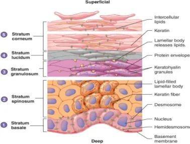
Epidermis is the outermost layer of the skin and the main responsible for the barrier property of this organ. Among its functions is the protection of the organism from mechanical injuries, microorganisms, chemicals, and radiation present in the environment, as well as the regulation of water and heat losses and production of vitamin D. The stratified squamous epithelium that characterizes this skin layer includes several types of cells, such as melanocytes, Langerhans cells and Merkel cells, however its major constituent are keratinocytes (90%). Keratinocytes are a type of cells named after the structural protein mixture that they produce called keratins, classified as intermediate filament, which hardens them, as well as the hair and nails, and provides them the strength needed to resist abrasion and to accomplish the functions of protection associated to this outermost skin layer. This system of production and accumulation of keratin by keratinocytes is called keratinization or cornification and is part of the process of cytodifferentiation, which the keratinocytes undergo to originate a stratified tissue. During this differentiation process, as new keratinocytes arise from mitosis in the deepest layer of epidermis, older ones are pushed towards the surface, continuously changing their shapes as well as chemical composition, until they slough off. In addition, as keratinocytes move farther from the basement membrane, which is the link between the epidermal cells and their oxygen and nutrients’ supplier (the dermal blood vessels), they receive fewer supplies, becoming less active and ultimately dying. Although this is a continuous development, 4 or 5 different strata with specific characteristics can be recognized in epidermis, varying the dimension of each stratum or even the number of strata with the body part from where the skin derives. From the deepest to the most superficial, those strata are the stratum basale, stratum spinosum, stratum granulosum, stratum lucidum and stratum corneum (Figure 1.2)9,11,12,14,17.
Figure 1.2 - Epidermal differentiation: The different strata that compose epidermis and their main constituents. (Adapted from12)
Stratum basale (SB) or Stratum germinativum is the innermost stratum of the epidermis and
consists of a single layer of columnar cells, mitotically active, which generate new keratinocytes. From the mitotic division, one of the two daughter cells remains as a keratinocyte stem cell in the stratum basale, and divides itself during the next mitotic cycle, whereas the other daughter cell is shoved towards the surface and starts its differentiation process. The cells from this stratum have incorporated melanosomes, transferred by melanocytes, conferring some pigmentation and protection against UV light to this layer, and are adhered to each other by desmosomes and anchored to the basement membrane by hemidesmosomes, both contain keratin fibers that diffuse into the cell’s cytoplasm. The keratins present in the cells from stratum basale are primary keratins with low molecular weight from type 5 and 1411,12,18.
Stratum spinosum (SS) is the stratum directly above the germinativum cell layer and consists
of a multiple layered stratum (8-10 cell layers) of many-sided cells. The name given to this stratum derives from an artifact produced during histological preparations, in which cells shrink increasing the intercellular spaces, except where they are linked by desmosomes, inducing a spiny appearance on cells. When arriving at this stratum, cells have already undergone a pressurizing process as they were pushed towards the surface, which led to their flattening and consequently to the break of the desmosomes previously formed, imposing the formation of new ones. In addition, envelope proteins and lamellar bodies (also known as keratinosomes), which are secretory organelles containing cholesterol, glycolipids and fatty acids as well as a battery of enzymes and antimicrobial peptides, are assembled in the cytoplasm. Later on, these membrane-coating granules will fuse with the plasma membrane of granular and stratum corneum cells and lipids will be released into the intercellular spaces, originating the extracellular matrix. The stratum spinosum presents not only K5/K14 keratins retained from the previous stratum, but also K1/K10 keratins, which are the keratins synthetized in this stratum and are accumulated as keratin fibers (tonofilaments). Therefore, the synthesis of type 1 and 10 keratins as well as lamellar bodies function as markers of the progressive differentiation11,12,19,20.
Stratum granulosum (SG) is the middle stratum of the epidermis and consists of 2-5 layers of
flattened, diamond-shaped cells that gradually start dying. The cells of this stratum exhibit a notorious specific feature, from which the stratum’s name derived, which is the presence of dark keratohyalin granules in the cytoplasm. These granules contain numerous proteins, such as profilaggrin, loricrin and involucrin, crucial both for aggregation of keratin filaments and formation of the cornified envelope. During this stage, lamellar bodies formed in the previous stratum move towards the plasma membranes, merge with these structures, and release their content into the extracellular spaces. Simultaneously a protein envelope arises beneath the plasma membrane of cells. In the outermost layer of stratum granulosum, the cell organelles as well as the nucleus begin to degenerate, leading to cells’ death, and tight junctions are established between adjacent cells, assisting in the epidermal structural barrier. This stratum marks the transition from the metabolically effective strata to the dead cells’ strata11,12,14.
Stratum lucidum (SL) is the stratum located between the stratum granulosum and stratum
corneum and is absent in the skin of most of the human body, being found only in areas where the skin is thicker, such as the palms of the hands, the soles of the feet and the fingertips. This stratum consists of 3-5 layers of flattened dead keratinocytes containing large amounts of keratin fiber yet with no visible keratohyalin granules. The keratohyalin instead of being accumulated in granules, have dispersed around the keratin fibers, causing a transparent appearance on the cells of this stratum12,14.
Stratum corneum (SC) is the outermost stratum of the epidermis and the final stage of
keratinocytes’ differentiation process. The thickness of this stratum is very variable, ranging from a few layers where the skin is thin and does not suffer from abrasion, to more than fifty in areas where the skin is thick and subject to a lot of abrasion. The stratum corneum consists of dead cornified squamous cells, called corneocytes, embedded in a lipid-enriched extracellular matrix, originating a “brick and mortar” organizational model. The corneocytes, due to the previous degeneration of all organelles, only incorporate a protein envelope, comprised by loricrin, involucrin, trichohyalin and small proline-rich proteins crosslinked, as well as keratin fibers enclosed in filaggrin. As to the lipidic extracellular matrix, it results from the processing of the pro-barrier lipids by the lipolytic enzymes, both previously enclosed on the lamellar bodies, and incorporates 13 species of ceramides, cholesterol, and free fatty acids, as well as proteolytic enzymes and antimicrobial peptides. Corneocytes, because of their composition and their constituents’ organization, confer strength and elasticity to this stratum along with hydration and UV protection, whereas the extracellular matrix provides antimicrobial protection as well as impermeability to water and water-soluble substances, avoiding transepidermal water loss and regulating selective transepidermal absorption. Additionally, stratum corneum presents an acidification of the environment relatively to the other strata of the epidermis, with pH values ranging between 4.5 and 5.5 instead of physiological pH values (7.35 - 7.45), derivative of endogenous mechanisms, which allows an effective enzymatic lipid processing for the assemble of the extracellular matrix, inhibits the early activity of the proteolytic enzymes and serves as a defense against pathogenic agents. The proteolytic enzymes have a crucial role in the upper layers of this stratum by degrading the corneodesmosomes, which are desmosomes that were modified during the terminal differentiation, allowing desquamation. According to the presence of corneodesmosomes and consequently the cellular cohesion, the stratum corneum can be divided in two sectors, which are SC compactum, in the deepest layers where cells are still strongly associated by corneodesmosomes, and SC disjunctum, in the most superficial layers where corneodesmosomes have already undergone proteolytic cleavage14,15,21–25.
Epidermis is a continually self-renewing epithelium as the desquamation rate of the cornified tissue and the proliferation rate in the basal layer are precisely balanced. As corneocytes slough off at the surface, new keratinocytes arise from the stratum basale, leading to a process of cellular ascension and transformation in chain, in which the cells below replace the cells directly above. A complete cycle of keratinocyte cytodifferentiation, since the cell is generated from the stratum basale until it reaches the epidermal surface and slough off takes approximately 40-56 days12,17.
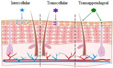
1.1.2.
P
ERMEATION ROUTES THROUGH SKINThe skin is the interface between the organism and its external environment, consequently it is permanently in contact with several substances that are either present in the environment or intentionally applied to the surface of the tissue. Any substance, in order to enter the system and have any impact on the organism, positive or negative, must transpose the stratum corneum and reach viable cells. Even though stratum corneum is the primary barrier to skin penetration, the viable epidermis as well as part of the dermis also provide a significant permeability barrier to any substance that needs to enter the systemic circulation through the dermal capillary bed, penetrating through these layers8,26.
The permeation of a substance through skin might occur either via transepidermal pathway, in which molecules diffuse through the intact epidermis, or via transappendageal pathway, in which molecules diffuse through skin appendages, such as hair follicles and sweat glands. Additionally, when it occurs
via transepidermal pathway, particles can adopt either an intracellular route or an intercellular route. (Figure 1.3)
The transappendageal pathway or ‘shunt’ route is considered to have a small contribution on the overall permeation process as skin appendages occupy only 0.1 % of the total skin surface, however, it is the only penetration pathway for particles larger than few nanometers. These appendages, due to their architectural structure, function not only as fast deliverers of compounds, that are capable of transfollicular penetration, to deeper skin layers and consequently into the systemic circulation, but also as reservoirs for substances which cannot overcome the transfollicular barrier8,27.
The transepidermal pathway is the main route for compounds penetration through skin, as it presents the major area available to the permeation process. The route through which compounds penetrate the stratum corneum and consequently the success of that penetration, differ according to the compound’s solubility and oil/water partitioning, usually described by the octanol-water partition coefficient (log P)15,28.
By the intracellular route, also called transcellular route, the penetrating particles must diffuse through the alternating sheets of corneocytes and extracellular matrix. Even though the migration distance to overcome the epidermal barrier is shorter through this pathway, the consecutive alternation between hydrophilic (keratinocytes’ interior) and lipophilic (extracellular matrix) domains makes it a very selective route. Generally, the intracellular route is selected by the hydrophilic (polar) compounds, with low log P values (log P < 1), which encounter greater energetic barriers while progressing through extracellular matrix due its lipidic nature8,28–31.
By the intercellular route, the penetrating particles, diffusing stratum corneum which are sized-restricted, progress through the extracellular matrix via passive diffusion, without intersecting any cells. When using this route, despite stratum corneum being a narrow layer, particles travel a much-extended pathway (sometimes 20 times longer than the thickness of the tissue), due to be a labyrinthine path. Generally, the intercellular route is preferred by the lipophilic (non-polar) compound, with intermediate/high log P values (log P > 3), which have a high affinity to the lipidic composition of the matrix8,28,29,31.
Figure 1.3 - Pathways for permeation through the skin: Molecules diffuse either through the intercellular spaces, through keratinocytes or even through skin appendages. (Adapted from8)
The permeation process of any substance may occur either by a specific route or a combination of any of the possible routes. As different substances penetrate through human skin by different pathways, when assessing the barrier property of the skin, all different routes should be taken in consideration. Therefore, permeability studies integrate drugs with different polarities in order to evaluate the barrier function against a greater range of penetrability behaviors.
1.2.
I
N VITROH
UMANS
KINM
ODELSSkin models have been designed to attend both basic and applied research demands, therefore enabling skin cellular and molecular studies, for the better understanding of skin and its processes, as well as providing a foundation for the screening of new ingredients and active principles for safety and efficacy along with formulation optimization. Nowadays, skin models are also used for medical purposes as skin replacements or implants, however this use remains limited due to the lack of some components, such as vasculature, innervation and immunity system. Due to different demands, from either researchers or industries, different models have emerged revealing different levels of biological complexity2,10,32–36. Monolayer cell cultures (2D models), which are cell cultures grown under contained flat conditions, have been the main approach for the study of both skin cell biology and pathophysiology in vitro as well as cellular responses to biophysical and biochemical stimulus. This method was the pioneer in the culture of human cells in vitro, with keratinocytes as the primary cell type, and since then has been widely used due to its simplicity, reproducibility and efficiency, allowing major advances in cellular knowledge37– 39.
Although these models provide foundations for many scientific progresses, they are a reductionist approach that do not reproduce the complexity and dynamism of the in vivo tissues, producing frequently inconsistent results with those produced in vivo. Thus, 3D cell cultures were conceived in order to better mimic in vivo conditions, enabling the study of a wider range of biological processes38,39.
Skin tissue engineering (3D models), emerging as early as 1975 to 1980, produces fully differentiated tissues, which recreate architectural, metabolic, cellular and functional aspects of native human skin. These models comprise both cells and extracellular matrices and allow cells to grow and interact with their surroundings in all three dimensions, generating a natural gradient of nutrient access along with a waste buildup. Currently, the study of skin barrier function, wound healing, biological skin interactions, such as cell-cell and skin-microbiome interactions, as well as regulation of proliferation and differentiation of skin cells, relies on these models. The existing human skin models can be divided into three categories: (i) epidermal equivalents, which consist only of keratinocytes that differentiate producing a fully stratified epidermis; (ii) dermal equivalents, which consist of fibroblasts and collagen that interact generating a uniform contraction distinctive of dermis; and (iii) full-thickness skin equivalents, which consist of both dermal and epidermal layers. All three categories have, nowadays, commercially available prototypes developed by various companies such as CellSystems, StrataTech, SkinEthic, MatTek, Advanced Tissue Sciences, Organogenesis and more. Even though the resemblance of these 3D models to in vivo skin suggests that this approach should be adopted whenever possible, disadvantages such as high manufacturing cost, low reproducibility due to high complexity and absence of a standard approach, as well as low-throughput screening, limit its use9,10,34,36,37.
Despite the major progresses in scientific models, the existing designs are still not capable to entirely mimic the skin’s natural environment, and further studies are required to develop improved models. Therefore, to assemble more physiological skin models, for both medical and screening purpose, intentions are to incorporate further skin components such as vasculature, immunity system, innervation,
appendages, pigmentation and adipose tissue into the pre-existing in vitro skin models, generating models of complete skin homeostasis. Additionally, to overcome the low reproducibility from the simplest to the most complex 3D models, 3D bioprinting presents itself as a promising method of model fabrication. Skin-on-a-chip is an existing revolutionizing method which incorporates skin equivalents in a device with a confined microenvironment and a microfluidic perfusion system. This technique has an enormous potential due to allowing a more controlled as well as automated tissue culture comprising dynamic perfusion, besides enabling a future integration on a multi-organ-chip10,40,41.
1.2.1.
H
UMANE
PIDERMAL MODELSDespite more complex models, such as full-thickness skin equivalents, generally resemble native skin more reliably than simpler 3D models, their laborious manufacturing along with their high maintenance cost, due to their complexity, makes their use less frequent, being mostly accounted for more accurate and conclusive studies. Therefore, simpler skin equivalents like in vitro human epidermis, even though mimicking less well some properties from in vivo skin, are still subject of great interest for screening purposes2,7,42.
In the year of 1975, James Rheinwald and Howard Green tried, for the first time, to reconstruct epidermis by culturing primary human keratinocytes on top of a feeder layer of lethally irradiated 3T3 fibroblasts in an immersed environment. This experiment resulted in a small sheet of squamous epithelium with only 2 or 3 keratinocyte layers. Even though this approach was a major scientific breakthrough demonstrating that keratinocytes were capable of breed and form stratified colonies, these first epidermal models didn’t present a normal differentiation pattern, lacking membrane-coating granules as well as a functional cornified layer, and consequently were far from resembling the native epidermal tissue. The first well-organized and fully differentiated epidermal model, evidencing both the inborn components that were missing, was only described in 1983 by Michel Pruniéras when human keratinocytes were seeded on top of a cell-free dermal substrate and were left at air-liquid interface for 2 weeks. The exposure of keratinocyte cultures to an air-liquid interface stimulated the synthesis of membrane-coating granules, which consequently led to the formation of the extracellular matrix and ultimately allowed the cornification of the cells, establishing the stratum corneum2,43,44.
Over the years, culture media industry has evolved and specific culture media for culturing isolated keratinocytes without requiring the use of fibroblast co-cultures or dermal substrates, were developed. Those media are chemically-defined culture media supplemented with pro-differentiation factors, such as keratinocyte growth factor (KGF), calcium and ascorbic acid, which play a similar role to the one produced by the factors released by the dermis. Thus, nowadays 3D keratinocyte cultures can be accomplished by plating keratinocytes both on (i) a collagen matrix with fibroblasts; (ii) a collagen matrix without fibroblasts or a de-epidermized dermis; or even on (iii) inert filters2,6,45.
Due to the high demand for epidermal in vitro models, as the years went by several designs were developed and submitted to the European Center for the Validation of Alternative Methods (ECVAM) for validation of their use and commercialization. The first validated models arose in the 1990s by EpiSkin and MatTek Corporation being commercialized as EpiSkin™ and EpiDerm™, respectively. Since then, many others emerged, resulting on the large variety of in vitro 3D reconstructed human epidermal (RHE) commercial models that are available nowadays2,6.
Although presently there is an immensity of RHE commercial kits available, which are of great value to research due to their standardized reproducibility and easy accessibility, their high cost and inflexible culture parameters force researchers to develop protocols for the conceiving of in-house RHE models, enabling a more cheaper and custom-made research2,36,46,47.
Currently, some standardized protocols stand for the reconstruction of in-house epidermal equivalents, which follow similar crucial steps (Figure 1.4). Firstly, keratinocytes are isolated from their source and cultured in monolayer cultures in order to increase the number of cells. In a second step, keratinocytes are seeded on a supports’ surface, which can either be a biological matrix or an inert filter, undergoing immersed conditions so that they expand and cover the all surface. Finally, once a keratinocyte monolayer is established, cells are exposed to air-liquid interface which allows them to differentiate and stratify, resulting ultimately on a fully differentiated epidermis after 11 to 14 days.
1.2.2.
A
TTEMPTS TO IMPROVE THE IN VITRO EPIDERMAL MODELSThe in vitro epidermal models produced so far, accurately resemble the morphological characteristics of the native human epidermis, however they do not entirely mimic the barrier function of the in vivo epidermal tissue. The barrier function of the in vitro models is reduced when compared to native epidermis, exhibiting much higher permeabilities (sometimes 10 times higher). Thus, many research studies have been developed in this field, aiming to identify the parameters which influence the formation of the skin barrier during epidermal tissue culture, in order to recreate an epidermal model with similar barrier properties to those found in the native skin2,7,37,48.
Studies have revealed that the lack of a normal barrier function by the epidermal models is related to deviations in lipid composition and packaging from in vivo human epidermis. Even though all major classes of human skin lipids are present in the in vitro RHE models and their arrangement into lipid lamellae is identical to the arrangement in the native epidermal tissue, some differences in the lipid profile may be the responsible for the existing differences on their packaging. Among the lipid profile discrepancies are the absence of ceramide 7 along with the low abundancy of ceramide 4, 5, 6 and glucosylceramides contrasting with their high abundancy in the native epidermis, the increased levels of monounsaturated free fatty acids (FFAs) although the levels of total FFAs are lower than the ones observed in vivo, the low values of cholesterol esters and high abundancy of both ceramide 2 and triglycerides. These disparities are reflected in the arrangement of the lipid lamellae, which are found in a larger number but less densely packed than in native epidermal tissue2,7,32,49,50.
To diminish or even extinguish these variations between in vitro models and in vivo human epidermis, several attempts of improving the methods of culture have been made so far. Regarding to ceramides 6 and 7 (C6 and C7), was discovered that the addition of vitamin C to the media facilitated the synthesis of both ceramides, since it stimulates the hydroxylation of sphingoids and long chain fatty acids which are the precursors of both C6 and C7. Additionally, was also discovered that the addition of essential fatty acids such as arachidonic, palmitic, oleic and linoleic free fatty acids promoted the assembly of lamellar bodies, ceramides and cholesterol esters, since fatty acids are part of their composition. Along with the previous discoveries which resulted in positive outcomes as to improving the lipid profile and consequently the barrier formation, other modifications to the culture methods were tested. Amid the ones that produced satisfactory effects are the lowering of the relative humidity (RH) to 50% - 75%
Figure 1.4 - Crucial steps for the reconstruction of epidermal equivalents.
instead of the 100% RH in which cultures were being incubated as well as the lowering of the temperature to 33 ̊C instead of the 37 ̊C (in order to better resemble the environment in which native skin grows), and also the supplementation of the culture media with vitamin D48–55.
Despite all the efforts, still no human epidermal equivalent has been able to fully mimic the barrier function of the in vivo epidermal tissue. Therefore, the search for a tissue culture recipe which produces an epidermal equivalent similar to the native human epidermis, in terms of morphology and barrier properties, continues2,48.
1.3.
N
EW ATTEMPTS TO IMPROVE THE IN VITRO EPIDERMAL MODELSEven though many attempts have already been made to achieve an epidermis with an optimized barrier function, a multitude of alternatives are yet to be tested. The factors to consider when making changes to culture conditions can either be of environmental or nutritional nature. Whenever the desired test factor is environmental, modifications are made to the equipment to be adopted, either by choosing a different culture material from the usual or just by changing equipments settings; while when the desired test factor is nutritional, changes are made to the cell culture media composition. To assess the impact of the new culture conditions, environmental or nutritional, on the morphology and barrier properties of the epidermal tissue, histology and permeability tests must be performed on the epidermis reconstructed under modified conditions.
1.3.1.
C
ONDITIONS INTENDED TO SCREENThe choice of culture conditions to be screened which could possibly improve the barrier properties of the in vitro skin, resulted from conflicting bibliography or divergent in vitro tissue culture protocols, from data previously acquired by the laboratory group through metabolomic analyzes performed on the culture media collected over the reconstruction of epidermal tissue or even from biological hints. The first variation on the culture conditions proposed to assess was regarding to the availability of
autocrine and homocrine factors during the air-liquid interface exposure. Many of the cellular
processes, such as cell proliferation, migration, differentiation and apoptosis, are regulated by cell signaling, which occurs mostly by diffusion. The soluble diffusible signals can either be endocrine, when travel through the circulatory system in order to reach cells from other tissues, or paracrine, when they don’t enter the bloodstream and only diffuse in between adjacent cells. Since in keratinocyte cultures there is no vasculature, only paracrine signaling is relevant in this type of in vitro cultures. Among the paracrine signals, some stimulate the cell that generated them, which are called of autocrine, other stimulate cells from the same cell type as the one that produced them, which are named homotypic paracrine or homocrine, and the ones that stimulate cells from a different cell type from the one in which the signal was created are termed heterotypic paracrine or simply paracrine. As in this in vitro cell culture only keratinocytes occur, just homocrine and autocrine signaling are present. During reconstruction of epidermal tissue, the media that supplies cells is renovated every other day, meaning that all signaling factors released by cells are disposed within a few hours leaving little time for action. In order to evaluate if an extended cellular exposure to homocrine and autocrine signals allows a greater proliferation and better differentiation of epidermal tissue, it was suggested to leave the medium unrenewed longer periods, while considering that the properties of the medium should be close to optimal. Thus, a preliminary study should be conducted to assess the acceptable extension of the media renewal period, without it losing its properties or accumulating too many toxic secreted compounds, and then the epidermal tissue grown under that same conditions would be analysed56,57.
The second variation on the culture conditions intended to assess was regarding to the nutrients’
availability during the air-liquid interface exposure. A long time ago, epidermal culture protocols began
to emerge in which the use of serum in culture media was eradicated, since it was a source of variability due to its uncleared composition and inconsistency from one lot to the other, as well as a probable source of contamination due to its animal derivation. However, this supplement contains several growth factors, cytokines, hormones, vitamins and proteins that stimulate cell growth and proliferation, some of which are still unidentified, and thus makes it difficult to formulate a serum-free medium which fully resembles the serum-containing medium. Until now, several experiments have evaluated both epidermises reconstructed with serum-containing media and serum-free media, nonetheless contradicting results become apparent between research studies. Therefore, it was suggested that the medium used for the reconstruction of human epidermis was supplemented with fetal bovine serum (FBS), in order to assess the epidermal tissue reconstructed with serum-containing medium33,58–60.
The third variation on the culture conditions proposed to assess was regarding to the temperature during the air-liquid interface exposure. Most of mammalian cells are grown at 37 ̊C, temperature in which their growth rate is most efficient as it is their host’s body temperature and both their enzymatic and protein machinery are adapted to operate optimally under those conditions. Yet skin cells are directly exposed to external temperatures that exhibit large variations, which causes average temperature at the surface of the skin to be close to 32 ̊C. Even though it is well-known that the temperature at the surface of the skin is lower than on the rest of the body, most of the studies on keratinocyte cultures have been conducted at normal body temperature, 37 ̊C, and only a few studies have been performed at 32 ̊C. Thus, it was suggested to culture epidermis at 32 ± 1 ̊C, which is referenced by oecd guidelines for the testing of chemicals as the normal skin temperature, to evaluate whether the shift in the temperature at which the tissue is reconstructed significantly influences its properties. Considering that at lower temperatures cell metabolism is substantially decreased, a preliminary study should be conducted to assess how long it takes the epidermis to reach the differentiation level equivalent to the one attained after 12 days at 37 ̊C, and then the culturing of epidermal tissue under those conditions for that time period would carried out61–64.
The last variation on the culture conditions intended to assess was regarding to the oxygen availability during the air-liquid interface exposure. Oxygen (O2) is crucial for cells to generate a chemical potential that allows them to produce energy and consequently perform their vital functions. Fluctuations in its concentration can significantly influence various cellular processes such as cell growth, differentiation, signaling and free radical production. According to a metabolomic study performed by the laboratory group on the culture medium collected over the reconstruction of epidermal tissue, cells produced lactate and formate during culture, which was suggested to be an indicator of limited oxygen conditions. Thus, it was proposed that the oxygen availability was increased in order to evaluate whether an improvement was verified in the reconstructed epidermal tissue, which among other ways could be achieved by expanding the contact surface between the culture media and the incubator atmosphere. By increasing the area of contact, the oxygen diffusion zone would also increase and consequently the diffusion of oxygen through the medium to the cells would increment65–67.
1.3.2.
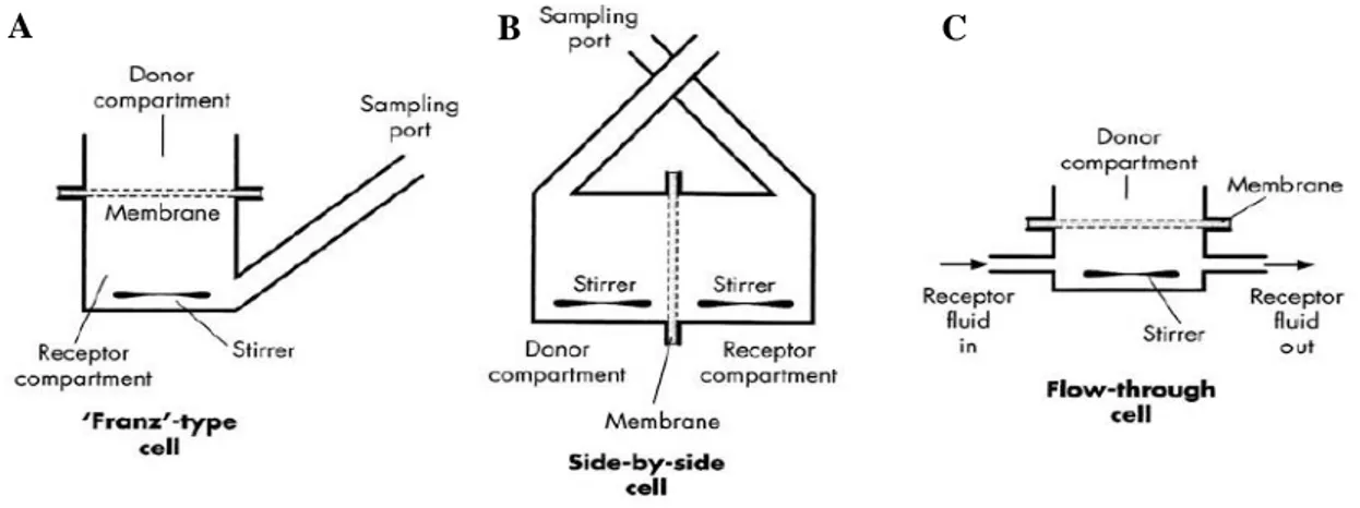
F
RANZ DIFFUSION CELLSFor the evaluation of skin permeation by measuring the amount of drug that passes across the skin, diffusion cells have become the industry standard methodology and consequently the most broadly used experimental equipment63,68.
The first diffusion cell was developed in 1970 by Thomas J. Franz, who design a simple experimental apparatus, the “Franz cell”, composed by a donor compartment, where the formulation was applied to a semipermeable membrane, and a receiver chamber, where samples could be withdrawn through a sampling port for drug release analysis. Over the years, various designs of diffusion cells have emerged in response to research needs, even though the fundamentals of the original template were always maintained. Nowadays, three main types of diffusion cells exist: Franz-type cell (vertical, one-chambered static diffusion cell), side-by-side cell (horizontal, two-one-chambered static diffusion cell) and flow-through cell (vertical, one-chambered non-static diffusion cell) (Figure 1.5)15,68,69.
Franz-type diffusion cells or upright static diffusion cells are the experimental apparatus used in the majority of in vitro percutaneous absorption studies, since it simulates the conditions most commonly encountered in the integumentary system of humans. In this device, the skin is hold between an upper opening to the ambient environment, as in vivo epidermis is exposed to the environment, and a lower compartment with a receiver solution. This layout allows the experiment of a wide range of formulations on skin surface, such as solutions, gels, creams, ointments, patches and other semi-solid materials of cosmetic or pharmaceutical nature. On the other hand, side-by-side diffusion cells or two-chamber diffusion cells are an experimental apparatus used in very limited situations, since it portrays circumstances in which stratum corneum is immersed in a liquid solution, such as while swimming or bathing, only allowing the experiment of compounds blended with that same liquid solution. In this device, the skin is hold between two horizontal compartments filled with aqueous solutions or other solvents, being one of them the donor solution where the permeant occurs and the other one the receiver solution. As to flow-through diffusion cells, they are an experimental apparatus used in the exact same circumstances as upright diffusion cells, since it follows the same basic principles of replicate the in vivo conditions. The difference between these two devices is that in the flow-through system the receptor fluid is continuously perfusing, mimicking the blood flow beneath the skin, which makes this device more suitable for full-thickness skin studies since in the integumentary system the bloodstream is never directly in contact with the epidermis. Due to this constant medium replacement, the solubilization of low solubility compounds is facilitated and sink conditions, which are an essential premise for the establishment of a proper in vivo-in vitro correlation, are maximized. Despite the advantages, the technical features associated with this mechanism represent not only a possible source of problems but also increase the commercial prize of the equipment15,68–70.
Currently, regulations as to in vitro skin absorption studies only partially standardize the experimental protocols as well as equipment design. Hence, researchers have a broad margin of flexibility to adapt
Figure 1.5 - Three main types of diffusion cells: (A, C) Franz-type diffusion cell and Flow-through diffusion cell, respectively, both mainly used for in vitro percutaneous absorption studies; (B) Side-by side diffusion cell, mainly used for evaluation of skin immersed conditions. (Adapted from15)