A rapid and simple high-performance liquid chromatographic method for the analysis of 1,3-dihydroxy-2-methylxanthone (DHMXAN) in biodegradable poly(D,L-lactide-co-glycolide) (PLGA) nanosphere and nanocapsule formulations is developed and validated. The method does not require any complex sample extraction procedure. Chromatographic separation is made with a reversed-phase C18column, using methanol–water (90:10, v/v) containing 1% (v/v) acetic acid as a mobile phase at a flow rate of 1 mL/min. Identification is made by UV detection at 237 nm. The isocratic system operates at ambient temperature and requires 7.5 min of chromatographic time. The developed method is statistically validated according to ICH guidelines and USP 29 for its specificity, linearity, accuracy, and precision. The assay method proposed in this study is specific for DHMXAN in the presence of nanosphere and nanocapsule excipients. Diode-array analyses confirm the purity of DHMXAN peak in stress conditions (> 99.0%). The method is shown to be linear (r≥ 0.999) over the concentration range of 0.25–3.0 µg/mL. Recovery ranges from 99.0% to 102.7% (RSD: 1.49%) and from 98.3% to 101.6% (RSD: 1.07%) for nanospheres and nanocapsules, respectively. Repeatability (intra-assay precision) and intermediate precision is acceptable with RSD values ranging from 0.6% to 1.9% and from 0.3% to 2.0%, respectively. The method is shown to be suitable for the evaluation of DHMXAN content entrapped in PLGA nanoparticles.
Introduction
Xanthones represent a large group of heterocyclic com-pounds, including natural, semisynthetic, and totally synthetic structures based on the dibenzo-γ-pyrone nucleus (1,2). Xanthonic derivatives constitute a group of compounds with a broad spectrum of biological activities (e.g., antitumor, hepato-protection, and modulation of enzyme protein Kinase C) (3). 1,3-Dihydroxy-2-methylxanthone (DHMXAN) (Figure 1) is a syn-thetic derivative that showed the highest inhibitory effect on the
growth of MCF-7 human breast cancer cell line among a set of 27 tested xanthones (4).
Poor aqueous solubility of many xanthonic derivatives is a major obstacle for the assessment of pharmacological activity of these compounds and for their use in the therapy. Besides the difficulty on the administration of water-insoluble drug sub-stances, this characteristic is often associated with poor bioavail-ability (5). Technology for micro- and nano-particulate systems has emerged as an alternative to overcome the difficulty of administration of poorly water-soluble compounds (6). The effi-ciency of this approach was successfully proven for different drugs (7–9). Nanoparticles are polymeric drug carriers of nanometer size (10). According to the preparation process either nanospheres or nanocapsules can be obtained. Nanospheres are matrix-type systems composed of an entanglement of oligomer or polymer units, in which the drug is dispersed throughout the polymer matrix. Nanocapsules are reservoir-type systems where the drug is confined to a cavity surrounded by a polymeric mem-brane (11,12). Poly(DL-lactide-co-glycolide) (PLGA) is an exten-sively studied polymer for pharmaceutical use due to its biocompatibility and biodegradability properties (13). Besides the improvement of delivery of water-insoluble drugs, nanopar-ticles have afforded several advantages for different drugs, such as reduce drug-associated adverse effects (11), protect the com-pound from inactivation before reaching its site of action (14), and increase the intracellular penetration (15). In a previous
Abstract
Development and Validation of an HPLC Method for
the Quantitation of 1,3-Dihydroxy-2-methylxanthone
in Biodegradable Nanoparticles
Maribel Teixeira1,2, Carlos M. M. Afonso1, Madalena M. M. Pinto1, and Carlos Maurício Barbosa3,*
1
Centro de Estudos de Química Orgânica, Fitoquímica e Farmacologia da Universidade do Porto e Laboratório de Química Orgânica -Faculdade de Farmácia do Porto, R. Aníbal Cunha, 164, 4050-047 Porto, Portugal;2Instituto Superior de Ciências da Saúde- Norte, R. Central de Gandra, 1317, 4585-116 Gandra, PRD, Portugal;3CTMUP/ Faculdade de Farmácia do Porto, R. Aníbal Cunha, 164, 4050-047 Porto, Portugal
* Author to whom correspondence should be addressed: email [email protected].
Figure 1. Chemical structure of 1,3-dihydroxy-2-methylxanthone (DHMXAN).
work, we have demonstrated that the encapsulation of xanthone and 3-methoxyxanthone in PLGA nanocapsules afforded a remarkable improvement of the inhibitory effect on the produc-tion of NO by the murine macrophage cell line J774 (16).
Through the incorporation of DHMXAN in nanoparticles, aqueous dispersions with concentrations higher than the max-imum aqueous solubility of this compound may be achieved for administration. Moreover, this approach may allow different ways of administration and may afford its in vivo protection and targeting.
In order to test the usefulness of PLGA nanoparticles as poten-tial carriers for xanthones, we routinely carry out in vitro studies with these colloidal formulations containing different xanthonic compounds. To facilitate the pharmaceutical development of xanthone-containing nanoparticle formulations an analytical method for the detection and quantitation of the analyte is nec-essary to guarantee the reliability of the results. The aim of the present work was to develop and validate a specific and simple high-performance liquid chromatographic method for the quan-titative analysis of DHMXAN, which was entrapped in PLGA nanoparticles for the first time. The procedures and parameters used for validation of the analytical method were those described in the International Conference on Harmonization (ICH) guide-lines (17,18), which are similar to the ones established by the United States Pharmacopoeia 29 (USP 29) (19).
Experimental
Reagents and chemicals
PLGA 50:50 (MW 50,000–75,000),Pluronic F-68, glucose, and
soybean lecithin (40% purity by TLC) were purchased from Sigma-Aldrich Química (Sintra, Portugal). DHMXAN (purity > 99.0% by HPLC; m.p. 141–142°C) was synthesised in our labora-tory according to the method already described (20).Myritol 318
was kindly supplied by Henkel (Lisboa, Portugal). HPLC-grade methanol and acetonitrile were obtained from Merck KGaA (Darmstadt, Germany). HPLC grade water was obtained by a MilliQ system (Millipore, Lisboa, Portugal). Other chemicals were of analytical grade.
Nanoparticle preparation and characterization
Nanospheres containing DHMXAN were prepared by the sol-vent displacement technique (21). Briefly, a solution of PLGA (50 mg) and DHMXAN (0.75 mg) in a mixture of acetone (9.5 mL) and methylene chloride (0.5 mL) was poured into 10 mL of an aqueous solution ofPluronic F-68 0.25 % (w/v), under moderate
stirring, leading to the formation of nanospheres. Then, acetone was removed under vacuum and the colloidal dispersion of nanospheres was concentrated to 5 mL by evaporation under reduced pressure. DHMXAN crystals were removed by filtration (0.8 µm mixed cellulose esters membrane filters from Millipore). Non-incorporated DHMXAN was removed by centrifugation at 2000× g for 2 h (Heraeus Sepatech Labofuge Ae centrifuge, Osterode/Harz, Germany) after solubilization of a certain amount of glucose for achieving a 5% (w/v) concentration. The supernatant was discarded and the pellet containing the
nanospheres was redispersed in water to complete the initial volume of nanosphere dispersion submitted to centrifugation. Empty nanospheres were prepared according to the same proce-dure but omitting the xanthone in the organic phase.
Nanocapsules containing DHMXAN were prepared by the interfacial polymer deposition method described by Fessi et al. (22). Briefly, 50 mg of polymer and 100 mg of soybean lecithin were dissolved in 10 mL of acetone. DHMXAN (2.5 mg) was dis-solved in 0.6 mL ofMyritol 318 and the obtained solution was
added to the acetone solution. The final solution was poured into 20 mL of an aqueous solution ofPluronic F-68 0.5% (w/v) under
moderate stirring, leading to the formation of nanocapsules. Then, acetone was removed under vacuum and the colloidal dis-persion of nanocapsules was concentrated to 5 mL by evapora-tion under reduced pressure. Non-encapsulated DHMXAN was separated by ultrafiltration/centrifugation using centrifugal filter devices Centricon YM-50 (Millipore, Lisboa, Portugal) at 4000× g for 2 h (Beckman UL-80 ultracentrifuge, Albertville, MN). Empty nanocapsules were prepared according to the same procedure but omitting the DHMXAN in the organic phase.
Particle size and zeta potential of nanoparticles were assessed by photon correlation spectroscopy (PCS) and by laser Doppler anemometry (LDA), respectively using a Zetasizer 5000 appa-ratus (Malvern Instruments, Malvern, UK).
Instrumental and chromatographic conditions
The HPLC analysis was performed with a JASCO liquid chro-matograph (Japan Spectroscopic, Ltd, Tokyo, Japan) equipped with a JASCO 880-PU pump and a JASCO 875-UV spectrophoto-metric detector. The separation was carried out on a 250× 4.6 mm i.d. Nucleosil C18column (5 µm) (Macherey-Nagel, Düren,
Germany). LC analysis was performed by isocratic elution. The mobile phase composition was 90:10 (v/v) methanol–water con-taining 1% (v/v) acetic acid and the flow rate was set at 1 mL/min. The injected volume was 20 µL, and the detection wavelength was set at 237 nm. CWS 1.7 software (DataApex Ltd, Prague, Czech Republic) managed chromatographic data.
Analysis of samples of DHMXAN subjected to thermal, acid, and alkaline stress conditions was also performed by HPLC using a different system equipped with an on-line diode-array detector (DAD). A Spectra System liquid chromatograph equipped with a Series II Digital pump (Science Marketing International, Gloucester, UK) with diode-array UV6000LP detector (Thermo Separation Products, Waltham, MA) was used. Samples were chromatographed using the same procedure described above, including the column, injected volume and detection wave-length. Chromquest for Windows NT software (ThermoQuest, Waltham, MA) managed chromatographic data.
Preparation of sample solutions for determination of DHMXAN in nanoparticles
Sample solutions were prepared by dissolving an aliquot of DHMXAN nanosphere or nanocapsule dispersion in acetonitrile (corresponding to a dilution of 1/50 and 1/500, respectively) and subjected to HPLC analysis. Considering 100% of entrapment of DHMXAN in nanoparticles, the obtained sample solutions had a maximum theoretical concentration (MTC) of 1.0 µg/mL and 3.0 µg/mL for nanocapsules and nanoparticles, respectively.
All analyses were performed in triplicate and the mean results (± SD) are reported.
Encapsulation efficiency (EE) was calculated as follows:
EE (%) = A/B× 100
where A is the drug concentration (µg/mL) in the nanoparticle dispersions and B is the theo-retical drug concentration (µg/mL).
Preparation of DHMXAN standard solutions Stock standard solutions of DHMXAN (20 µg/mL) were prepared in acetonitrile. Standard solutions were obtained by dilution of a freshly prepared stock standard solution with acetoni-trile to give seven different concentrations over the range of interest (0.25 to 3.0 µg/mL). Method validation
Specificity
The specificity of the analytical method was determined either in samples of DHMXAN sub-mitted to thermal, acidic, and alkaline stress conditions or in samples containing the xan-thonic compound and nanoparticle excipients
(i.e., spiked with empty nanoparticles). For the evaluation of thermal degradation, a known amount (1 mg) of the compound was placed in an oven at 120°C for 2 h. Afterwards, the sample was dissolved in methanol and subjected to the HPLC analysis. For the evaluation of the DHMXAN degradation in acidic and alkaline conditions, a known amount (1 mg) of the compound was mixed with 25 mL of 1N HCl and with 1N NaOH. Following 36 h of stirring at room temperature, samples were filtered. The pH of the alkaline solution was adjusted to 1.0 with 1N HCl. Both solutions were submitted to extraction with chloroform. The obtained organic solutions were evaporated until dryness and the residues dissolved in methanol and subjected to HPLC anal-ysis. Control samples (without DHMXAN) were also prepared and assayed.
Linearity and range
Linearity of the method over the concentration range of 0.25–3.0 µg/mL was examined. This range corresponds to 8.5–100% and 25–300% of the MTC of DHMXAN in nanospheres and nanocapsules, respectively. Linear regression analysis was carried out by plotting peak area (y) versus analyte
concentra-tion (x). Calibration curve was constructed at seven
concentra-tion levels using the linear squares regression procedure. The overall procedure was repeated three times on different days.
Accuracy
According to ICH guidelines (17,18) and USP 29 (19), the accuracy of an analytical method expresses the closeness of agreement between the value that is accepted either as a con-ventional true value or an accepted reference value and the value found. Accuracy is often calculated as percent recovery by the assay of known, added amounts of analyte to the sample.
Figure 2. Representative chromatograms obtained following injection of: (A) DHMXAN standard solution (1.5 µg/mL), (B) DHMXAN subjected to thermal degradation (120°C, 2 h), (C) acid degrada-tion (HCl 1N, 36 h) and (D) basic degradadegrada-tion (NaOH 1N, 36 h), (E) DHMXAN standard soludegrada-tions (1 µg/mL) spiked with empty nanospheres, and (F) empty nanocapsules.
Figure 3. UV spectra of DHMXAN samples submitted to acid (A) and alkaline (B) degradation conditions.
Accuracy should be assessed using a minimum of nine determi-nations over a minimum of three concentration levels.
Accuracy was determined by spiking known amounts of DHMXAN to aqueous dispersions of empty nanospheres or nanocapsules in order to obtain concentrations of 0.5, 1.0, and 2.0 µg/mL (corresponding approximately to 17%, 33%, and 67% of MTC in nanospheres) and of 0.25, 0.5, and 1.0 µg/mL (corre-sponding approximately to 25%, 50%, and 100% of MTC in nanocapsules).
Precision
Repeatability (intra-assay precision) of the chromatographic method was determined by the analysis on the same day of seven standard solutions of DHMXAN in the concentration range of
0.25–3.0 µg/mL (three replicates each). Intermediate precision of the chromatographic method was determined by the analysis of the same standard solutions on three different days.
The repeatability of the total analytical method was investi-gated by performing six replicates samples of the same batch of nanospheres and of nanocapsules containing DHMXAN.
Results and Discussion
Method development and optimization
The methanol–water (90:10, v/v) was initially evaluated as mobile phase for the HPLC determination of DHMXAN based on our previous experience with other xanthones (23). Nevertheless, in this case, the use of this mobile phase resulted in asymmetric and tailed peaks. In order to overcome peak tailing, sample ionization was suppressed by adding 1% (v/v) of acetic acid to the selected mobile phase (24). Therefore, a mobile phase of 90:10 (v/v) methanol–water containing 1% (v/v) acetic acid was selected. A flow rate of 1 mL/min gave an optimal signal-to-noise ratio and a reasonable separation time. Retention time of DHMXAN was 5.3 min, and the total time required for analysis was 7.5 min. The maximum absorption of the compound in the experimental conditions was found to be 237 nm, which was the selected wavelength for the analysis.
Acetonitrile was used for preparation of sample solutions of nanosphere and nanocapsule dispersions containing DHMXAN. Different dilutions were evaluated in order to achieve a complete dissolution of nanoparticle aqueous dispersions. Upon a 50- and 500-fold dilution of aliquots of nanosphere and nanocapsule dis-persions, respectively, a complete disso-lution was observed.
Method validation
Specificity
Figure 2A shows a representative chro-matogram of a DHMXAN standard solu-tion (1.5 µg/mL) with a retensolu-tion time of 5.3 min. Another peak with a retention time of approximately 2.5 min, due to the solvent front, was observed. Chro-matograms corresponding to xanthone samples subjected to stress treatment in thermal, acidic, and alkaline conditions are also shown in Figure 2 (2B, 2C, and 2D). No degradation was observed under the studied temperature conditions (Figure 2B). In acidic and alkaline condi-tions, no interfering peaks with retention times similar to the one of the DHMXAN were observed.
The same samples were also analyzed using an HPLC apparatus equipped with an on-line diode-array detector (HPLC– DAD). In samples submitted to acidic and alkaline stress, additional peaks, besides the one correspondent to DHMXAN, Table I. Summary of Standard Curve Results*
Concentration Average peak area response RSD
(µg/mL) (mV) (%) 0.25 35.28 4.44 0.5 73.13 4.28 0.6 81.87 4.20 1.0 142.79 3.22 1.5 206.42 4.37 2.0 285.31 2.51 3.0 427.04 1.75
* Y-intercept = –1.09 ± 4.56 and Slope = 142.3 ± 2.93, with confidence limits (P = 0.05); correlation coefficient (r) = 0.99908; coefficient of determination (R2) = 0.99816.
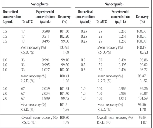
Table II. Results of Accuracy Determinations
Nanospheres Nanocapsules
Theoretical Experimental Theoretical Experimental
concentration concentration Recovery concentration concentration Recovery
(µg/mL) % MTC (µg/mL) (%) (µg/mL) % MTC (µg/mL) (%)
0.5 17 0.508 101.60 0.25 25 0.250 100.00
0.5 17 0.511 102.20 0.25 25 0.251 100.56
0.5 17 0.495 99.00 0.25 25 1.250 100.00
Mean recovery (%) 100.93 Mean recovery (%) 100.19
R.S.D. (%) 1.69 R.S.D. (%) 0.323
1.0 33 0.991 99.10 0.5 50 0.494 98.86
1.0 33 0.995 99.50 0.5 50 0.495 99.02
1.0 33 1.027 102.70 0.5 50 0.494 98.72
Mean recovery (%) 100.43 Mean recovery (%) 98.87
R.S.D. (%) 1.96 R.S.D. (%) 0.152
2.0 67 2.039 101.95 1.0 100 0.983 98.26
2.0 67 2.034 101.70 1.0 100 0.989 98.87
2.0 67 1.989 99.45 1.0 100 1.016 101.59
Mean recovery (%) 101.3 Mean recovery (%) 99.56
R.S.D. (%) 1.36 R.S.D. (%) 1.78
Overall mean recovery (%) 100.80 Overall mean recovery (%) 99.54
were found (Figures 2C and 2D). The UV-spectra recorded for these samples by the HPLC–DAD system are shown in Figure 3. Regarding acidic samples (Figure 3A), the UV-spectrum of peak no. 2 (tr = 7.04 min) clearly deviate from the DHMXAN spec-trum, indicating modifications in the chromophore of the molecule, probably due to an alteration of molecular structure (e.g., the hydrogen bond disruption). The UV-spectrum of peak no. 1 (tr = 4.68 min) was not representative, probably due to the small amount of the formed product. Regarding alkaline samples (Figure 3B), the UV-spectra of peaks no. 3 and no. 4 were also not representative. Obtained results indicate a high resistance to degradation of the DHMXAN under the tested alkaline condi-tions.
The UV-spectra of the peak corresponding to xanthonic com-pound remained identical after subjecting the samples to thermal, acidic, and alkaline stress conditions. The peak purity was, in all cases, higher than 99.0%. This indicates that there were not degradation products co-eluting with DHMXAN and demonstrates the stability-indicating capability of the analytical method.
In order to evaluate the specificity of the method concerning the presence of nanoparticle excipients (i.e., the potential inter-ference of the excipients), a comparison of the results from the analysis of DHMXAN standard solutions spiked with empty nanospheres or nanocapsules with those correspondent to DHMXAN standard solutions was carried out. Obtained chro-matograms (Figures 2E and 2F) show the absence of any peak in the region where xanthone elute, which indicates that the method is specific concerning to nanoparticle excipients.
Linearity and range
The assay was linear with a relative standard deviation of 2.4% in the response factors (area divided by concentration) in the tested concentration range and correlation coefficients (r)≥
0.999 and coefficients of determination (R2) > 0.9982 (i.e., over
99.82 % of relationship betweenx and y) were found for all three
calibration curves (Table I).
Accuracy
Table II summarizes the accuracy results, expressed as percent recovery and relative standard deviation (RSD). Recovery data were within the range of 99.45–102.20% (RSD = 1.49%) and 98.26–101.59% (RSD = 1.07%) for nanospheres and nanocap-sules, respectively. Overall mean recovery values were 100.80%
(n = 9) for nanospheres and 99.54% (n = 9) for nanocapsules.
Because the mean recovery results were within an acceptable ± 3% range, according to Segall et al. the method was deemed to be accurate (25).
Precision
Tables III and IV summarize precision of the chromatographic and of the total analytical method, respectively. Obtained RSD values ranged from 0.6% to 1.9%. Results are greater than 2%, indicating that both the chromatographic and the total analyt-ical method show acceptable precision, in agreement with the criteria proposed by Shabir (26).
Application of the developed method for the quantitation of DHMXAN in nanoparticles
The analytical method is effective, fast, and meets all criteria for method validation and can be applied for the quantification of DHMXAN encapsulated in different batches of nanoparticle for-mulations. Figures 4A and 4B show representative chro-matograms of nanospheres and nanocapsules containing DHMXAN. None of these chromatograms show any interfering peaks. Furthermore, peak purity was higher than 99%, indi-cating that the preparation method did not produced degrada-tion of xanthonic compound.
Table III. Results of Precision of the Chromatographic Method
Repeatability (intra-assay precision) Theoretical Mean experimental
concentration concentration (µg/mL) (µg/mL) [n] SD RSD (%) 0.25 0.248 [3] 0.001 0.6 0.5 0.522 [3] 0.008 1.6 0.6 0.587 [3] 0.009 1.6 1.0 0.997 [3] 0.019 1.9 1.5 1.480 [3] 0.019 1.3 2.0 2.016 [3] 0.034 1.7 3.0 2.999 [3] 0.026 0.9
Intermediate precision (different days) Theoretical Mean experimental
concentration concentration (µg/mL) (µg/mL) [n] SD RSD (%) 0.25 0.246 [3] 0.003 1.2 0.5 0.508 [3] 0.010 2.0 0.6 0.574 [3] 0.011 1.9 1.0 1.000 [3] 0.009 0.9 1.5 1.476 [3] 0.005 0.3 2.0 2.018 [3] 0.029 1.5 3.0 2.996 [3] 0.057 1.9
Figure 4. Representative chromatograms of DHMXAN nanospheres (A) and nanocapsules (B).
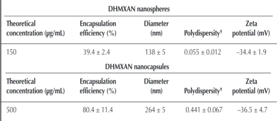
Table V shows encapsulation parameters, mean particle size, polydispersity, and zeta potential of PLGA nanoparticle formula-tions. Nanospheres and nanocapsules showed mean diameters around 140 nm and 265 nm, respectively, and zeta potential values > –40 mV. Encapsulation efficiency is the percentage of drug incorporated in nanoparticles with respect to the initial amount of drug used. Higher encapsulation efficiency was observed for nanocapsules (80.4%) than for nanospheres (39.4%). This result is in accordance with the data we have obtained in a previous work for other xanthonic derivatives (27). Several factors
influence the amount of drug associated with nanosystems, such as drug affinity for the polymer, type of polymer, type of oil, and drug solubility in the oily core (28–30). Our results suggest that the encapsulated xanthonic compound, which presents a lipophilic character (logP value = 2.6, calculated using the
CSLogP™ program of Chemsilico), has low affinity for the polymer and is soluble in the oily core of nanocapsules.
Conclusions
A simple isocratic reversed-phase HPLC method was devel-oped for the determination of DHMXAN in PLGA nanosphere and nanocapsule formulations, which was entrapped for the first time in this type of nanocarriers. The method was validated according to ICH guidelines and USP 29 for its specificity, lin-earity, accuracy, and precision. Obtained results showed that the proposed method is specific, linear, accurate, and precise within the established range.
No degradation of the compound was found upon nanoparticle preparation by the adopted solvent displacement methods and high encapsulation efficiencies were obtained for nanocapsule formulations. Results clearly demonstrate the suitability of the selected methods for incorporating DHMXAN in PLGA nanopar-ticles.
The developed and validated HPLC method was successfully applied to the quantification of DHMXAN content in nanopar-ticle formulations, affording an important tool for the quality control of finished products. Obtained nanosystems are under study in vitro on a tumour cell line.
Acknowledgments
To Fundação para a Ciência e a Tecnologia (FCT) (Unidade de I&D nº226/94), POCTI, FEDER and Praxis XXI (grant to Maribel Teixeira) for financial support.
References
1. L.M.M. Vieira and A. Kijjoa. Naturally-occur-ring xanthones: recent developments. Curr. Med. Chem. 12: 2413–46 (2005).
2. M.E. Sousa and M.M.M. Pinto. Synthesis of xanthones: an overview. Curr. Med. Chem. 12: 2447–79 (2005).
3. M.M.M. Pinto, M.E. Sousa, and M.S.J. Nascimento. Xanthone derivatives: new insights in biological activities. Curr. Med. Chem. 12: 2517–38 (2005).
4. M. Pedro, F. Cerqueira, M.E. Sousa, M.S.J. Nascimento, and M. Pinto. Xanthones as inhibitors of growth of human cancer cell lines and their effects on the proliferation of human lymphocytes in vitro. Bioorg. Med. Chem. 10: 3725-3730 (2002).
Table V. Encapsulation Parameters, Mean Diameter, Polydispersity and Zeta Potential of PLGA Nanoparticle Formulations Containing DHMXAN*
DHMXAN nanospheres
Theoretical Encapsulation Diameter Zeta
concentration (µg/mL) efficiency (%) (nm) Polydispersity† potential (mV)
150 39.4 ± 2.4 138 ± 5 0.055 ± 0.012 –34.4 ± 1.9
DHMXAN nanocapsules
Theoretical Encapsulation Diameter Zeta
concentration (µg/mL) efficiency (%) (nm) Polydispersity† potential (mV)
500 80.4 ± 11.4 264 ± 5 0.441 ± 0.067 –36.5 ± 4.7
* Values express the mean results ± SD values of three different batches.
†Varies from 0.0 corresponding to a perfect homogeneous dispersion to 1.0 corresponding to a complete heterogeneous
dispersion.
Table IV. Results of Precision of the Entire Analytical Method of DHMXAN in Nanospheres and Nanocapsules
Repeatability (Intra-assay precision) DHMXAN nanospheres
Sample Peak area response (mV) Concentration (µg/mL)
1 163.7 56.3 2 162.4 55.9 3 166.3 57.2 4 164.6 56.6 5 169.8 58.4 6 168.3 57.9 Mean 165.9 57.1 S.D. 2.83 0.972 R.S.D. 2.00 1.70 DHMXAN nanocapsules
Sample Peak area response (mV) Concentration (µg/mL)
1 144.0 495.3 2 139.5 479.8 3 142.1 488.8 4 140.4 481.9 5 139.9 482.9 6 141.1 485.3 Mean 141.2 485.7 S.D. 1.65 5.64 R.S.D. 1.17 1.16
5. P. Speiser. Poorly soluble drugs, a challenge in drug delivery. In Emulsions and Nanosuspensions for the Formulation of Poorly Soluble Drugs, R. H. Müller, S. Benita and B. Böhm, Eds. Scientific Pub., Stuttgard, 1998, pp. 15–19.
6. S. Sweetana and M. J. Akers. Solubility principles and practices for parenteral drug dosage form development. PDA Pharm. Sci. Technol. 50: 330–342 (1996).
7. A. Sanches, J.L. Villa-Jato, and M.J. Alonso. Development of biodegradable microspheres and nanospheres for the controlled release of cyclosporin A. Int. J. Pharm. 99: 263–73 (1993). 8. Y.I. Kim, L. Fluckiger, M. Hoffman, I. Lartaud-Idjouadiene,
J. Atkinson, and P. Maincent. The antihypertensive effect of orally administered nifedipine-loaded nanoparticles in spontaneously hypertensive rats. Brit. J. Pharmacol. 120: 399–404 (1997). 9. C. Mallard, J. Coudane, I. Rault, and M. Vert. In vitro delivery of a
sparingly water soluble compound from PLA50 microparticles. J. Microencapsul. 17: 13–28 (2000).
10. B. Magenheim and S. Benita. Nanoparticle characterization: a com-prehensive physicochemical approach. S.T.P. Pharma Sciences 1: 221–41 (1991).
11. P. Couvreur, C. Dubernet, and F. Puisieux. Controlled drug delivery with nanoparticles: current possibilities and future trends. Eur. J. Biopharm. 41: 2–13 (1995).
12. M.J. Alonso. Nanoparticulate drug carrier technology. In Microparticulate Systems for the Delivery of Proteins and Vaccines. S. Cohen and H. Bernstain, Eds. Marcel Dekker, New York, 1996, pp. 203–42.
13. R. Jain, N.H. Shah, A.W. Malick, and C. Rhodes. Controlled drug delivery by biodegradable poly(ester) devices: different preparative approaches. Drug. Dev. Ind. Pharm. 24: 703–27 (1998).
14. P.J. Lowe and C.S. Temple. Calcitonin and insulin in isobutyl-cyanoacrylate nanocapsules: protection against proteases and effect on intestinal absorption in rats. J. Pharm. Pharmacol. 46: 547–52 (1994).
15. G. Barrat, G. Couarraze, P. Couvreur, C. Dubernet, E. Fattal, R. Gref, D. Labarre, P. Legrand, G. Ponchel, and C. Vauthier. Polymeric micro- and nanoparticles as drug carriers. In Polymeric Biomaterials, S. Dumitriu, Ed. Marcel Dekker, New York, 2002, pp. 753–81.
16. M. Teixeira, F. Cerqueira, C. M. Barbosa, M.S.J. Nascimento, and M. Pinto. Improvement of the inhibitory effect of xanthones on NO production by encapsulation in PLGA nanocapsules. J. Drug Target. 13: 129–35 (2005).
17. European Comission, Ditectorate General III- Industry Pharmaceuticals and cosmetics. Validation of Analytical Procedures: Methodology. The Rules Governing Medicinal Products in European Union, vol. 3A: 107–17 (1998).
18. European Comission, Ditectorate General III- Industry Pharmaceuticals and cosmetics. Validation of Analytical
Procedures: Definition and Terminology. The Rules Governing Medicinal Products in European Union, vol. 3A: 119–25 (1998). 19. United States Pharmacopeia 29/NF 24. Validation of Compendial
Methods, Section (1225), United States Pharmacopeial Convention, Rockville, Maryland, USA, 2005, pp. 3050–52.
20. M. Pinto and J. Polónia. Synthesis of new xanthones. I. Helv. Chim. Acta 57: 2613–17 (1974).
21. H. Fessi, J.P. Devissaguet, F. Puisieux and, C .Thies. Procédé de pré-paration de sytémes colloidaux dispersibles d’une substance, sous forme de nanoparticules. French Patent 2608988-A1 (1988). 22. H. Fessi, F. Puisieux, J. Ph. Devissaguet, N. Ammoury, and S. Benita.
Nanocapsule formation by interfacial polymer deposition following solvent displacement. Int. J. Pharm. 55: R1–R4 (1989).
23. M. Teixeira, C.M.M. Afonso, M.M.M. Pinto, and C.M. Barbosa. A validated HPLC method for the assay of xanthone and 3-methoxyxanthone in PLGA nanocapsules. J. Chromatogr. Sci. 41: 371–76 (2003).
24. L.R. Snyder and J.J. Kirkland. Introduction to Modern Liquid Chromatography, 2nd ed. John Wiley & Sons, New York, 1979, pp. 781–23.
25. A. Segall, F. Hormaechea, M. Vitale, V. Perez, and M. T. Pizzorno. Development and validation of a reversed-phase liquid chromato-graphic method for analysis of estradiol valerate and medroxypro-gesterone acetate in a tablet formulation. J. Pharm. Biomed. Anal. 19: 803–808 (1999).
26. G.A. Shabir. HPLC method development and validation for phar-maceutical analysis. Pharm. Technol. Eur. Mar. 37–49 (2004). 27. M. Teixeira, M.J. Alonso, M.M.M. Pinto, and C.M. Barbosa.
Development and characterization of PLGA nanospheres and nanocapsules containing xanthone and 3-methoxyxanthone. European Journal of Pharmaceutics and Biopharmaceutics 59: 491–500 (2005).
28. J.M. Barichello, M. Morishita, K. Takayama, and T. Nagai. Encapsulation of hydrophilic and lipophilic drugs in PLGA nanoparticles by the nanoprecipitation method. Drug Dev. Ind. Pharm. 25: 471–76 (1999).
29. S. R. Schaffazick, S.S. Guterres, L.L. de Freitas, and A. R. Pohlmann. Physicochemical characterization and stability of the polymeric nanoparticle systems for drug administration. Quim. Nova 26: 726–37 (2003).
30. S.S. Guterres, H. Fessi, G. Barratt, J-P Devissaguet, and F. Puisieux. Poly(D,L-lactide) nanocapsules containing non-steroidal anti-inflammatory drugs: gastrointestinal tolerance following intra-venous and oral administration. Int. J. Pharm. 113: 57–63 (1995).
Manuscript received January 31, 2007; revision received June 18, 2007.