Universidade de Lisboa
Faculdade de Ciências
Departamento de Biologia Animal
Unravelling the regulation of Dll4 expression
and its function during embryonic
neurogenesis
Pedro Miguel Branco Barbacena
Dissertação
Mestrado em Biologia Evolutiva e do Desenvolvimento
Universidade de Lisboa
Faculdade de Ciências
Departamento de Biologia Animal
Unravelling the regulation of Dll4 expression
and its function during embryonic
neurogenesis
Dissertação orientada por:
Catarina Ramos, PhD, Instituto de Medicina Molecular, Faculdade
de Medicina da Universidade de Lisboa
Maria Gabriela Rodrigues, PhD, Departamento de Biologia Animal,
Faculdade de Ciências da Universidade de Lisboa
Pedro Miguel Branco Barbacena
Dissertação
Mestrado em Biologia Evolutiva e do Desenvolvimento
Em primeiro lugar quero agradecer ao Domingos Henrique, por me ter proporcionado a possibilidade de desenvolver a minha tese de mestrado na Unidade de Biologia do Desenvolvimento. Por todos os conselhos, todas as reuniões, toda a disponibilidade que teve durante este ano. Por todas as discussões, científicas ou não, e pela transmissão de conhecimentos técnicos, científicos e gastronómicos do nosso belo país à beira mar.
Catarina, bem a ti, tenho tanta coisa para agradecer. Obrigado por teres sido minha mentora e acima de tudo, amiga, durante este ano. Obrigado pela partilha de conhecimento, pelas n explicações, pela paciência, pela conversa e por teres estado sempre lá. Obrigado pelas dicas, pelos incentivos, mas também pelos cafés e pelos jantares e pelo acompanhamento ao longo do ano, mesmo quando te mudaste para outras bandas. Spinal cord for the win!
À Professora Gabriela Rodrigues, o meu sincero obrigado por ter aceitado orientar-me, pela paciência e acompanhamento ao longo do ano e pela compreensão que teve com pedidos de declarações em cima da hora.
À Ana e à Elsa, o que tinha sido de mim sem vocês, durante este ano! Obrigado às duas, pela paciência, amizade e companheirismo. Obrigado pelas pausas, pelas conversas, pelas opiniões e dicas, pelas noções de estética que tanto contribuíram para o sucesso desta tese. Obrigado Elsa pelos “puxões de orelhas”!
Um obrigado especial ainda à Cláudia, pelos troubleshooting, pelas discussões científicas, pelas opiniões e pelas perguntas científicas de algibeira, que tanto me puseram a matutar. Um obrigado gigante à Sara, pelas ajudas e orientações no laboratório e pela grande ajuda no trabalho que passa despercebido a quem lê uma tese ou uma publicação, como algum dia me disseram é gigante fazer o trabalho de backstage e tu fá-lo como ninguém.
Obrigado a todos os restantes colegas da UBD, por me terem recebido tão bem e por terem feito da minha adaptação, a melhor possível.
Aos “Mais bonitos”, ao nosso grupo fantástico, bem meus caros a vocês tenho que agradecer muito! Obrigado pela companhia e amizade, já lá vão uns bons quatro anos e continuamos todos na mesma, cada vez mais parvos mas não há milagres. Se pudesse partilhar a aventura que é fazer uma tese com outras pessoas não seria a mesma coisa, acreditem. Obrigado pela compreensão da minha recorrente falta de tempo, obrigado por estarem sempre interessados e por me fazerem rir uma e outra vez. Particularmente ao core fortíssimo de BED (Matos) por todo o companheirismo e as outsider opinions, aos conservacionistas fantásticos que há muito me acompanham
e me aturam (Filipa, Margarida, Joana, David, Rita e Mauro), à aventureira do Gorongosa (Márcia), aos marinhos e aos polares (Maria, Hilário e Miguel), aos “imigrantes” (Nuno, Toga e Inês) e aos que estão lá sempre (Denise e Chambel). A vocês obrigado por todos os momentos que temos passado nestes últimos anos e que isto continue por muito mais tempo.
Ao pessoal do mestrado, principalmente à Margarida, por toda a companhia durante este ano, por todas as viagens de comboio e todos os serões passados no IMM, o meu obrigado.
Ao pessoal da Damaia obrigado por estarem sempre lá, obrigado pela compreensão de algumas ausências e pela força. Em particular à Ana e ao Ricardo que especialmente durante este ano tiveram de ser ainda mais compreensivos para lidarem com a minha recorrente impossibilidade de alinhar nos planos, um agradecimento sentido pela paciência e por, apesar de tudo, estarem sempre no sítio do costume, muito perto.
E por último, o agradecimento mais importante, à minha família. Obrigado pela compreensão, por terem levado com todos os minhas angústias e os meus problemas, sempre com um sorriso nos lábios. Obrigado por se manterem sempre interessados, ainda que os meus termos técnicos, nada façam parte do vosso background, obrigado por terem respeitado o meu cérebro sempre a mil e as minhas ausências durante este ano. Obrigado Pai e Mãe! A ti João, obrigado pela companhia, pelas palhaçadas, por nos fazer sempre rir, pelo interesse e as opiniões sinceras.
Obrigado a todos, Pedro Barbacena
During spinal cord development, Notch signalling regulates both the maintenance of progenitors and neuronal specification. In most spinal cord domains, a single Notch ligand is expressed however in the V2 domain two ligands (Dll1 and Dll4) are expressed. It is not known how Dll4 expression is regulated and how Dll4-mediated Notch signalling has a role in the specification of V2 interneurons (V2a, V2b and V2c) that are generated from V2 progenitors.
To investigate how Dll4 expression is regulated, we have focused on three proneural bHLH genes (Mash1, Ngn1 and Ngn2), expressed in V2 progenitor cells. We characterized Dll4-expressing cells, by double RNA in-situ hybridization with each of the proneural genes. Our results revealed that Dll4+ cells can coexpress all three proneural genes but to different extents. Next, we have overexpressed each proneural gene (singly and in combination) in the chick neural tube, using in ovo electroporation. Embryos were tested in terms of: Dll4 expression and V2 cell-type specification.
Although electroporation of either Mash1 or Ngn1 leads to downregulation of
Dll4 expression in the V2 domain, only NGN1 is capable of inducing ectopic Dll4
expression. Moreover, both double electroporation of Mash1 and Ngn1, or Mash1 and
Ngn2, represses Dll4 expression in the V2 domain, while leading to ectopic expression
in other ventral spinal cord domains.
Concerning cell-type specification, electroporation of Mash1 represses both V2a and V2b cell fate. Electroporation of either Ngn1 or Ngn2 increases the number of V2a INs, while repressing V2b INs. Double electroporation of Mash1/Ngn1, Mash1/Ngn2 or
Ngn1/Ngn2 increases the number of V2a INs and represses V2b fate.
Together, the results presented in this thesis show that MASH1 and NGN1 can repress Dll4 expression and that this repression correlates with alterations in V2 IN specification. Our results indicate that NGN2 can affect V2 specification, although it does not regulate Dll4 expression.
Neurogenesis, Spinal Cord, V2 domain, Notch Signalling, Delta-like 4,
Um dos mais complexos sistemas biológicos é o Sistema Nervoso Central que é constituído por um elevado número e variedade de células, como neurónios e células da glia. Estas células são produzidas durante o desenvolvimento embrionário no momento e posição correctos de modo a interagirem entre si e formarem circuitos funcionais. Durante o desenvolvimento embrionário da espinal medula, vários interneurónios excitatórios e inibitórios são gerados, sendo o balanço entre eles estritamente regulado. No entanto os mecanismos moleculares subjacentes a esta diversidade celular ainda não são totalmente conhecidos.
A via de sinalização Notch desempenha um papel essencial durante o desenvolvimento do Sistema Nervoso Central. Esta via de sinalização é um sistema de comunicação célula-a-célula, de contacto directo entre duas células vizinhas, através de duas proteínas membranares: o receptor Notch e os seus ligandos (Delta, Serrate ou Lag-2, dependendo do organismo). Sempre que um ligando de uma célula se liga ao receptor de outra célula, uma série de clivagens proteolíticas são desencadeadas, o que, em última instância, leva à libertação do domínio intracelular do receptor Notch (NICD). O NICD é translocado para o núcleo onde se associa ao CSL e ao
Mastermind, activando a transcrição de genes alvo, como os genes HES. Na ausência
de NICD, a proteína CSL actua como repressor transcricional dos mesmos genes. Uma das funções da via de sinalização Notch é a capacidade de manutenção de progenitores neurais, durante a neurogénese, impedindo uma diferenciação prematura massiva de todos os progenitores. A decisão de uma célula permanecer como progenitor, ou de se diferenciar, é controlada pelo balanço de dois tipos de factores de transcrição: as proteínas proneurais, que promovem a diferenciação; e as proteínas HES, que reprimem a diferenciação neural. Uma célula que, estocasticamente expresse elevados níveis de proteínas proneurais vai diferenciar-se. Normalmente as proteínas proneurais activam a expressão dos ligandos do Notch, o que leva a que esta célula expresse elevados níveis de ligandos, activando a via de sinalização Notch nas células vizinhas. Esta activação leva à expressão de elevados níveis de proteínas HES, sendo que estas células são mantidas como progenitores.
Assim, a célula que expressa o ligando diferencia-se em neurónio mas simultaneamente assegura que as células vizinhas se mantenham como progenitores. Visto que a neurogénese ocorre durante uma larga janela temporal, a manutenção de uma população de progenitores permite que estas células sejam expostas a diferentes estímulos e, como tal, se diferenciem em diferentes neurónios durante o
desenvolvimento, assegurando uma grande variedade celular ao nível do Sistema Nervoso Central.
A espinal medula, ao longo do seu eixo dorso-ventral, pode ser dividida em onze domínios molecularmente distintos (seis deles dorsais e cinco ventrais). Estudos recentes mostram que a via de sinalização Notch está envolvida na especificação de células neurais, nomeadamente de interneurónios no domínio V2 da espinal medula. Neste domínio, três tipos de interneurónios diferentes: V2a, V2b e V2c, são gerados a partir de progenitores comuns. Na ausência da sinalização Notch, apenas os interneurónios V2a são gerados, o que significa que a sinalização Notch é necessária para a produção dos interneurónios V2b. Os interneurónios V2c foram recentemente identificados. Curiosamente, nesta fase do desenvolvimento, este é o único domínio onde se sabe que diferentes interneurónios são simultaneamente produzidos e o único domínio onde dois ligandos da via Notch são expressos: Dll1 e Dll4. Isto é uma excepção ao que ocorre em todos os outros domínios da espinal medula, onde apenas um ligando da via Notch é suficiente para regular a neurogénese. Possivelmente o ligando Dll1 está envolvido na activação da via Notch e consequentemente na manutenção de progenitores, enquanto o ligando Dll4 está envolvido na especificação dos interneurónios.
Como é que a expressão de Dll4 é regulada no domínio V2 da espinal medula e como é que a especificação de interneurónios é controlada neste domínio são os principais temas investigados nesta tese. Os organismos modelo utilizados para estudar estas questões foram o embrião de ratinho e de galinha.
Recorrendo à técnica de hibridação in-situ comecei por mapear a expressão de
LFng (lunatic fringe) e Hes6 de modo a verificar de que modo a sinalização Notch via Dll1 ou Dll4 poderiam ser diferentes. Verifiquei que LFng é expresso em células que
expressem Dll4 mas não em células que expressem Scl (marcador de V2b). Hes6 é expresso em células que expressem Dll1 ou Dll4 mas não em células Scl+, o que parece sugerir que, de facto, a sinalização Notch através destes dois ligandos é diferente.
De modo a verificar como o Dll4 é regulado no domínio V2, foquei-me em três proteínas proneurais (MASH1, NGN1 e NGN2) que são expressas em progenitores do V2 e que, segundo abordagens bioinformáticas anteriores, são boas candidatas a regular a sua expressão.
Primeiro foram caracterizadas as células que expressam Dll4 e cada um dos genes proneurais através de hibridações in-situ duplas. As células Dll4 podem co-expressar os três genes proneurais testados mas a diferentes níveis.
Para testar se estas proteínas podem regular a expressão de Dll4, plasmídeos contendo os genes que codificam cada uma destas proteínas foram injectados (sozinhos ou em combinação) na espinal medula de embriões de galinha no estádio HH16-17 pela electroporação in ovo, de modo a sobre-expressar estas proteínas. Todos os embriões foram testados em termos de expressão de Dll4 e especificação de interneurónios.
No que diz respeito à expressão de Dll4, a electroporação de Mash1 ou de
Ngn1 leva a uma repressão de Dll4 no interior do domínio V2, mas apenas Ngn1 é
capaz de induzir ectopicamente a expressão de Dll4. A sobreexpressão de NGN2, não altera a expressão endógena de Dll4, indicando que esta proteína proneural não será um factor importante no controlo da expressão deste gene. A electroporação dupla de
Mash1 com Ngn1 ou de Mash1 com Ngn2 reprime também a expressão de Dll4 no
domínio V2 mas leva à sua expressão ectópica noutros domínios ventrais da espinal medula. A sobreexpressão de NGN1 e NGN2 não parece afectar a expressão de Dll4 na espinal medula.
Visto que as proteínas proneurais controlam a especificação de neurónios durante o desenvolvimento de Sistema Nervoso Central, foi analisado se a sobreexpressão destas proteínas afectaria o número de interneurónios V2a. Após a sobreexpressão de MASH1, uma diminuição no número de interneurónios V2a foi observada, indicando que esta reprime, directa ou indirectamente, a produção destes interneurónios. Contrariamente e após a sobreexpressão de NGN1 ou NGN2, o número de interneurónios V2a aumenta, o que sugere que estas proteínas promovem directamente a diferenciação de interneurónios V2a. A sobreexpressão em diferentes combinações também leva a um aumento de interneurónios V2a no domínio V2.
Foi ainda analisado se o número de progenitores de V2a através da análise de BHLHB5 e LIM3. Verifiquei que MASH1 e NGN1 reprimem o número destes progenitores enquanto Ngn2 não os afecta.
Por hibridação in situ foi verificado se o número de interneurónios V2b também seria afectado e verifiquei que em todas as condições o número de V2b sofre uma diminuição, indicando que todas as proteínas proneurais reprimem, directa ou indirectamente a produção destes interneurónios.
Além disto foi analisado o que acontecia à especificação DV da espinal medula aquando da sobreexpressão das proteínas proneurais. Para isso foi analisado o domínio MN que mantem interacções antagónicas com o domínio V2, através da expressão de OLIG2. Verificou-se que o número de células OLIG2+ aumentou quando MASH1 é o expressa ou quando as combinações de proneurais são sobre-expressas.
Assim verificou-sei que MASH1 e NGN1 regulam a expressão de Dll4 e, consequentemente a especificação de interneurónios enquanto NGN2 não parece regular a expressão de Dll4, afectando apenas a especificação de interneurónios.
Este estudo apresenta novas evidências sobre a função que diferentes proteínas proneurais têm na regulação da expressão de Dll4 e na especificação de interneurónios no domínio V2.
Neurogénese, Espinal medula, Domínio V2, Sinalização Notch,
AP – Anterior-Posterior AP – Alkaline Phosphatase ASC - Achaete-Scute ATO - Atonal
bHLH – basic Helix-Loop-Helix BMP - Bone Morphogenetic Protein CNS – Central Nervous System
CSL - CBF1, Suppressor of Hairless, Lag-1 DIG - Digoxigenin
Dll – Delta-like
DNA – Deoxyribonucleic acid DSL - Delta/Serrate/Lag-2 DV – Dorsal-Ventral E – Embryonic day
EDTA – Ethylenediamine tetraacetic acid FBS – Fetal Bovine Serum
FGF – Fibroblast Growth Factor FP – Floor Plate
HES - Hairy and Enhancer of Split homologues HH stage – Hamburger and Hamilton stage IN – Interneuron
IVZ – Inner Ventricular Zone LB – Luria Bertani bacterial médium MN – Motor Neuron
MZ – Mantle Zone NC – Notochord NGN – Neurogenin
NECD – Notch Extracellular Domain NICD – Notch Intracellular Domain NLS – Nuclear localization signal o/n – Overnight
PCR – Polymerase Chain Reaction PFA – Paraformaldehyde
POD – Peroxidase RA – Retinoic Acid
RBPJ – Recombination signal binding protein for immunoglobulin kappa J RNA – Ribonucleic acid
RP – Roof plate
RT – Room Temperature SD – standard deviation Shh - Sonic Hedgehog SOB – Super Optimal Broth
TSA – Tyramide Signal Amplification VEGF – Vascular endothelial growth factor VZ – ventricular zone
ACKNOWLEDGEMENTS ... i ABSTRACT ... iii RESUMO ... iv ABBREVIATIONS ... viii INDEX ... ix INDEX OF FIGURES ... xi INDEX OF TABLES ... xi CHAPTER I – INTRODUCTION ... 1
I. 1. VERTEBRATE NEUROGENESIS AND PATTERNING ... 1
I.2. NOTCH SIGNALLING IN VERTEBRATE NEUROGENESIS ... 2
I.2.1. Notch Targets ... 3
I.2.2. Notch Signalling Pathway during Vertebrate CNS Development ... 4
I.2.2.1. Maintenance of neural progenitors through lateral inhibition ... 4
I.2.2.2. Cell fate specification ... 6
I.3. SPINAL CORD NEUROGENESIS – THE V2 DOMAIN ... 6
I.4. REGULATORS OF Dll EXPRESSION AND CELL TYPE SPECIFICATION ... 8
I.4.1. bHLH Proneural Proteins ... 8
I.4.2. Regulation of Dll4 Expression ... 8
I.5. AIMS ... 9
CHAPTER II – MATERIALS AND METHODS ... 10
II.1. MOLECULAR BIOLOGY TECHNIQUES ... 10
II. 1.1. Plasmid Transformation of chemically competent E.coli bacteria ... 10
II. 1.2. DNA Constructs – Plasmid DNA purification... 10
II. 1.3. DNA Quantification ... 10
II. 1.4. Analytical digestions ... 10
II.2. CHICK EMBRYO MANIPULATION ... 10
II.2.1. Chicken embryos ... 10
II.2.2. In ovo chick embryo electroporation ... 11
II.2.3. Tissue embedding and preparation of cryostat sections ... 11
II.3. MOUSE EMBRYO MANIPULATION ... 11
II.4. IN SITU HYBRIDIZATION ... 12
II.4.1. Antisense RNA probe synthesis ... 12
II.4.1.2. Probe synthesis ... 12
II.4.2. Fluorescent In situ hybridization ... 12
II.5. IMMUNOFLUORESCENCE ... 13
II.6. FIXED TISSUE IMAGING ... 13
II.7. STATISTICAL ANALYSIS ... 14
CHAPTER III – RESULTS ... 15
III.1. Notch Signalling in the V2 Domain ... 15
III.2. Characterization of Dll4-expressing Cells ... 18
III.3. Mash1 or NGN1 overexpression represses Dll4 expression in V2 domain ... 19
III.3. Mash1 overexpression leads to a decrease in V2a INs whereas NGN1 and NGN2 overexpression has the opposite effect ... 21
III.4. Mash1 and Ngn1 overexpression leads to a decrease in the number of prospective V2a . 23 III.5. Mash1, NGN1 and NGN2 overexpression leads to a decrease in V2b IN fate ... 25
III.6. Single Mash1 overexpression affects DV specification of the spinal cord ... 27
CHAPTER IV – DISCUSSION ... 29
IV.1. NOTCH SIGNALLING IN THE V2 DOMAIN ... 29
IV.2. Dll4 REGULATION ... 30
IV.3. V2 INTERNEURON SPECIFICATION ... 32
IV.4. PRONEURAL GENES MISEXPRESSION AND NEUROGENESIS ... 34
CHAPTER V – REFERENCES ... 377
ANNEX A – MATERIALS AND METHODS ... 422
Figure 1 - Diagram of a transverse section of the spinal cord. ... 2
Figure 2 – Notch signaling pathway. ... 3
Figure 3 – Lateral Inhibition mechanism. ... 5
Figure 4 – V2 domain of the developing spinal cord. ... 7
Figure 5 – Cell dynamics inside V2 domain. ... 15
Figure 6 – LFng expression in the prospective V2 INs. ... 16
Figure 7 – Hes6 expression in the V2 domain. ... 17
Figure 8 – Characterization of Dll4+ cells. ... 18
Figure 9 – Dll4 expression after electroporation of Mash1, Ngn1 and Ngn2, single or in combinations. ... 20
Figure 10 – CHX10 expression after electroporation of Mash1, Ngn1 and Ngn2, single or in combinations. ... 22
Figure 12 – bHLHB5 and LIM3 expression after electroporation of Mash1, Ngn1 and Ngn2. ... 26
Figure 13 – Scl expression after electroporation of Mash1, Ngn1 and Ngn2, single or in combinations. ... 26
Figure 14 - JAG1 and OLIG2 expression after electroporation of Mash1, Ngn1 and Ngn2, single or in combinations. ... 29
Figure 15 – Summary data regarding % of analysed cells for each condition of proneurals overexpressed... 32
Figure 16 – Controls for Dll4 probe specificity. ... 43
Table 1 – Results summarized regarding Dll4 expression and IN specification in different conditions (single or simultaneous overexpression of proneural proteins). .... 31
Table 2 – Probes synthetized with respective linearization site and RNA polymerase used. ... 42
The vertebrate nervous system contains a great diversity of neurons and glial cells interconnected to form highly complex neural networks. These cells are generated in the embryonic neural tube at specific time and position in order to accurately interact with each other and to assemble into a functional network.
For the central nervous system (CNS) to be functionally assembled not only the balance between progenitors and differentiating neurons must be controlled but also the position where specific neurons are generated must be tightly regulated.
After neural tube formation, neural progenitors acquire different fates according to their relative positions along the anterior-posterior (AP) and dorsal-ventral (DV) axes of the embryo. The neural tube will originate the brain anteriorly (from which the forebrain, midbrain and hindbrain will be formed) and the spinal cord posteriorly 1.
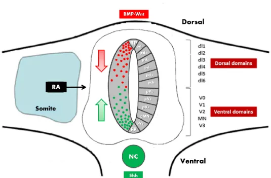
Neural progenitors of the neural tube normally develop anterior identity and differentiate into forebrain neurons. While some neural progenitors along the AP axis of the neural tube need to be kept in an undifferentiated state in order to gradually acquire different identities, others will differentiate into midbrain, hindbrain and spinal cord neurons. The anterior secretion of retinoic acid (RA), which promotes neuronal differentiation, and the posterior secretion of FGF, which represses neuronal differentiation, by surrounding mesodermal tissues, is responsible for the generation of the CNS in a rostral-to-caudal sequence, allowing progenitors to gradually differentiate in the correct moment and position (Figure 1) 2, 3.
Along the dorsal-ventral axis of the developing spinal cord, neural progenitors can be subdivided into eleven molecularly distinct domains (six dorsal and five ventral)4. Within each of these domains, populations of progenitor cells are defined by distinct combinations of transcription factors, induced by different levels of morphogens, namely by the bone morphogenetic proteins (BMPs) and WNT’s produced dorsally (either by roof plate or dorsal epidermis), and Sonic hedgehog (Shh), secreted ventrally by notochord and floor plate (Figure 1) 4, 5, 6, 7. These morphogens confer specific positional identities, activating region-specific differentiation programmes and, therefore, specifying the identity of neurons that derive from individual progenitor populations 8, 9, 10.
Notch signalling is one of the most conserved pathways in the regulation of metazoan development. It is involved in different processes like body segmentation (e.g. segmentation clock), skeletal development, embryonic haematopoiesis and neural development, including neuronal differentiation and glial determination controlling several biological functions such as apoptosis, cell proliferation and cell fate decisions, (Reviewed in 11).
The Notch pathway is a cell-cell communication system that results from the interaction of surface proteins: a Notch receptor and its ligand. The Notch receptors are a type I transmembrane heterodimeric proteins present at the plasma membrane. These receptors are composed by an ectodomain called NECD (Notch Extracellular Domain) and a membrane bound intracellular domain, which are conserved between species 12.
Notch receptors are activated by transmembrane ligands of the DSL family (Delta and Serrate from Drosophila and Lag-2 from C.elegans). The DSL ligands are type I transmembrane proteins with an extracellular domain that is conserved across species and an intracellular domain that present a small conservation even between different types of ligands 12.
Despite being a highly conserved pathway, different organisms have different receptors and ligands, for example, in Drosophila, there is only one Notch receptor and
Figure 1 - Diagram of a transverse section of the spinal cord. Different populations of
neural progenitors acquire different fates according to their position along the dorsal-ventral axis. The patterning is established by gradients of Shh, secreted by the floor plate (FP) and the notochord (NC), and Wnts and BMPs, produced by the roof plates (RP) and the dorsal epidermis. The Retinoic Acid (RA) produced by the adjacent somites is also involved in the patterning (AP and DV) of the spinal cord.
two ligands (Delta and Serrate), while in chick two Notch receptors (Notch1 and
Notch2) and four ligands (Delta1, Delta4, Serrate1 and Serrate2) have been described.
In mammals there are four receptors (Notch1, Notch2, Notch3 and Notch4) and five ligands (Delta-like1 (Dll1), Delta-like3 (Dll3), Delta-like4 (Dll4), Jagged1 (Jag1) and
Jagged2 (Jag2)) 13, 14. This thesis will focus on Dll4.
In this pathway a cell expressing the Notch ligand (signal-sending cell), either
Delta or Serrate, signals to the neighbour cell, which expresses a Notch receptor
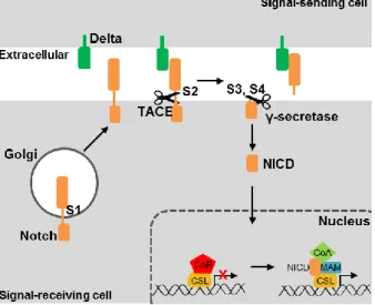
(signal-receiving cell). Notch receptors are formed in the trans-Golgi as a result of a proteolytic cleavage at the site S1 (Figure 2). Whenever a ligand protein binds to the extracellular domain of the Notch receptor, this one undergoes successive proteolytic cleavages. The first cleavage occurs at the extracellular S2 site and it is mediated by extracellular proteases of the ADAM/TACE family. The S2 cleaved form of Notch receptor is then processed at the endomembrane S3 site by the γ-secretase. After the proteolytic cleavages there is a release of the NICD (Notch Intracellular Domain) that is translocated to the nucleus where it associates with a DNA-binding protein called CSL (in human, CBF1; in Drosophila, Suppressor of Hairless; and in C.elegans, Lag-1) and to Mastermind co-activator, forming a nuclear complex which recruits other factors to regulate the expression of its target genes (Figure 2). In the absence of NICD, the transcription factor CSL is part of a transcriptional repressor complex that represses genes containing promoters with CSL-binding sites, whereas in the presence of NICD it activates the transcription of the same genes (Figure 2) 11, 15,16.
There are several CSL binding sites throughout the genome but the best described Notch transcriptional targets are genes encoding bHLH (basic Helix-loop-helix) transcriptional repressors. These repressors include the Enhancer of Split
(E(spl)) complex in Drosophila and the Hairy and Enhancer of Split homologues (HES) Figure 2 – Notch signaling pathway.
Whenever a ligand (Delta) expressed in the signal-sending cell binds to the extracellular domain of Notch receptor in the signal-receiving cell, it leads to a conformational change in the receptor and, as a consequence three successive proteolytic cleavages occur (at sites S2, S3 and S4) mediated by TACE/ADAM and γ-secretase, respectively. There is a release of NICD that is translocated to the nucleus where it associates with CSL and Mastermind, forming a complex that recruits other factors and activate the transcription of target genes.
or Hes-related (Hesr/Hey) families of proteins in vertebrates. All these proteins are bHLH-Orange (bHLH-O) as they contain a conserved amino acid sequence – Orange domain – located C-terminal to the bHLH domain 17, 18, 19.
The HLH domain allows homo- and heterodimerization of these bHLH-O proteins, which can then exert their function by DNA-binding and transcriptional repression. These proteins can bind DNA in consensus sequences called E-boxes (CANNTG) or N-boxes (CACNAG) by their basic domain. After DNA binding, the C-terminal motif WRPW recruits co-repressor factors, like Groucho, and this leads to the transcriptional repression of target genes, like proneural genes. Moreover, bHLH-O proteins can repress transcription by directly interact and form heterodimers with bHLH activator proteins, through the HLH domain, and this prevents binding of the activators to DNA, thereby stopping their transcriptional activity 18, 20.
During CNS development, the Notch pathway has a critical role in neuronal differentiation and also in glial determination through different mechanisms. Regarding the neural tube, Notch receptors are expressed in uncommitted neural progenitor cells, which are arranged in a polarized neuroepithelium in the ventricular zone (VZ) 21.
During vertebrate neurogenesis, Notch signalling has been shown to mediate cell fate decisions by maintaining neural progenitor identity, while supressing neuronal differentiation. In the vertebrate neural tube, the walls are composed by neuroepithelial cells that can differentiate into neurons. These neuroepithelial cells are polarized, with the basal region in contact with the basal lamina at the periphery of the tube, whereas the apical end is next to the lumen of the tube. Cells are in close contact to one another in the apical region by a variety of specialized junctions (e.g. tight junctions) 21. The neural progenitors are bound at the apical and basal surfaces of the neuroepithelium but their nuclei migrate along the axis of the cell accordingly to the cell cycle phase: M-phase nuclei are at the apical side while cells in S-M-phase have their nuclei close to the basal surface. During G1 and G2 phases, the nuclei migrate between these two opposing positions, in a movement known as interkinetic nuclear migration. However, after division, each of the daughter cells either repeats or exits the cell cycle. When the cell exits the cell cycle, it loses the apical attachment and migrates out of the ventricular zone to the mantle zone (MZ) where it starts to differentiate 21, 22.
Notch signalling can act to promote cell diversity through a process called lateral inhibition. In this process, a group of cells with a similar developmental potential
can give rise to different cell types and it ensures that two interacting cells do not acquire the same fate 9, 23.
Regarding the maintenance of neural progenitors, two neighbouring cells signal to each other and one starts the process of neuronal differentiation while the other is kept as a progenitor. The decision of becoming a neuron or remaining as a neural progenitor is controlled by the balance between two different sets of transcription factors: proneural bHLH proteins, which instruct progenitors into neuronal differentiation, and HES proteins, which repress neuronal differentiation and therefore maintain cells as progenitors. Nevertheless, due to lateral inhibition, the decision of each neural progenitor is influenced by the fates of the neighbour cells. Neural progenitors in the VZ of the neural tube express proneural proteins, but stochastic variations in gene expression causes one cell to express higher levels of proneural genes and therefore start the differentiation process 24, 25. It is known that proneural proteins positively regulate the expression of Notch ligands, so the expression of these ligands will be higher in the differentiating cell (Figure 3) 26. By lateral inhibition, this cell will then signal to neighbouring cells expressing Notch receptors, leading to an increase in HES expression, which will repress proneural genes and prevent premature differentiation of the cells into neurons (Figure 3) 9.
Lateral inhibition mediated by Notch signalling provides a feedback mechanism
Figure 3 – Lateral Inhibition mechanism. Notch signalling amplifies small or weak differences
within roughly equivalent populations of cells. Cells in the proneural cluster express proneural proteins. One cell expresses higher levels of proneural proteins and start differentiating. This will regulate positively the expression of Delta, which by lateral inhibition causes this cell to signal to its neighbours (Notch+).Notch signalling is activated in the surrounding cells, increasing the expression of HES genes, which repress proneural proteins. These cells will remain as progenitor cells, while the other cell will differentiate. Adapted from Gilbert, 2006.
to control the production of neurons 23. Also it maintains a pool of neural progenitors throughout neurogenesis, which allow these cells to be exposed to different environmental cues and to differentiate into different neurons during development. Furthermore, as neurons and glia cells are produced from the same progenitor pool, the maintenance of these cells by Notch signalling allows the later production of glia cells, after neurogenesis 11.
Notch signalling is also important in the specification of neuronal fates. Several studies indicate a role of Notch signalling not only in progenitor maintenance but also as an instructive cue to the production of glial cells 11, 27, 28.
In the vertebrate CNS, neurons are generated before glial cells. The shift from neurogenesis to gliogenesis involves a decrease in gene expression required for neurogenesis and an increase in expression of genes required to gliogenesis 11. Although Notch signalling might induce the formation of glial cells, like astrocytes, it may also contribute to inhibition of the oligodendrocyte fate 29.
Notch signalling is also proposed to regulated cell-type specification in ear, retina and spinal cord development 28, 30. In the retina, the differentiating retinal neurons strongly express the Notch ligand, Dll4, and it is known that Notch-Dll4 signalling is involved in cell-fate choice steps during retinogenesis 14. The next section of this thesis will focus on the spinal cord case.
In the developing spinal cord, along the DV axis, there are eleven molecularly distinct domains in the spinal cord: six dorsal and five ventral (Figure 1) and each one of them express at least one Notch ligand. The most widespread ligand is Dll1, which is expressed in the majority of dorsal and ventral domains, being only absent in dl6 and V1 domains where Jag1 expression occurs 13. However, in the V2 domain, a second ligand is expressed, Dll4, together with Dll1 13, 14.
This is an exception to what seems to be a general rule exhibited in other domains of the developing spinal cord, where only one Notch ligand (Dll1 or Jagged1) is sufficient to regulate neurogenesis 13, 14. Remarkably, in the V2 domain, Dll4 is only expressed in a small number of cells that are starting neuronal differentiation 31. Another unique feature of the V2 domain is that the p2 cells (progenitor cells from the V2 domain) co-express different proneural bHLH proteins (MASH1, NGN1 and NGN2), suggesting that combinations of these transcription factors may regulate the expression
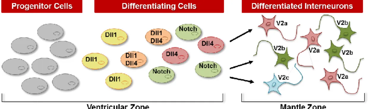
of Dll1 and Dll4 in this domain 32. The V2 domain is also particular as it gives rise to three functionally distinct classes of interneurons: V2a, V2b and V2c 33, all generated from a pool of molecularly similar progenitors with a specific molecular identity (e.g.
Nkx6.1, Irx3 and Pax6) 5, 34, 35. V2a and V2b interneurons are specified over the same time window but they have different physiological functions: V2a are excitatory glutamatergic INs, V2b are inhibitory GABAergic INs and V2c function is still unknown
33, 36.
Notch-Delta interactions have been suggested to play an important role in the initiation of a binary cell fate choice in immature V2 interneurons 30, 37, 38. Within the VZ of the V2 domain, cells that start the differentiation process express high levels of proneural proteins, and therefore high levels of Dll1, activating Notch in the neighbouring cells (signal-receiving cells), keeping them as progenitors (Figure 4) 32, 33. Some V2 cells that are committed to differentiation will express high levels of Dll4 and will differentiate into V2a INs (Ramos, C. et al, unpublished). These Dll4+ signal-sending cells have been shown to downregulate Gata2 39, while maintaining the expression of Lim3 (LIM homeobox 3) 38, 40, 41. These cells will then downregulate Lim3 and activate the expression of Bhlhb5 and Chx10, adopting the V2a cell fate 37, 42. The remaining cells committing to differentiation, do not express Dll4 and, will downregulate
Lim3 40, while maintaining the expression of Gata2 39, and activate the expression of
Scl (Stem cell leukaemia) and Gata3 to adopt V2b cell fate (Figure 4) 37, 42, 43.
Even though it is known that Notch signalling is crucial in V2 cell fate specification, how Dll1 and Dll4 ligands mediate this process is still under study. Previous analyses of Dll1-mutant mice (conditional knock-out) reveal that both V2a and V2b INs are generated, although in different numbers (increased number of V2a INs), when compared to control littermates 14, 30, 42. In Notch1 mutants all V2 progenitors acquire V2a fate, at the expense of the V2b fate 42. Furthermore, overexpression of DLL4 in chick spinal cord increases the number of V2b INs at the expense of V2a INs, whereas overexpression of DLL1 doesn’t affect significantly the number of V2a vs. V2b30. These results suggest that Notch signalling is required for the generation of V2b
INs and that Dll1 is not the key ligand for the V2a-V2b binary switch, a decision that might be controlled by Dll4-mediated Notch signalling 13, 14.
Proneural transcription factors contain a Helix-Loop-Helix (HLH) domain that allows these proteins to dimerize and, subsequently, to bind DNA through their basic domain. The proneural proteins were first identified in Drosophila and were divided into two families: the Atonal (ATO) and Achaete-scute (ASC) families (Reviewed in 9). The vertebrate Achaete-scute (ASC) family includes Ash1 (e.g. MASH1 in mouse, CASH1 in chick and ZASH1 in zebrafish) and other genes that are specific for vertebrate classes (MASH2 in mammals, XASH3 in Xenopus and CASH4 in chick) 9. The number of vertebrate proteins related to Drosophila ATO family is larger, but only two of them (MATH1 and MATH5 in mouse) have a bHLH domain similar enough to that of ATO to be considered as orthologues (reviewed in 9). Other vertebrate ATO-related proteins can be grouped into distinct families, e.g., the Neurogenin (NGN) family, the NeuroD family and the Olig family 9.
Some of the above mentioned proneural proteins are expressed in the V2 domain, specifically MASH1, NGN1 and NGN2, making them good candidates to regulate neurogenesis in this domain 32. Previous data suggest that Mash1 is expressed in V2 progenitors but how it is involved in interneuron specification is still controversial: Parras et al.reported a decrease in V2a interneurons in Mash1-mutant mice while the opposite result is reported by Li et al. Ngn1 and Ngn2 are also expressed in V2 cells but how their expression might influence V2 cell fate is still under study. In fact, in zebrafish, expression of DeltaD (Notch ligand) in the brain and in the spinal cord has been shown to be regulated by NGN and ZASH1 44 and, in mouse spinal cord and brain, MASH1 and NGN1/2 have been reported to directly regulated
Dll1 expression 26. The regulation of Dll4 expression during spinal cord development is one of the aims of this thesis.
Dll4 might have an important role for interneuron specification in the V2 domain.
Therefore, Dll4 expression must be tightly regulated for the correct number and type of V2 INs to be produced.
Regulation of Dll4 expression has been extensively studied in endothelial cells, since Notch signalling mediated by Dll4 has been implicated in vascular growth 31.
Here, vascular endothelial growth factor (VEGF) induces Dll4 expression as part of a negative regulatory loop, in which Dll4 act as an inhibitor of vascular sprouting. Also
Dll4 is likely to be involved in the modulation of diverse forms of pathological
angiogenesis, like in tumour angiogenesis. The blockade of Dll4 was found to delay tumour growth by enhancing the abnormal vascular sprouting, characteristic of tumour angiogenesis 45. Arterial specification is regulated by the combinatorial function of Notch signalling and SoxF transcription factors, via direct transcriptional gene activation. Arterial Dll4 expression requires the direct binding of both RBPJ (CSL)/NICD and SoxF transcription factors and this occurs downstream of the action of VEGF. The combinatorial role may contribute to more precise spatial and temporal control of gene expression within the differentiating vasculature 46. In endothelial cells, the coordinated activation of Notch signalling produces a wave of Dll4 expression. However in the V2 domain of the developing spinal cord, Dll4 is expressed in a salt-and-pepper pattern, with cells expressing high levels of Dll4 surrounded by cells that express low levels of this ligand. Hence, the mechanism behind Dll4 expression in the V2 domain is most likely different from the mechanism controlling Dll4 expression in endothelial cells.
Due to their expression in the V2 domain, good candidates to regulate Dll4 expression are the proneural proteins MASH1, NGN1 and NGN2.
Published work demonstrates that, in the chick developing spinal cord, the proneural protein CASH1 is involved in the regulation of Dll4 expression 30. In mouse,
Dll4 expression has been suggested to be regulated by both MASH1 and FoxN4, a
winged-helix forkhead protein, expressed in some V2 progenitor cells 42, 47, 48.
The aim of this thesis is to unravel: i) how Dll4 expression is regulated in the V2 domain; ii) how is Dll4 biased expression involved in V2 cell type specification. Particularly, I investigated how the asymmetric expression of Dll4 emerges from initially identical progenitor cells by testing whether there is an induction of Dll4 in some V2 cells or if is there a repression of Dll4 in some cells in the V2 domain, using in ovo electroporation of regulator candidates. The chicken embryo was used as a model organism in this approach due to its accessibility and easy handling.
Furthermore I characterized Dll4-expressing cells and unraveled if there is a difference in Dll1 and Dll4-mediated Notch signaling in the V2 domain, using mouse embryo as a model due to its genetic tools availability.
For plasmid DNA transformation, 100μL of competent E. coli bacteria were used. Competent bacteria were first thawed on ice and then incubated with plasmid for 20 min on ice. The mixture was heat-shocked for 45 seconds in a water bath at 42°C and then incubated on ice for 2 min. After adding 900μL of Super Optimal Broth (SOB) medium supplemented with 10mM MgCl2 and 10mM MgSO4, bacteria were incubated
at 37°C for 1 hour, shaking. 100μL of the mixture were plated on the appropriate selective LB agar media and incubated at 37°C o/n.
Plasmid constructs were stored at -80ºC as bacterial stocks (400µl of an o/n grown bacterial culture carrying the plasmids and 400µl of LB containing 30% glycerol). For large scale preparation of plasmid DNA (200-400µg), 100mL of the selective LB medium was inoculated with 1mL of plasmid bacterial culture (previously grown o/n). Bacteria were grown at 37ºC and processed using Genopure Plasmid Midi Kit (Roche), according to the given instructions.
DNA concentration was determined by spectrophotometry using the NanoDrop spectrophotometer (Thermo Scientific) – see details in Annex A.
Analytical digestions were performed to confirm gene size and identity (by fragment profile analysis). DNA was digested for 1-2h using 5-10U of commercially available enzymes and respective buffers. Restriction analysis was then performed in 1% agarose gel.
Fertilized chicken eggs were stored for a maximum of one week at 15ºC and incubated ate 37ºC in a humidified incubator (SMA 60) until the required developmental stages.
Embryos were injected with plasmid DNA using capillary needles made from borosilicate glass capillaries. The plasmid DNA was injected in spinal cord of chicken embryos at HH16-18 using the Inject+Matic (Genève®) injector (1 µg/µl). Each plasmid DNA was co-injected and co-electroporated with mCherryNLS@pCAGGS so that the electroporated cells can be visualized and act as a positive control for electroporation efficiency. Each plasmid DNA was injected with filtrated Fast green in order to visualize the injected solution. Platinum electrodes were placed 4mm apart of each other and parallel to the neural tube and the embryos were pulsed 5 times (25V/50 ms) using the Electro Square Porator™ ECM830 (BTX). Embryos were incubated again and after 36 hours were harvested and fixed in a 4% paraformaldehyde (PFA) in PBS solution at 4ºC o/n.
After fixation, embryos were washed twice in PBS and then transferred to a 15% and then 30% sucrose in PBS solution for cryoprotection. The embryos were embedded in a solution containing 7.5% gelatin and 15% sucrose in PBS and frozen in cold isopenthane (-75ºC). The frozen embryos were stored at -80ºC until sectioned in a cryostat (Leica CM 3050). Embryonic tissue was sectioned (12 µm) and collected on Superfrost® slides.
Mouse embryos, both wild-type and mutant (single and double mutants for Dll1 and Dll4) were collected at embryonic day 11.5. For the generation of these embryos, males with Cre-recombinase under regulation of Nestin promoter (specifically expressed in neural cells) and floxed for Dll1 and/or Dll4 were crossed with floxed females for Dll1 and/or Dll4. Crosses were set and vaginal plugs checked routinely to confirm pregnancy and, when confirmed, E11.5 embryos and the corresponding yolk sacs were collected. Pregnant females were sacrificed and a caesarean section performed to collect the embryos. All animals were fed freely and housed in SPF facilities. Animal experiments were approved by the Animal Ethics Committee of Instituto de Medicina Molecular and according to National Regulations.
After embryo collection, the yolk sacs were denatured and digested and the isolated DNA was used for genotyping with PCR for the presence of Cre in a first step and then for Dll1 and Dll4, followed by gel electrophoresis. Embryos without Cre were treated and used as wild-types control embryos. Tissue embedding and preparation of cryostat sections was the same as for chick embryos (previously described).
Several RNA antisense probes were used to perform in situ hybridization on whole-mount or cryostat sections of both mouse and chick embryos. Digoxigenin- (DIG) and Fluorescein- (FLUO) labeled RNA antisense probes were synthesized in
vitro by T3 or T7 RNA polymerase, from several plasmid templates (see Table 2 –
Annex A).
Different Plasmid DNA constructs (10µg) were linearized using 100U of the specific restriction enzyme in a final volume of 100 µl for 1 hour at 37ºC. After checking complete digestion by running 1 µl in an agarose gel 1%, DNA template was subjected to column purification and cleanup using Wizard Plus SV Gel and PCR Clean-up
System (Promega). DNA was quantified (as previously described).
Antisense probes were produced using 1µg of linearized plasmid DNA and 20U of RNA polymerase (T3 or T7) with 30mM DTT, 1x DIG-NTP mix (1mM ATP, CTG, GTP, 0.65mM UTP and 0.35mM DIG-UTP), 40U of RNase inhibitors (Roche), 1x Transcription Buffer (Stratagene) and RNase-free water in a final volume of 25µl. After 3 hours of incubation at 37ºC, the sample was precipitated by adding 20.5µl of RNase-free water, 2µl 0.5M EDTA, 2.5µl of 8M LiCl, 150µl of 100% ethanol and 1µl of glycogen and incubated o/n at -20ºC. The samples were centrifuged and the supernatant was discarded, RNA precipitate was washed with 70% ice-cold ethanol and then ressuspended in 100µl of 10mM EDTA and stored at -20ºC. To check for the quality of the probe, 2µl were mixed with RNA loading buffer containing formamide and, after a denaturation step of 5 min at 70ºC, run in agarose gel along with a probe of known concentration.
Fluorescent In situ hybridization on cryostat section was done by hybridizing DIG- or Fluo-labelled antisense RNA probes o/n at 68ºC in a humidified chamber wetted in 1x salts/50% formamide. Probes were diluted (1:100) in hybridization buffer and denatured at 70ºC for 10 min. After o/n hybridization, sections were washed for 10 min with pre-warmed washing solution at 68ºC to remove coverslips and then washed twice with the same solution and temperature for 15 min. Sections were washed three times for 15 min with TBST at RT and blocked with a solution of 2% blocking reagent and 20% heat inactivated sheep serum in TBST for more than 1 hour at RT in a
humidified chamber. Section were then incubated with antibodies DIG AP or anti-FLUO AP (Roche, 1:2000) o/n at 4ºC. After o/n incubation, sections were washed three times with TBST for 10 min and then twice with 0.1M Tris for 10 min. The staining reaction for AP (alkaline phosphatase) was performed using Fast Red (Roche) for 1-3h at 37ºC. After the development of the probe, sections were washed twice in PBS and incubated with the antibody that recognizes the second probe (FLUO-POD, anti-DIG-POD or anti-DNP-POD, Roche, 1:500). After 1 hour of incubation in a humidified chamber at RT, sections were washed with 1x TNT three times for 10 min and the staining reaction for POD (peroxidase) was performed using Tyramide Signal Amplification (1:50, TSA) for 20 min at RT. Sections were then washed four times with 1x TNT for 5 min, counterstained with DAPI and mounted with Mowiol mounting medium.
Gelatin was removed from selected regions with pre-warmed PBS1x at 37ºC, sections were treated with H2O2 in methanol for 30 min at RT to reduce background by
blocking endogenous peroxidases. Sections then, went through a 0.1M Glycine in PBS1x treatment for 10 min at RT to quench paraformaldehyde and stop cross-linking reactions and were permeabilized with 0.5% Triton in PBS1x for 10 min at RT. After this, sections were blocked with 10% Fetal Bovine Serum (FBS) in TBST for 1 hour at RT and then incubated with primary antibodies, previously diluted in 10% FBS in TBST, o/n at 4ºC in a humidified chamber. After primary antibody binding, sections were washed three times with TBST for 10 min at RT and incubated with secondary antibodies (diluted in 10% FBS in TBST) for 1 hour at RT in a humidified chamber. Sections were washed again three times with TBST for 10 min at RT and then counterstained with DAPI for 10 min. After this sections were mounted with Mowiol mounting medium.
The H2O2 pre-treatment was not performed on electroporated chick embryos
and on mice embryos when fluorescence was to be preserved.
The antibodies used during this project are described in Annex A.
Images of fixed sections with fluorescence were acquired using Leica DM5000B microscope with a Leica DC350F digital camera. Co-expression analysis was made using Confocal Laser Point-Scanning Microscope – Zeiss LSM 510 META. The acquired images were then treated using Adobe Photoshop software or ImageJ.
Cell counts were performed using Adobe Photoshop software and were expressed as the number of positive cells for the relevant marker, as a percentage of total DAPI (mean ± SD) from the electroporated or non-electroporated side of the neural tube. Data were analysed using the unpaired t-test to compare two data sets (GraphPad Prism v.6.0). Values of p<0.05 were considered significant.
Within the VZ of the V2 domain, progenitors that start the differentiation process express high levels of Dll1, activating Notch in surrounding cells to maintain them as progenitors (these cells express Hes5 and LFng) 17, 49 (Fig. 5A). The Dll1-expressing cells can subsequentially express Dll4 and became prospective (Bhlhb5+) V2a INs, activating Notch in the surrounding cells that will differentiate into prospective (Scl+) V2b INs (Fig. 5B). Previous data from our lab showed that Dll4-expressing cells will differentiate into V2a INs and activate Notch signalling in neighbouring cells (Dll4- and
Scl+) that, in turn, differentiate into V2b INs (Ramos C. et. al unpublished) (Figure 5B).
In the developing spinal cord, the V2 domain is the only one where two Notch ligands are expressed (DLL1 and DLL4), therefore a question arises: Is Dll1-mediated Notch signalling different from Dll4-mediated Notch signalling? One hypothesis is that, indeed, Dll1 and Dll4-mediated Notch signalling are different due to the presence of
LFng, a potentiator of Notch signalling 50, in signal-receiving cells. It is known that Dll1 is keeping the surrounding cells as progenitors, which express LFng 49, however concerning Dll4-mediated Notch signalling, the expression of LFng have not yet been mapped, so our hypothesis is that LFng might be involved in Dll1-mediated Notch signalling but not in Dll4-mediated Notch signalling.
To test this hypothesis, we analysed simultaneously the expression of LFng,
Dll4 and Scl in the V2 domain of E11.5 mouse spinal cord. The results show that LFng
is expressed in the VZ of most spinal cord domains, being only absent in the dl6 and V1 domains, in agreement with previous results (Fig.6 A and B). We did not observe cells expressing both LFng and Scl (Fig.6 A’-A’’’) indicating that LFng is not expressed in prospective V2b INs. However, LFng is co-expressed with Dll4+ in cells within the V2
A B
Figure 5 – Cell dynamics inside V2 domain. (A) - Cells that express high levels of Dll1 will keep their
neighbours as progenitors.(B) - Differentiating cells can express Dll4, upregulate the expression of Bhlhb5 (prospective V2a IN marker) and became V2a INs (Chx10+ cell). Their neighbours (Dll4- Notch+ cells) upregulate the expression of Scl (prospective V2b marker) and differentiate into V2b INs (Gata3+ cell).
progenitor domain (Fig.6 B’-B’’’ – indicated by arrow), but not in cells outside this region, where only Dll4 expression can be detected.
These data point out that Notch is activated by Dll4 in Scl+ cells without LFng potentiator and that Dll4-mediated Notch signalling is independent of LFng (Fig.5A).
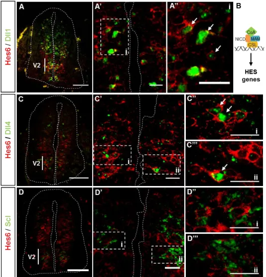
We next analysed the expression of Hes6, to try to correlate its expression to the possible differential activity of Dll1 or Dll4 ligands. Hes6 is a downstream target of Notch signalling, expressed in differentiating neurons, in which it functions to release proneural gene expression from the inhibitory effects of downstream targets of Notch signalling (Hes5) 51.
To test whether Hes6 might be activated by Dll1 and/or Dll4 we analysed simultaneously the expression of Hes6, Dll1, Dll4 and Scl in the V2 domain of E11.5 mouse spinal cords. Our data show that Hes6 is expressed throughout all domains of the spinal cord (Fig.7 A, B, C), in the VZ. However in the V2 domain, Hes6 expression is detected in cells located next to the lumen in the inner ventricular zone (IVZ), an exception to what is observed in all the other domains. Cells co-expressing both Hes6 and Dll1 can be observed in the V2 domain (Fig.7 A’, A’’ – indicated by arrows). Hes6 and Dll4 were also co-expressed (Fig.7 C’-C’’’ – indicated by arrows) in cells of the V2 domain, while coexpression of Hes6 and Scl was not detected (Fig.7 D’-D’’’). This last
*
Figure 6 – LFng expression in the prospective V2 INs. (A-B) – LFng is expressed in the VZ
of V2 domain, in progenitor cells. (A’) – LFng is not coexpressed with prospective V2b marker
Scl. (A’, A’’) – magnification of the selected regions from confocal image A’. (B’) – LFng can be
coexpressed with Dll4. In the ventricular zone, Dll4+ cells express also LFng (B’’ – white arrow) but when they migrate outside this region the co-localization of both mRNAs is no longer detected (B’ - indicated by asterisk). (B’, B’’) – magnification of the selected regions from confocal image B’. Scale bars: A, B - 50µm; A’-A’’’, B’-B’’’ - 10µm.
finding suggests that Hes6 is not a Dll4 target, as it is not expressed in prospective V2b INs (Scl-expressing cells) where Notch signalling is activated via Dll4. Another possibility is that Hes6 mRNA is no longer expressed when Scl expression starts. Concerning Dll1, it is known that cells activated through Dll1-mediated Notch signalling (Hes5+LFng+) can also express Hes6 17, therefore it is possible that Hes6 is a target of
Dll1-mediated Notch signalling.
Figure 7 – Hes6 expression in the V2 domain. (A, C, D) – Hes6 is expressed in the VZ
of V2 domain, in progenitor and differentiating cells. (B) – Schematic representation of activation of HES genes transcription. (A’) – Hes6 is coexpressed with Dll1 and Dll4 (C’) as the magnifications of the selected regions indicate (A’’, C’’, C’’’ – indicated by arrows). (D’) – Hes6 is not coexpressed with Scl, a marker for prospective V2b cells. (D’’, D’’’) – magnification of selected regions from confocal image D’.
Previous results from our lab showed that several regions in the Dll4 promoter, conserved between mouse and chicken, contain regulatory information for Dll4 expression. In these regions, several E-boxes are present and are putative binding sites for proneural proteins like MASH1 and Neurogenins (NGNs), which are therefore good candidates to regulate the Dll4 expression in the V2 domain. Also, it has been reported that Mash1, Ngn1 and Ngn2 are expressed in the V2 domain, although the relationship between their expression and that of Dll4 is still unclear.
In order to see if Dll4-expressing cells also express Mash1, Ngn1 or Ngn2, double in-situ hybridization was performed in neural tubes of E11.5 mice embryos.
As previously published, Mash1 is expressed in all dorsal spinal cord domains, except in dl6. Ventrally, it is only expressed in V2 domain (Fig.8 A). Both Ngn1 and
Ngn2 are expressed in all ventral domains, being absent in the dorsal most domains
(Fig.8 C, E).
Figure 8 – Characterization of Dll4+
cells. (A, C, E) – Mash1, Ngn1 and Ngn2 are expressed in the V2 domain. (B) – Mash1 is expressed in the majority of Dll4+ cells (white arrows). (D) – Ngn1 is also expressed in Dll4+
cells (white arrow) but some of these cells don’t express Ngn1 (asterisk). (F) – Ngn2 is the proneural less co-expressed with Dll4 as many Dll4+ cells don’t express Ngn2 (asterisk). (G) – Graphic showing the percentage of Proneural+
Dll4+ cells as a fraction of the Dll4 population. 79% of Dll4+ cells are also Mash1, 39% are also Ngn1 and 17% are also Ngn2. Average from three different sections of five different embryos. Scale bars: A, C and E - 20µm (Fluorescence Microscope); B, D and F - 10µm (Confocal Microscope).
G
*
In the V2 domain, the population of Mash1+ Dll4+ cells (Fig.8 B – white arrows) corresponds to 79% of the total Dll4 population (Fig.8 G). Regarding Ngn1, less than half of Dll4+ cells co-express Ngn1 – 39% (Fig.8 D – arrow, G). Cells that express Dll4 but do not express Ngn1 could also be identified (Fig.8 D – asterisk). When analysing results for Ngn2, the results show that the majority (83%) of the Dll4+ cells do not express Ngn2 (Fig. 8 F – asterisks) with only 17% being Ngn2+ (Fig.8 F – white arrow,
G).
Overall, these results indicate that the three proneural genes are expressed in
Dll4-expressing cells but to a different extent, with Mash1 being the most co-expressed
gene, followed by Ngn1 and finally by Ngn2. The fact that Mash1 is the proneural gene that shows higher co-expression with Dll4 raises the hypothesis that Mash1 is the major regulator of Dll4 expression, when compared to both Ngn1 and Ngn2.
To test possible role of Mash1, Ngn1 and Ngn2 in regulating Dll4 expression, these transcription factors were overexpressed, single or in combination, in the chick embryonic spinal cord, and their effect in eliciting Dll4 expression was evaluated. In these experiments, a plasmid encoding the CHERRY fluorescent protein was co-electroporated with the vectors encoding proneural genes, in order to allow the identification of electroporated cells in the spinal cord (see Materials and Methods). Embryos were harvested 36 hours after electroporation and Dll4 expression analysed, using fluorescent in-situ hybridization
Two different positive controls for Dll4 probe specificity were made. It is well known that endothelial cells express high levels of Dll4 (Fig. 16, Annex B – white arrows), and our results show also strong Dll4 expression in these cells. In embryos electroporated with Hes6-2:VP16 (dominant-negative form of Hes6-2 protein that act as an activator of Dll4 expression) we observed Dll4 expression in the electroporated cells, accordingly to previous reports (Fig. 16, Annex B).
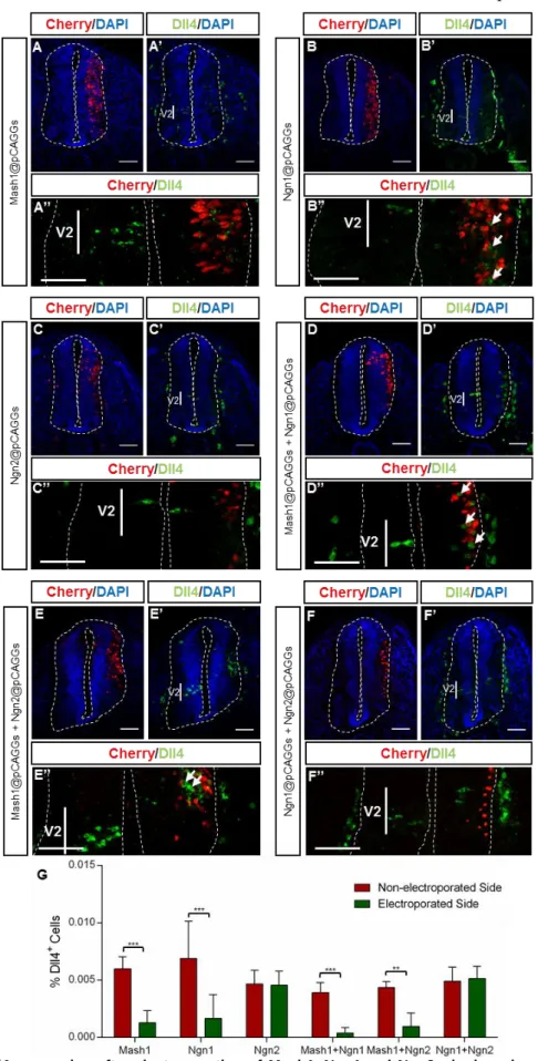
Our results show that MASH1 overexpression represses Dll4 expression in the V2 domain (Fig. 9 – A’, A’’, G). This repression is significant and consistent across different embryos (n=8, p=2.512702e-006).
79%
Figure 9 – Dll4 expression after electroporation of Mash1, Ngn1 and Ngn2, single or in combinations.
(A-A’’) – MASH1 represses Dll4 expression in the V2 domain. (B-B’’) – NGN1 overexpression also represses
Dll4 in the V2 domain but it induces ectopic Dll4 expression along DV axis (White arrows). (C-C’’) – NGN2
overexpression does not change Dll4 expression. (D-D’’) – MASH1 together with NGN1 repress Dll4 within V2 domain while induce ectopic Dll4 expression along the DV axis of the spinal cord. (E-E’’) – MASH1 and NGN2 also repress Dll4 expression in the V2 domain and induce it, in the electroporated cell region. (F-F’’) – Both NGNs overexpressed do not alter Dll4 expression in the developing spinal cord. (G) – Percentage (%) of Dll4+
Overexpression of NGN1 also represses Dll4 expression in the V2 domain (Fig.9 – B’, B’’, G; n=8, p=0.00383657) however it induces ectopic Dll4 expression in other domains along the DV axis of the spinal cord (Fig. 9 – B’’, white arrows). The observed induction of Dll4 expression occurs mostly in a non-cell autonomous manner (cells next to the electroporated ones).
Overexpression of NGN2 does not alter Dll4 expression in the V2 domain, neither causes ectopic expression in other domains (Fig. 9 – C-C’’’, G; n=8, p=0.89066). These results show that MASH1 and NGN1 are able to regulate Dll4 expression, while NGN2 is not.
To test for the presence of combinatorial effects in regulation of Dll4 expression, MASH1, NGN1 and NGN2 were overexpressed in different combinations.
Simultaneous overexpression of MASH1 and NGN1 causes a repression of Dll4 expression in the V2 domain (Fig. 9 – D-D’’’, G; n=3, p=0.00356786), while inducing ectopic expression of Dll4, dorsally to V2, (Fig. 9 D’’’ – white arrows), in a non-cell autonomous manner.
Simultaneous overexpression of MASH1 and NGN2 also represses Dll4 expression (Fig. 9 – E-E’’’, G; n=3, p=0.0103982) while induces ectopic expression (Fig. 9 – E’’’, white arrows).
When both NGNs are overexpressed, there are no significant differences in the number of Dll4-expressing cells (Fig. 8 – F-F’’’, G), comparing both sides of the spinal cord (n=3, p=0.828263).
In conclusion, our data, from simultaneous overexpression of proneural genes, suggest that there are no additive effects in regulation of Dll4 expression by proneural proteins. However NGN2 can overcome NGN1 function, as simultaneous overexpression of both NGNs mimics the effect of single overexpression of NGN2 -
Dll4 expression is not altered.
Parras et. al and Li et. al reported that proneural proteins can affect IN specification inside V2 domain. To determine whether overexpression of proneural proteins affects V2a interneuron specification, embryos that were electroporated with plasmids encoding these proteins, were analysed for the expression of CHX10, a protein specifically expressed in V2a INs, using immunofluorescence with an appropriate antibody.