Synthesis, Functionalization and
Bioconjugation for Biological Applications
Ana Sofia da Cunha Miguel
Dissertation presented to obtain the Ph.D degree in Engineering Sciences
and Technology
Instituto de Tecnologia Química e Biológica | Universidade Nova de Lisboa
Ana Sofia da Cunha Miguel
Dissertation presented to obtain the Ph.D degree in Engineering Sciences
and Technology
Instituto de Tecnologia Química e Biológica | Universidade Nova de Lisboa
Thesis Supervisors:
Professor Dr Christopher David Maycock
(Head of the Organic Synthesis Laboratory at Instituto de Tecnologia Química e Biológica)
Dr Abel Gonzalez Oliva
(Head of the Biomolecular Diagnostic Laboratory at Instituto de Tecnologia Química e Biológica)
The research work described in this thesis was performed at Organic Synthesis and Biomolecular Diagnostic Laboratories, Instituto de Tecnologia Química e Biológica – Universidade Nova de Lisboa, Oeiras, Portugal. This work was supported by Fundação para a Ciência e Tecnologia (FCT) through a PhD grant to Ana Sofia Miguel (SFRH/BD/40303/2007) and the grant # PEst-OE/EQB/LA0004/2011. This work was also supported by the national funded project NTec/SQA/0131/2007 from FCT
I would like to express my gratitude to all the people who, in one way or
another, have helped me throughout my Doctoral work:
A special thanks to Professor Dr Christopher Maycock, for accepting me in
his lab 9 years ago, for believing in me and my work, for trusting in my
capabilities when I decided to accept this project. More particularly, thank you
for the guidance and advice throughout these years, for his invariable efforts
to keep the laboratory in excellent condition which allowed me to proceed with
my research work unhindered, for the fruitful ideas and discussions during my
chemistry problems, for his great sense of humour and for the opportunity to
work with him.
I would like to thank Dr Abel Oliva, for giving me the opportunity to work with
him, for his availability and help, for all the financial efforts that he made to
support my work during the past few years as well as all the research
collaborations that he established giving me the opportunity to expand my
work.
I thank all my colleagues (past and present members) from Organic Synthesis
laboratory (ITQB-UNL), for their help, especially to Eva Lourenço, Dr Rita
Ventura, Paula Rodrigues and Osvaldo Ascenso for their support, patient,
enthusiasm, friendship and for all the relaxing lunches, afternoon talks and for
the good moments we passed together.
I thank my colleagues (past and present members) from the Biomolecular
Diagnostic laboratory (ITQB-UNL) especially Ana Raquel Santos and Joana
Campos for their support and help in my work and for all the good moments
that we shared together.
To Elisa de Campos, a deep and sincere thank you for all the support during
my Doctoral work, especially for the discussions on my biological data. Thank
you for sharing all your knowledge and expertise with me and for your
I would like to thank to Dr Pedro Fevereiro from ITQB-UNL for his sympathy,
availability and for the great work resulting from our research collaboration.
A special acknowledgement to Dr Ana Domingos from IHMT-UNL for her
help with the antibody production and helpful discussions related to biological
data and for her availability and friendship.
I also thank to Analytical Services Unit (ASU) (ITQB-UNL and IBET),
especially to all the members of the analytical and microbiology laboratories
located in the chemistry building. I am grateful for the opportunity that was
given to me to use their equipment which contributed largely to the
performance and success of my work during the last four years.
I would also like to thank the Instituto de Tecnologia Química e Biológica of
the Universidade Nova de Lisboa (ITQB-UNL) for the excellent conditions
provided to perform my work.
I would like to express my gratitude to the Fundação para a Ciência e
Tecnologia (FCT) for the PhD fellowship that was conceded to me
(SFRH/BD/40303/2007).
To all my friends, a big thanks for the encouragement, support and friendship
all these years! You know who you are.
I want to thank my parents for their endless affection, support, patience and
for always believing in me and my capabilities. Thank you for making possible
what I am today.
Finally, I especially thank Filipe Alvito for his love, support and patience.
Thank you for your excellent advice, for your guidance to be always better
Acknowledgments ... v
Table of Contents ... ix
Dissertation Outline ... xvii
Abbreviations... xxiii
Abstract ... xxix
Resumo ... xxxv
Chapter 1 General Introduction ... 1
Chapter 2 Synthesis and Characterization of CdSe/ZnS QDs ... 27
Chapter 3 Ligand Synthesis and Design for Water-Soluble CdSe/ZnS QDs ... 61
Chapter 4 Conjugation of Luminescent QDs with Antibodies ... 141
Chapter 5 Q u an tum D ot Ap p l ic a t i ons ... 189
PART I Detection of RAP-1 Antigenic Protein in Infected Babesia bovis Erythrocytes Using QDs as Fluorescent Reagents ... 193
PART II Work Resulting From Collaboration with Other Research Groups ... 219
Chapter 6 G e ner a l D is c us s i o n an d F u tur e P er s p ec t i v es ... 235
Chapter 7 A pp e nd ix ... 251
Published papers related with this dissertation: ... 253
Book chapters by invitation related with this dissertation: ... 253
List of Equations:
Equation 1| Expression applied to calculate the quantum yield. ... 39
Equation 2 | Expression of Lambert-Beer’s Law given to calculate the concentration of QDs solutions. ... 39
Equation 3 | Stokes-Einstein equation for the hydrodynamic diameter. ... 49
Equation 4 | Expression given to estimate the number of functional amine groups available on a single QD surface. ... 100
Equation 5 | Expression used to estimate the number of sugar molecules available on a single QD surface... 101
Equation 6 | Smoluchowski eaquation used to convert electrophoretic mobility of nanoparticles measured in zeta potential values. ... 126
Equation 7 | Expression used to determine the concentration in mg/mL of the immobilized antibody in QD-antibody complexes. ... 155
Equation 8 | Expression given to determine the QD/Ab ratios. ... 155
List of Figures:
Figure 1 | Electronic Energy Levels Depending on the Number of Bound Atoms. ... 6Figure 2 | Quantum Dots.. ... 8
Figure 3 | Surface Modification of QDs. ... 13
Figure 4 | Bioconjugation Strategies.. ... 15
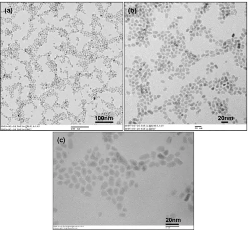
Figure 5 | Transmission Electron Microscopy Analysis. ... 47
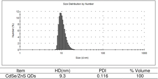
Figure 6 | Size determination by dynamic light scattering. ... 48
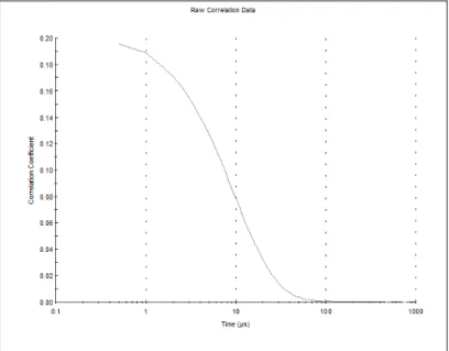
Figure 7 | Correlation Data. ... 50
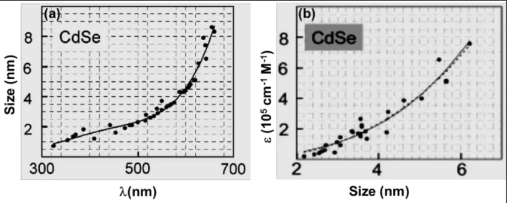
Figure 8 | Determination of the extinction coefficient of QDs. ... 52
Figure 9| Optical Measurements:. ... 53
Figure 10 | 1H-NMR spectra of DHLA (1) and DHLA-PEG400-OH (3). ... 104
Figure 11 | Infrared Analysis of dichloride and diazide from PEG400. ... 106
Figure 12 | Optical Measurements of Hydrophilic QDs. ... 122
Figure 13 | HD Measurements of Hydrophilic QDs. ... 124
Figure 14 | Size Determination by Dynamic Light Scattering. ... 125
Figure 15 | -potential Measurements of Hydrophilic QDs. ... 126
Figure 17 | Ratio of detected amine concentration vs. % of amine-terminated
ligand in ligand mixture. ... 129
Figure 18 | Estimation of the Number of Sugar Molecules per QD. ... 130
Figure 19 | Structure of an IgG Molecule. ... 146
Figure 20 | Biological Activity of the Antibody-Bioconjugated QDs.. ... 158
Figure 21 | pH Stability of MPA and DHLA-QDs.. ... 159
Figure 22 | Electrophoretic Mobility of MPA and DHLA-QDs.. ... 161
Figure 23 | Comparison of Electrophoretic Mobility of MPA-QDs in different buffers.. ... 162
Figure 24 | EDC Activation Step Analysed by Agarose Gel Electrophoresis. ... 166
Figure 25 | Analysis of Bioconjugation Protocol by Agarose Gel Electrophoresis.. ... 169
Figure 26 | Separation of the Functional Components of MPA-Bioconjugated QDs. ... 174
Figure 27 | Separation of the Functional Components of DHLA-Bioconjugated QDs.. ... 176
Figure 28 | Testing the Functional Activity of QD-Ab complexes. ... 178
Figure 29 | Erythrocyte Invasion by Babesia Parasites. ... 197
Figure 30 | Apicomplexan main features for Toxoplasma gondii tachyzoite and Plasmodium falciparum merozoite. ... 198
Figure 31 | Immunofluorescence Assays on Slide.. ... 208
Figure 32 | Immunofluorescence Assays in Solution. ... 209
Figure 33 | Immunofluorescence Assays on Slide with Vortex Stirring. ... 210
Figure 34 | QDs Uptake by the Plant Cells.. ... 223
Figure 35 | Oxidative Stress Dose Response Assay... 224
Figure 36 | DNA Damage in Medicago sativa cells in Suspension Cultures.. ... 227
Figure 37 | Expression of Tdp1 β, Top1.β, Fpg, SOD and APX genes in Medicago sativa cells treated with MPA-QDs. ... 228
List of Schemes:
Scheme 1| Reaction scheme for the synthesis of TOPO/HDA capped-CdSe
QDs. ... 34
Scheme 2 | Reaction scheme for the surface passivation of CdSe/ZnS core-shell QDs with 5 monolayers of ZnS based on SILAR method. ... 37
Scheme 3 | DHLA-PEG Hydrophilic Ligands. ... 67
Scheme 4 | DHLA and DHLA-PEG400-OH Synthesis. ... 102
Scheme 5 | Modular ligands with functional terminal groups.. ... 105
Scheme 6 | General reaction for the synthesis of ditosyl compound 20 from PEG400. ... 107
Scheme 7 | Amino-PEGn-DHLA and Carboxy-PEGn-DHLA Synthesis. ... 107
Scheme 8 | LA-NHS Synthesis. ... 108
Scheme 9 | Sugar-capped QDs. ... 110
Scheme 10 | Synthesis of 3-maleimido propionic acid NHS ester. ... 111
Scheme 11 | Thiol linker synthesis. ... 112
Scheme 12 | Carbohydrate Hydrophilic Ligands. ... 114
Scheme 13 | DHLA-TEG-Mannose Synthesis. ... 115
Scheme 14 | Structure of collateral product TEG-N3 (38) formed from degradation of initial compound 31 under hydrolysis conditions. ... 117
Scheme 15 | DHLA-PEG400-Mannose Synthesis. ... 118
Scheme 16 | Structure of collateral product PEG400-N3 (47) formed from degradation of initial compound 42 under hydrolysis conditions. ... 119
Scheme 17 | Functionalization Procedure.. ... 120
Scheme 18 | Schematic Diagram of Covalent Conjugation. ... 148
Scheme 19 | Bioconjugation Protocol Proposed.. ... 164
Scheme 20 | Conjugation of Antibodies to Carboxy-QDs using EDC or EDC/NHS. ... 165
Scheme 21 | Antibody Reduction with -Mercaptoethanol.. ... 173
Scheme 22 | Structure of an Individual Amino Acid Present in Proteins. .... 180
List of Tables:
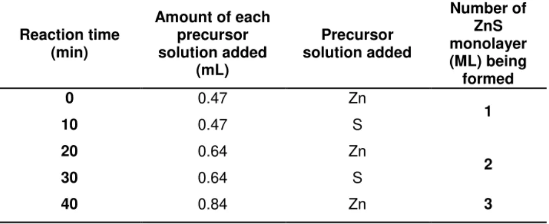
Table 1| Amount of each precursor solution added for the ZnS shell growth
procedure around the CdSe cores. ... 36
Table 2| Glossary of terms related with quantum dots. ... 55
Table 3| Solvent and reagent purification. ... 71
Table 4 | Graphical index of compounds and experiments of ligand synthesis
and design. ... 131
Table 5 | QD/Ab molar ratios obtained for the different bioconjugation
reactions performed to link antibodies onto QD surface using the EDC/NHS
strategy. ... 172
Table 6 | Glossary of terms related with some molecular biology techniques
applied in this work . ... 182
The main goal of this Doctoral work was to develop a fluorescent biomarker to
identify antigenic proteins associated with specific parasites. Although it is
known that fluorescent techniques making use of standard organic dyes are
widely used for this purpose, the development of a new method was proposed
using nanotechnology; in particular the use of nano optical reporters also
called quantum dots (QDs). In order to achieve this goal, the research work
was divided into four main tasks: (1) synthesis and characterization of
CdSe/ZnS core-shell QDs; (2) design of dihydrolipoic acid (DHLA) ligands
appended to oligo and poly (ethylene) glycols (PEG) with different functional
groups to generate biocompatible QDs; (3) bioconjugation of QDs to
monoclonal antibodies based on sophisticated protocols and (4) some
chemical and biological applications of the synthesized non-conjugated and
conjugated nanoparticles.
Chapter 1 of this thesis is a general introduction related to nanotechnology,
particularly on the subject of quantum dots. This includes a general overview
of reported work related to the synthesis, functionalization, bioconjugation,
toxicity and possible applications of these nanoparticles.
Chapter 2 describes protocols for the synthesis of CdSe cores and surface
passivation with a ZnS multishell. Moreover, the structural and optical
characterization of these nanoparticles making use of diverse techniques is
also presented. Data shown in this chapter has been published in a book
chapter called “Synthesis and Functionalization of CdSe/ZnS QDs using the
Successive Ion Layer Adsorption Reaction and Mercaptopropionic Acid Phase
Transfer Methods” within the “Nanoparticles in Biology and Medicine. Methods and Protocols.”from Springer.Ana S. Miguel is the first author of this
book chapter.
Chapter 3 describes a simple and versatile scheme for the preparation of a
family of heterobifunctional ligands incorporating i) dihydrolipoic acid as QD
ligand, ii) a hydrophilic spacer such as polyethylene glycol with different
molecular weights and iii) different functional end termini. With these ligands,
for posterior conjugation with biomolecules. In parallel, the production of
hydrophilic nanoparticles with shorter ligands such as 3-mercaptopropionic
acid by the phase transfer method was also carried out and discussed. The
hydrophilic nanoparticles were characterized in terms of size, surface charge
and fluorescence making use of light scattering techniques and quantum yield
measurements respectively. When appropriate, colorimetric assays were also
used to characterize the presence of some functional groups in the QD
surface. The overall data resulting from this characterization is also described
and discussed in this chapter. Part of the data obtained in chapter 3 has also
been published in the book chapter previously mentioned and edited by
Springer.
Chapter 4 focuses on the conjugation of luminescent water-soluble QDs with
monoclonal antibodies. The conjugation of MPA and DHLA-QDs with
monoclonal antibodies against a recombinant antigenic protein was performed
based on EDC/NHS amide bond forming chemistry. To better understand the
experimental conditions necessary to carry out this reaction, the
bioconjugation assay was proven by studies of the electrophoretic mobility of
carboxy-QDs. Confirmation of the success of the bioconjugation process,
characterization of the binding properties of the bioconjugated-QDs as well as
the capacity that these bioconjugated complexes had to recognize their
antigen, were analysed and discussed. The antibody/QD molar ratio was also
determined.
Biological and chemical applications of the conjugated and non-conjugated
QDs respectively are described in Chapter 5. This chapter is divided into two
sections. The first part is related to the use of bioconjugated samples as
fluorescent biomarkers to detect the RAP-1 antigenic protein in erythrocytes
infected with Babesia bovis. The second part comprises the work with QDs
developed in collaboration with other research groups. For example, the
cytotoxic and genotoxic effects caused by the non-conjugated nanoparticles in
higher plant cells was studied and is discussed. In addition, application of the
in Journal of Nanobiotechnology and in the book chapter entitled “Evaluation
of Cytotoxicity of 3-Mercaptopropionic Acid-Modified Quantum Dots on
Medicago sativa Cells and Tissues”, edited by Springer (Nanoparticles in
Biology and Medicine. Methods and Protocols). The work related to the
chemical applications of the native CdSe/ZnS QDs has been published in
Journal of Physical Chemitry C and is also submitted in Physica Status Solidi
A. The study of genotoxic effects of the non-conjugated nanoparticles in
higher plant cells has been submitted in Particle and Fibre Toxicology.
Chapter 6 gathers all the results/findings obtained in this Doctoral work
together to form general conclusions along with suggestions for future work.
A printed version of the published, in press or submitted papers/book chapters
related to the work described in this thesis can be found in Chapter 7
AAS atomic absorption spectroscopy
Ab antibody
ADH adipic acid dihydrazide
AFM atomic force microscopy
AMA-1 apical membrane antigen 1
A/N alkaline/neutral
a.u. arbitrary units
BCA bicinchoninic acid
BME-mercaptoethanol
br s broad singlet
BSA bovine serum albumin
BTTS bis (trimethylsilyl) sulphide
CAT catalase
CCD charge couple device
CT C-terminal
Cys cysteine chemical shift
J coupling constant
DAB3, 3’-diaminobenzidine
DAPI4′, 6-diamidino-2-phenylindole dihydrochloride
DCC dicyclohexylcarbodiimide
DHLA dihydrolipoic acid
DMAP 4-(dimethylamino) pyridine
DMF dimethylformamide
DLS dynamic light scattering
DSBs double strand breaks
DTNB5, 5’-dithiobis (2-nitrobenzoic acid)
DTT dithiothreitol
EDC N-Ethyl-N′-(3-dimethylaminopropyl) carbodiimide
EDX dispersive x-ray spectroscopy
Endo III endonuclease III
ELISA enzyme-linked immunosorbent assay
ESI-MS electrospray ionization mass spectrometer
FITC fluorescein isothiocyanate
FPG formamidopyrimidine DNA glycosilase
FRET Förster resonance energy transfer
FTIR Fourier transform infrared spectroscopy
GPI glycosyl-phosphatidylinositol
GR glutathione reductase
HD hydrodynamic diameter
HDA 1-hexacedylamine
HOMO highest molecular orbital occupied
HRTEM high resolution transmission electronic microscopy
HRP horseradish peroxidase
H2DCFDA2’, 7’-dichlorodihydrofluorescein diacetate
IFAT immunofluorescence antibody test
IgG immunoglobulin G
IHC immunohystochemistry
LA-NHS lipoic acid N-hydroxysuccinimide ester
LEDs light emitting devices
LUMO lowest molecular orbital unoccupied
MAA mercaptoacetic acid
MAPLE matrix-assisted pulsed laser evaporation
MBP maltose binding protein
MES 4-morpholineethanesulfonic acid
ML monolayer
MPA 3-mercaptopropionic acid
MW molecular weight
Na-P sodium phosphate buffer
NBT/BCIP nitro-blue tetrazolium / 5-bromo-4-chloro-3'-indolyphosphate
NC’s nanocrystals
NHS N-Hydroxysuccinimide
Ni-NTA nickelnitriloacetic acid complex
NMR nuclear magnetic resonance
NT N-terminal
PCR polymerase chain reaction
PCS photon correlation spectroscopy
PDI polydispersity index
PBS phosphate buffer saline
PEG poly (ethylene) glycol
pI isoelectric point
PL photoluminescence
PMAO maleic copolymer
PTFE polytetrafluoroethylene p-TsCl tosyl chloride
PVDF polyvinylidene fluoride
QD-Ab quantum dot-antibody conjugate
QDs quantum dots
QRT-PCR real time quantitative polymerase chain reaction
QY quantum yield
RAP-1 rophtry associated protein
RBC red blood cell
Rf retardation factor
RLB reverse line blotting
ROS reactive oxygen species
r.t. room temperature
g relative centrifugal force
SBP spherical body protein
SDS sodium dodecyl sulphate
SILAR successive ion layer adsorption and reaction
SMCC 4-(maleimidomethyl) cyclohexanecarboxylic acid
N-hydroxysuccinimide ester
SOD superoxide dismutase
SSBs single strand breaks
Sulfo-SMCC 3-sulfo-N-hydroxysuccinimide ester sodium salt
TA thioctic acid
TBE tris borate EDTA
TBP tributylphosphine
Tdp tyrosyl-DNA phosphodiesterase
TEG tri (ethylene) glycol
TEM transmission electronic microscopy
THF tetrahydrofuran
TLC thin layer chromatography
TMSOTf trifluoromethanesulfonic acid trimethyl silylester
TOP trioctylphosphine
Top topoisomerase
TOPO trioctylphosphine oxide
TRAP thrombospondin-related anonymous protein
Trp tryptophan
TTBS tris buffered saline with Tween
Tyr tyrosine
During the past few decades, technology has made great improvements to
enable visualisation, identification and quantitation in biological systems. In
recent years, nanoparticles have become the center of attention of many
researchers. The integration of inorganic synthetic methods with techniques to
produce nano-sized particles of semiconducting materials has led to the
creation of a class of fluorescent optical reporters, called quantum dots (QDs).
These nanometer-sized crystalline particles are composed of elements of
periodic groups of II-VI or III-V elements but so far the CdSe/ZnS core-shell
QDs are the most used. These nanoparticles possess unique optical and
electronic properties, compared to organic dyes such as size-tuneable light
emission, superior signal brightness, superior resistance to photobleaching
and simultaneous excitation of multiple fluorescence colours.
One of the main goals of this doctoral work was to synthesize core-shell
CdSe/ZnS QDs and design ligands that permit the preparation of compact
hydrophilic functionalized QDs amenable to conjugation with a variety of
biomolecules via simple covalent and non-covalent binding strategies. In
order to achieve this objective, detailed protocols for the preparation of CdSe
nanocrystals coated with a multishell structure of ZnS were developed and
described. For the controlled formation of the ZnS shells the Successive Ion
Layer Adsorption and Reaction (SILAR) method was used. To analyse the
size, size distribution, shape and fluorescence of the nanoparticles, a
combination of optical and structural characterization techniques was used
including electron transmission microscopy, dynamic light scattering and
quantum yield measurements. To promote the hydrophilicity of the native QDs
in aqueous environments a variety of dihydrolipoic acid (DHLA) ligands were
synthesized appended to a hydrophilic spacer, such as polyethylene glycol, of
various lengths. These ligands also included a functional terminus such as an
amine, a carboxylate or a sugar moiety for the posterior covalent attachment
of the biomolecules. Similarly 3-mercaptopropionic acid (MPA), a short
hydrophilic ligand, was attached using the phase transfer method for ligand
exchange thus forming compact water soluble QDs.
Having available surface functionalized QDs, a further interesting step was
their bioconjugation with antibodies and other proteins. QD/antibody
and some of them involve the subtle modification of the antibody presumably
without reduction of bioactivity. The most used procedures are covalent
antibody-QD-conjugation based on cross linking reactions, between the amino
groups that decorate the stem of the antibody and carboxylic acid groups
which terminate the ligand chains connected to the QDs (random
bioconjugation, amide bond formation catalysed by carbodiimide). Taking
advantage of this methodology, the coupling of MPA and DHLA-QDs with
antibodies using EDC/NHS coupling chemistry was carried out. Monoclonal
antibodies against recombinant RAP-1 Babesia bovis protein were attached
using this method. The bioconjugated nanoparticles were characterized by
comparing their molecular weight and electrophoretic mobility with
non-conjugated nanoparticles as well as their functional and binding properties,
using SDS-PAGE, agarose gels and western blotting techniques.
Finally, the application of the developed bioconjugated nanoprobes in the
biological field, namely in the detection of recombinant RAP-1 antigenic
protein in infected Babesia bovis erythrocytes was carried out. Babesia bovis
is an intraerythrocytic protozoan parasite transmitted by Ixodid ticks and
responsible for great economic losses in cattle industry in many tropical and
subtropical regions worldwide. Over the years many different methods have
been applied to identify early infections in whole blood of the animals, in
particular immunofluorescence antibody test (IFAT). However, one of the
significant limitations presented by this method is photobleaching. Using the
QDs conjugated with anti-RAP 1 antibody for detection of rhoptry associated
antigenic protein (RAP-1) present in the apical complex of different B. bovis
parasites in infected blood, these nanoparticles proved to be a good
alternative to traditional IFAT and also useful tools in biosensing applications
since they are much less prone to photodegradation.
Concerns about the toxic and genotoxic effects of these nanoparticles in
biological systems have been raised since they contain toxic elements such
as cadmium and selenium. Another interesting area of development is for
possible applications in electronics for the creation of biosensors, lasers or
high performance devices. This interest has led to the creation of
genotoxic effects in Medicago sativa cells (higher plant cells) when exposed
to CdSe/ZnS core-shell QDs coated with 3-mercaptopropionic acid as well as
the use of a recent laser-based deposition technique to immobilize the native
QDs onto solid substrates for further application in electronic devices. For the
cytotoxicity and genotoxicity studies, in general, the resulting work
demonstrated that these effects are dependent on a number of factors
including the QD’s properties, dose and environmental conditions of
administration. It was concluded that, QD concentrations between 1 and 5nM
should not be exceeded for such biological applications. On the other hand,
the native CdSe/ZnS core-shell QDs could prove to be useful tools in the
electronics field since they are resistant to laser deposition techniques
creating a specific network-like surface morphology of the deposited material
and after the immobilization process, their functional properties (fluorescence)
were not altered.
This doctoral work has demonstrated the numerous advantageous properties
and some of the potentialities of the chosen QDs not only in the biological
field but also in other areas.
Key-words: Quantum dots, SILAR method, biocompatible, bioconjugation, immunofluorescence assays, Babesia bovis, cytotoxicity, genotoxicity and
Durante as últimas décadas, a tecnologia tem produzido notáveis avanços
para permitir a identificação, a visualização e a quantificação em sistemas
biológicos. Nos últimos anos, as nanopartículas tornaram-se o centro das
atenções de muitos investigadores. A integração de métodos sintéticos
inorgânicos com técnicas de produção de nanopartículas, constituídas por
materiais semicondutores, levou à criação de uma classe de repórteres
ópticos fluorescentes, denominados de quantum dots (QDs). Estas partículas
cristalinas de tamanho nanométrico podem ser compostas por elementos dos
grupos II-VI ou III-V da tabela periódica, mas até agora as que possuem um
núcleo de seleneto de cádmio (CdSe) e uma cobertura de sulfureto de zinco
(ZnS) têm vindo a ser as mais utilizadas. Estas nanopartículas possuem
propriedades ópticas e electrónicas únicas, quando comparadas com
corantes orgânicos, tais como um tamanho definido de acordo com a
emissão de luz pretendida, um superior sinal de brilho, uma elevada
resistência à foto-degradação e podem ser excitadas em diferentes
comprimentos de onda permitindo obter múltiplas cores de fluorescência.
Um dos principais objectivos deste trabalho Doutoral foi sintetizar
nanopartículas semicondutoras com a estrutura núcleo-cobertura CdSe/ZnS
e criar ligandos orgânicos que permitissem a obtenção de nanopartículas
compactas, funcionalizadas e hidrofílicas de forma a serem posteriormente
conjugadas com diversas biomoléculas fazendo uso de ligações simples
covalentes e não-covalentes. De forma a alcançar este objectivo, foram
desenvolvidos e descritos protocolos detalhados para a preparação destes
nano-cristais. A formação controlada das coberturas de sulfureto de zinco foi
efectuada com base no método SILAR (successive ion layer adsorption and
reaction). Para analisar o tamanho, a distribuição de tamanho, a forma e a
fluorescência das nanopartículas, foi utilizada uma combinação de técnicas
de caracterização estrutural e ópticas, nomeadamente microscopia de
transmissão electrónica (TEM), dynamic light scattering (DLS) e medições do
rendimento quântico. Para promover a hidrofilicidade das nanopartículas
semicondutoras nativas em ambientes aquosos, foi sintetizada uma
variedade de compostos derivados do ácido dihidrolipóico (DHLA) contendo
anexada uma molécula hidrofílica, como o caso do polietileno glicol (PEG), de
um terminal funcional que pode ser constituído por diferentes grupos
funcionais, como por exemplo, uma amina, um ácido carboxílico ou um
glúcido para uma posterior ligação covalente das biomoléculas. Da mesma
forma, embora se trate de um ligando hidrofílico de pequeno tamanho, o
ácido 3-mercaptopropiónico (MPA) foi também acoplado à superfície das
nanopartículas, usando o método de transferência de fase, originando assim
QDs compactos e solúveis em meios aquosos.
Após a superfície dos QDs se encontrar funcionalizada, um passo posterior
interessante foi a sua conjugação com anticorpos e proteínas. A conjugação
QD-anticorpo pode ser efectuada utilizando diferentes métodos sofisticados e
envolvendo, alguns deles, uma modificação suave do anticorpo, sem que
haja uma presumível redução da sua actividade biológica. Um dos
procedimentos utilizados para a conjugação covalente QD-anticorpo é
normalmente baseado em reacções cruzadas entre os grupos amino, que
decoram a região Fcdo anticorpo, e os ácidos carboxílicos, que terminam as
cadeias dos ligandos que se encontram ligados à superfície dos QDs
(bioconjugação aleatória em que a formação da ligação amida é catalizada
por uma carbodiimida). Tirando partido desta metodologia, neste trabalho, a
reacção de conjugação entre os QDs funcionalizados com MPA e DHLA e
anticorpos monoclonais contra a proteína antigénica recombinante RAP-1 de
Babesia bovis foi realizada usando a via química de acoplamento EDC/NHS.
As nanopartículas bioconjugadas foram posteriormente caracterizadas
utilizando técnicas de biologia molecular. Nesse sentido, foi efectuada uma
comparação do peso molecular e da mobilidade electroforética, entre as
partículas conjugadas e não conjugadas, usando géis de agarose.
Adicionalmente, usando SDS-PAGE e immunoblotting, as partículas
bioconjugadas foram também analisadas de forma a avaliar as propriedades
de ligação dos anticorpos à superfície dos QDs assim como a sua actividade
biológica.
As nanopartículas bioconjugadas desenvolvidas neste trabalho foram
aplicadas no campo biológico, nomeadamente na detecção da proteína
antigénica recombinante RAP-1 B. bovis, em eritrócitos provenientes de
responsável por elevadas perdas económicas na indústria bovina em
diversas áreas tropicais e sub-tropicais a nível mundial. Ao longo dos anos,
diferentes métodos têm sido aplicados para identificar infecções precoces no
sangue dos animais, em particular, IFAT (immunofluorescence antibody test).
No entanto, uma das limitações significativas apresentadas por este método
é a foto-degradação. De forma a ultrapassar este problema, os QDs
conjugados com o anticorpo anti-RAP 1 foram testados na detecção da
proteína antigénica RAP-1 presente no complexo apical de diferentes
parasitas B. bovis, em sangue animal infectado. Os resultados obtidos
demonstraram que estas nanopartículas, quando conjugadas com o anticorpo
específico, podem ser uma boa alternativa ao método tradicional IFAT. Para
além disso, também revelaram ser ferramentas úteis em aplicações como
biosensores uma vez que são muito menos propensas ao processo da
foto-degradação.
Actualmente um dos temas que tem vindo a ser desenvolvido na área da
nanobiotecnologia e que tem levantado alguma preocupação são os efeitos
citotóxicos e genotóxicos que estas partículas semicondutoras podem
provocar em sistemas biológicos, uma vez que são constituídas por
elementos tóxicos, como o cádmio e o selénio. Outro campo de aplicação, e
que também tem sido alvo de estudo, é a possível utilização deste tipo de
partículas na área da electrónica, nomeadamente para a criação de
biossensores, lasers ou dispositivos electrónicos de elevado desempenho.
Estes dois tópicos levaram à criação de colaborações com outros grupos de
investigação de diferentes institutos/universidades localizados em Portugal,
Espanha e Itália. Um dos principais objectivos destas colaborações foi
estudar os efeitos citotóxicos e genotóxicos destas nanopartículas em células
vegetais superiores de Medicago sativa quando expostas a QDs de
CdSe/ZnS revestidos com ácido 3-mercaptopropiónico (MPA-QDs). Outro
dos objectivos foi testar a imobilização das nanopartículas hidrofóbicas em
substratos sólidos usando uma técnica recente de deposição com laser para
posterior aplicação em dispositivos electrónicos. Relativamente aos estudos
de citotoxicidade e de genotoxicidade efectuados, de uma forma geral,
demostrou-se que estes efeitos são dependentes de uma série de factores
das condições em que são administradas. Com estes estudos foi possível
concluir que, para este tipo de aplicações biológicas, concentrações de QDs
entre 1 e 5nM não deverão ser excedidas. No que se refere às aplicações
electrónicas, os QDs nativos de CdSe/ZnS provaram ser ferramentas úteis
neste tipo de área, uma vez que são resistentes a técnicas de imobilização
com laser. Estas partículas demonstraram ter a capacidade de criar na
superfície uma morfologia de rede específica, as quais após o processo de
imobilização mantêm as suas propriedades funcionais (fluorescência)
inalteradas
Com este trabalho Doutoral foi possível demonstrar, tanto no campo biológico
como em outras áreas, as inúmeras propriedades vantajosas e algumas das
potencialidades das nanopartículas semicondutoras escolhidas para a
realização deste trabalho.
Palavras-chave: Quantum dots, método SILAR, biocompatível, bioconjugação, ensaios de imunofluorescência, Babesia bovis, citotoxicidade,
General Introduction
General Introduction
Introduction ... 5
Synthesis of QDs ... 8
Surface Modification of QDs for Stability and Biocompatibility... 11
Conjugation of Biomolecules to QDs ... 13
Some Applications of QDs... 16
QDs Toxicity ... 19
Concluding Remarks ... 20
Introduction
The term Nanotechnology has been employed to describe the creation and
exploitation of materials with structural dimensions between those of atoms
and bulk materials, with at least one dimension in the nanometer range
(1nm=10-9m)[1]. During the last decade, engineered nanoparticles have become an important class of new materials with several properties that make
them very attractive for commercial development [2]. Among the various classes of nanoparticles, Quantum Dots (QDs) have attracted much attention
in biotechnology and biomedical fields due to the wide availability of
precursors, straightforward synthesis and the unique optical and electronic
properties. In particular, biosensing, drug delivery, and in vitro and in vivo
imaging are the areas which have most benefitted from bioconjugated QDs [3]. Core/shell QDs are nanocrystals composed of a core of a semiconductor
material, coated within a shell of another semiconductor material with a larger
spectral band gap. They were initially prepared in colloidal solutions in 1982
[4]
, but the systematic advancement in the science and technology of these
nanoparticles was fuelled only after 1984 when Louis Brus [5, 6] derived a relationship between size and band gap for semiconductor nanoparticles by
applying a particle in a sphere model approximation to the wave function for
bulk semiconductors [3]. QD cores are usually composed by elements from groups II and VI such as CdSe (most common) or groups III and V like InP,
while the shell is typically a high band gap material such as ZnS [7]. The best available QD fluorophores for biological applications are made of CdSe cores
coated with a layer of ZnS because their chemistry is the most refined. The
ZnS passivates the core surface, protects it from oxidation, prevents leaching
of the Cd/Se into the surrounding solution and also produces substantial
improvement in the photoluminescence (PL) yield [8, 9]. These nanoparticles possess distinct properties that give them their unique capabilities when
compared with standard organic dyes. Quantum dots in general are very
small with a diameter in the range between 4 and 12nm and their unique
optical and electronic properties originated from a combination of bulk
semiconductor properties (e.g. band gap of the semiconductor material) and
structure of a bulk solid, due to the confinement of the electron wave function
within the physical dimensions of the particle. This phenomenon is called
quantum confinement and this is why nanocrystals are known as QDs[1]. In these particles, the highest occupied atomic levels of the atomic (or ionic)
species interact with each other to form the valence band of the nanoparticle.
Similarly, the lowest unoccupied levels combine to form the conduction band
of the particle. The energy gap between the valence and conduction bands
results in the band gap of the nanoparticle[10].
Figure 1 | Electronic Energy Levels Depending on the Number of Bound Atoms.
By binding more and more atoms together, the discrete energy levels of the atomic orbitals merge into energy bands (here shown for a semiconducting material). Adapted from reference [10].
The most striking evidence of quantum confinement in semiconductor
nanocrystals is their size-tuneable optical properties. In other words, the shift
in the optical absorption and emission spectra of QDs is dependent on their
size. When a semiconductor absorbs a photon, an electron is promoted from
the valence band to the conduction band, creating an exciton (electron-hole
pair) or leaving behind a region of positive charge (hole) in the valence band.
During this process, both electron and hole can move around in the bulk
relatively localized within a nanometer-sized crystal. This means that due the
small size of the nanocrystals, the electron-hole separation is confined and
smaller than the Bohr radius of the semiconductor [11, 12]. Hence, when core nanocrystals are made progressively smaller, more energy is required to
confine the exciton, and the energy of the emitted photons increases.
Therefore, larger nanocrystals absorb and emit in the red while smaller
nanocrystals absorb and emit in the blue. By controlling the core size it is
possible to develop a range of nanocrystals with distinct emission spectral
characteristics. The spectral characteristics of QDs offer other distinctive
advantages over those of organic dyes and fluorescent proteins, particularly
with regard to implementation of spectral multiplexed detection schemes (e.g.
multicolour imaging). The broad absorption spectra of QDs and the fact that
the absorption coefficients increases towards shorter wavelengths facilitates
single wavelength excitation of multiple QD nanocrystal types. In addition to
large molar absorptivity coefficients (~105-106 M-1cm-1), the emission spectra of core-shell QDs are narrow, symmetric (Gaussian in shape) and can span a
broad range of wavelengths by a careful choice of particle size and
composition. QDs also possess high quantum yields due the large capacity
for photon absorption which is propagated into emission of fluorescence
photons [13].
Another property of QDs is their high photostability when not exposed to
extreme conditions. This is because, generally, the core material with a
narrow band gap is enclosed within a shell coating, comprised of a different
semiconductor material with higher band gap. This core-shell architecture not
only confines excitation and emission of the core but also enhances quantum
yield and protects the core from photobleaching [14].
These unique properties show why these nanomaterials of excellence have
been applied in a variety of biological applications and have been allowing
investigators to unravel biological function at the molecular level [3, 12].
Quantum Dots, despite their many advantages, possess also some issues
that must be considered, prior to their widespread adoption, including blinking,
biocompatibility, larger relative size as compared to standard fluorophores
and conjugation. However, all of them have been overcome to some extent
ongoing efforts in synthetic methods, surface modification and bioconjugation
will be addressed as well as general applications of these nanoparticles.
Figure 2 | Quantum Dots. (a) Absorption and Emission spectra of a standard organic dye (rhodamine red) and a genetically encoded DsRed2 protein. (b) Absorption and Emission spectra of six different QD dispersions, showing how multiple narrow and symmetric QDs emission can be used in the same spectral window when compared with an organic or genetically encoded dye. (c) Photo demonstrating the size-tuneable fluorescence properties of colloidal CdSe quantum dots dispersed in hexane. (d) Structure of a lipophilic CdSe/ZnS core-shell QD with TOPO and HDA as surrounding organic ligands. Images (a) and (b) adapted by permission from Macmillan Publishers Ltd: Nature Materials[15] , copyright 2005.
Synthesis of QDs
Semiconductor nanoparticle synthesis was reported for the first time in the
1980’s by Alexei Ekimov[16] and Louis Brus[4], but it was Murray et al[17] who in 1993 brought radical changes to the basic research and applications of QDs.
This protocol produced QD samples that were highly monodisperse, regular in
core structure, size-tuneable and possessed a surface capping. Briefly, the
CdSe core-QDs were synthesized using dimethyl cadmium (CdMe2) and
trioctylphosphine selenide (TOPSe) as cadmium and selenium precursors
respectively in a coordinating solvent mixture composed of trioctylphosphine
(TOP) and its oxide (TOPO). The synthesis was carried out in an inert
the colloidal synthesis of QDs, different semiconductor nanocrystals were also
obtained such as CdS or CdTe by simply replacing TOPSe by bis
(trimethylsilyl) sulphide (BTSS) or TOPTe [3]. Although this approach produced narrow size-distributed QDs with individual luminescence colours, the quality
of the core-shell nanocrystals was not high in terms of size and size
distribution control and the use of extremely toxic, pyrophoric, explosive
and/or expensive substrates such as CdMe2 represented a drawback. For
these reasons, since that date many modifications have been made to this
procedure in order to overcome these issues. The hazardous CdMe2 was
substituted by non-volatile cadmium precursors as demonstrated by
Vossmeyer et al[18] who accomplished the synthesis of colloidal QDs by replacing the CdMe2 with cadmium perchlorate (CdClO4). However, it was
Peng and co-workers in 2001 that more significantly contributed to the
synthesis of CdX semiconductor nanocrystals eliminating the use of CdMe2
with cadmium chloride (CdCl2) [19]
, cadmium acetate (CdAc2) [20]
or cadmium
carbonate (CdCO3) [21]
. More recently the synthesis of QDs was simplified and
optimized by the use of metal oxides such as cadmium oxide (CdO) [22, 23]. Another important point that has also been developed in the synthesis
process of colloidal QDs is the choice of coordinating solvents. This type of
solution has the ability to stabilize the bulk semiconductors and prevent
aggregation as the QDs grow [17, 24]. Originally the preparation method of high quality QDs include the use of TOPO and TOP, but the synthesis of CdX
nanocrystals was improved when the mixture TOPO/TOP was supplemented
by alkylphosphoric acids such as dodecylphosphoric acid or more importantly
by alkylamines such as 1-hexadecylamine (HDA) which proved to be very
good for the production of core nanocrystals with much smaller size
distribution and high PL [24, 25]. It is important to point out that in the search for green synthetic methods for colloidal nanocrystals, non-coordinating solvents
such as 1- octadecene (ODE) [22, 26, 27] was also implemented. Among other advantages, the lower reactivity of ODE with the precursors and the excellent
solvation power, made it very useful for the growth of high quality QDs in
general. With the many developments made over the years, it is clear that the
central synthetic methodology to provide protocols that enable core-QDs with
the semiconductor core material needs to be protected from degradation and
oxidation to maintain optimum QD performance.
Shells on core QDs, in general, possess many functions including the
protection of the core against oxidation and leaching and serve as a platform
for ligand exchange and bioconjugation reactions. In addition, these shells are
also made up of suitable semiconductor materials which preserve and
improve the optical properties of the core [3]. The CdSe cores coated with a ZnS shell have been the most investigated. The ZnS shell does not
incorporate into the core or alter the core structure but increases the quantum
yield of the CdSe core [14]. As has happened with the core-QDs, the core-shell nanocrystals have also undergone some modifications regarding the
semiconductor precursors. Dabbousi et al[8] accomplished the preparation of core-shell QDs by the epitaxial growth of ZnS shells using diethyl zinc (Et2Zn).
Zinc stearate and zinc oxide (ZnO) were introduced by Reiss and co-workers
[25]
to avoid the use of pyrophoric dialkyl zinc precursors. In general,
semiconductor shells grown epitaxially by addition of shell precursors into the
reaction mixture containing the core nanocrystals. This shell growth process
was traditionally carried out by drop-wise addition [25], but the search for homogeneous monolayer growth of the shell precursors onto all core
nanocrystals in solution led to the introduction of the successive ion layer
adsorption and reaction (SILAR) method. This method was introduced in 2003
by Peng et al [22] and was developed for the growth of the shell where each monolayer is grown one at a time by alternating the injection of air-stable and
inexpensive cationic and anionic precursors into the reaction mixture. The
shell thickness may be controlled by various parameters including growth
temperature, concentration and rate at which the reagents are added. The
temperature is a crucial factor because if the shell growth temperature is too
close to that of the core synthesis temperature, core seeds can continue to
grow and negatively affect the size distribution. On the other hand, if the shell
growth proceeds at lower temperatures, the cristallinity of the shell decrease
leading to imperfect passivation of the core surface. The concentration and
rate of addition of the shell precursors are also important to promote the
produced under controlled conditions and usually does not exceed more than
five.
Although semiconductor shells have been demonstrated to provide important
protection, contributing to the stability of the core against degradation and
photobleaching, they are poorly soluble in aqueous solutions. Thus, surface
modification of QDs is needed for further stability, biocompatibility and
bioconjugation.
Surface Modification of QDs for Stability and Biocompatibility
The inorganic core-shell semiconductor nanoparticles such as CdSe/ZnS
QDs, once prepared, are covered and are initially only soluble in organic
solvents. To be useful in biological applications, these nanoparticles must be
soluble in aqueous solutions. The three main strategies used to make QDs
biocompatible are: ligand exchange with bifunctional molecules, silica coating
and encapsulation with amphiphilic polymers and phospholipids micelles.
The ligand exchange method is a surface exchange reaction which results in
the addition of a heterobifunctional ligand and so far seems to be the
methodology most applied. This ligand employs a hydrophobic and chelating
end to displace the native organic ligands such as TOPO, TOP or HDA from
the QD, while the hydrophilic end extends out into the aqueous phase, aiding
solubility [14]. Common heterobifunctional ligands applied for this procedure are mono and dithiols which complex with the metal ions at the surface, and
having a hydrophilic group such as a carboxylic acid at the other end of a
carbon chain. Since 1998, various methods for the synthesis of thiol-capped
QDs have been developed. The first work was reported by Warren et al [28] where CdSe/ZnS core-shell QDs were solubilized with mercaptoacetic acid
(MAA). These mono-mercapto liganded QDs are obtained either commercially
or by simple synthesis, but have short shell lives due to dynamic thiol-ZnS
interactions [15]. The substitution of the mono by the dithiol dihydrolipoic acid (DHLA) emerged with Mattoussi and co-workers to improve the long-term
stability of the hydrophilic nanoparticles [29]. Although DHLA allows QD dispersion in basic buffer solutions, the aggregation of DHLA-QDs under
acidic conditions was registered. To overcome these limitations a coating with
stability and biocompatibility of the QDs. These polymers were introduced by
several groups [30-33] and offer several advantages including protection against hydrolysis and biochemical reactions, improved solubility and adapt the
surface of QD for bioconjugation due to their ability to append different
functional end groups (e.g. COOH, NH2 or sugar moieties) for further coupling
with biomolecules.
Silica shells and amphiphilic polymers/phospholipids micelles constitutes
other alternatives to provide solubility/stability to the QDs in aqueous
environments. Silane derivatives have been used to displace the coordinating
ligands on the QD surface which resulted in a layer of silica around the QD.
This approach is more complicated when compared for example with
monothiol-capped QDs; however the difficult nature of their synthesis is
countered by considerable advantages. QDs coated with silica are more
stable due to the high degree of crosslinking between the silane molecules.
Moreover, the experimental procedures do not change if a different type of
siloxane is used. In the case of amphiphilic polymers such as
octylamine-modified polyacrylic acid [34, 35], the non-polar QD shell is used to interact with the hydrophobic region of the polymer while the hydrophilic portion of the
polymer become free to increase solubility. The process of growing an
amphiphilic polymer shell around quantum dots is similar to that used for silica
coating but instead of forming the shell by displacing the coordinating ligands
(e.g. TOPO) from QD surface, the amphiphilic polymer takes advantage of the
hydrophobic nature of these ligands and creates a network with them. This
methodology provides higher stability over a broader pH range however the
resulting water-soluble QDs are significantly larger in terms of size which can
be a limitation for certain biological applications [14, 15].
It is clear that whatever be the surface capping methodology, the introduction
of a shell is very important, not only for the improvement of the solubility but
also for protection against chemical/physical/biochemical damage and
ultimately for the appendage of functional groups for further bioconjugation.
However, it’s important to point out that the hydrodynamic size of the QDs is a
parameter that has to be controlled for the optimum efficiency of the
Figure 3 | Surface Modification of QDs. Schematic diagram with some selected surface coatings applied for surface modification of QDs[15, 36]. Common surface modification strategies include: (a) monothiols, (b) bidentate thiols, (c) silane derivatives, (d) PEG-phospholipids micelles and (e) amphiphilic polymers.
Conjugation of Biomolecules to QDs
For biological applications, QDs must be linked to biomolecules without
altering the biological activity of the conjugated form. Various chemistries are
available to conjugate biologically active molecules onto the surface of the
nanocrystals, including covalent and non-covalent attachment methodologies.
Specific conjugation methods include direct adsorption on the QD surface, the
use of inert polymers coatings or biotin-streptavidin linkages. In general the
choice depends on the features of the biomolecule of interest which can be
divided into three main classes: antibodies, peptides and small molecules
(oligonucleotides or serum albumins). If an antibody exists for an extracellular
epitope of the target, normally the simplest and quickest labelling route is to
use an antibody-QD conjugate whereas if the ligand-target interactions are
facilitated by a peptide epitope of a protein or a known peptide, the logical
normally used when ligands with high affinity and selectivity can carry the QD
directly to the binding site of the protein of interest. A variety of small
molecules have already been conjugated to QDs (e.g. PEGylated serotonin
derivative, dopamine, etc.), however, this approach could represent a
disadvantage because sophisticated organic chemistry is required to
synthesize an optimized ligand [12, 14].
The antibody-conjugates are often the method of choice to modify the QD
surface in order to direct it to a target. For the labelling of QDs with antibodies
many different strategies have been applied. The covalent attachment of
biomolecules to QDs is achieved through direct linkage to the QD surface
coating containing reactive groups such as an amine or carboxylic acid or
using cross-linker molecules. The most used procedures for this purpose are
based on cross linking reactions, (1) between carboxylic acid (-COOH) coated
QDs and primary amines (-NH2) (random bioconjugation, catalyzed by
carbodiimide), (2) between amine coated QDs and sulfhydryl groups from
antibody fragments obtained via disulphide reduction (coupling between
Michael acceptor and sulphydryl groups), (3) between a hydrazide (or amine)
function and an aldehyde formed by oxidation of carbohydrate groups located
on the antibody’s Fc region (reductive amination)[37]. Other usual conjugation chemistry is the strong biotin-streptavidin linkage. This method could be
advantageous because not only a variety of antibodies but also proteins can
be biotinylated for further coupling with streptavidin-coated QDs, however the
increased hydrodynamic diameter could represent a limitation for certain
biological applications [12, 14]. Non-covalent biomolecular attachment has also been employed and the simple electrostatic interactions and direct adsorption
are commonly used.
Electrostatic interactions’ coupling was introduced by Mattoussi and co -workers. In this strategy the bifunctional recombinant protein of interest
consisting of positively charged attachment domains interacts electrostatically
with the negatively charged surface of the QD [29]. The bioconjugated QDs formed in an electrostatic fashion are less stable than those formed by
covalent attachment and the method is limited to nanocrystals with charged
been demonstrated, in particular, for chemically modified peptides which
adsorb spontaneously on the QD surface. This conjugation strategy permits
the coupling of multiple peptides to one QD leading to an increase in the
binding capacity of the resulting complexes through multivalent interactions.
The reagents used are usually N-hydroxysuccinimidyl iodoacetate (SIA) or
2-iminothiolane-HCl commonly known as Taut’s reagent[38]. Non-specific binding has also been used to couple small biomolecules to QD surface. This kind of
strategy normally depends of the ionic strength, pH, temperature and surface
charge of the biomolecule. However, this approach although observed in
several systems, could negatively impact the results of an experiment
because nonspecific attachment of unintended molecules and aggregation is
possible to occur [7, 12]. For this reason, several alternatives have been reported to address this issue, including the coating of QDs with an inert
hydrophilic polymer such as polyethylene glycol (PEG)[39].
With the variety of methods available and described above for the conjugation
of biomolecules on QD surfaces it is easy to understand the amount of works
that has been reported over the years to facilitate biolabeling. However, it is
important to point out that bioconjugation is directly related to the
development of new caps and work in this field continues in order to improve
and expand the application of QDs to biological labelling.
Some Applications of QDs
Core-shell QDs possess physical and chemical properties that provide
advantages for a number of different life science applications when compared
with the standard fluorophores currently employed [14]. These nanoparticles are mainly used for controlled drug delivery, bioimaging, cell labelling,
biosensing, diagnosis, histochemistry and tissue engineered applications[41]. Cellular labelling applications of QDs have been most successful and much
progress has been made in the last decade. Many reports works [42-44] has shown that QD labelling permits extended visualization of cells under
continuous illumination as well as multicolour imaging, highlighting the
advantages offered by these nanoparticles related to the standard
fluorophores[15]. The first labelling experiments were performed in 1998 by Alivisatos and Warren groups separately using protein interactions to
specifically label 3T3 fibroblast and HeLa cells respectively to QDs[28, 45]. These early studies demonstrated a lack of efficacy and specificity which are
required requisites for QD labelling in live cell studies. However, then many
developments have been made especially in surface coatings to improve
aqueous solubility and the use of bioconjugation approaches such as
avidin-biotin, antibody-antigen and ligand-receptor interactions to provide specificity
in fixed and live cells [46]. In general the labelling of fixed cells preparates is a harsh treatment to facilitate the entry of the QD reagent while in live cells the
process must be handled carefully to maintain cellular viability. One issue of
cellular labelling is the entry of the relatively large QD into the cell across lipid
bilayer of the cellular membranes. Some strategies have been developed to
(e.g. uptake of nutrients or viruses) and can be stored in granular
compartments around the nucleus, without penetrating the nucleus (endocytic
compartments). The micro/nano-injection of micro/nano-volumes of QDs into
the cytoplasm and nucleus of individual cells was demonstrated[48], but this strategy is a time consuming approach and the number of cells labelled
limited. The electroporation process uses charge to physically deliver QDs
through the membrane, but this method result in aggregation of QDs in the
cytoplasm and widespread cell death[49]. Summarizing, the labelling of cells with QDs can be carried out by methods applied by man or by methods
adopted by cells and several works have been developed in this way to
improve the specificity of these nanoparticles for the study of relevant
biological problems.
Another important biological application of the QDs is the molecular imaging
in vitro and in vivo. Due to their photostability and great resistance to
photobleaching, QDs allow the acquisition of images over a longer period of
time without damaging the specimen. When QDs are employed in biological
imaging, some biochemical factors should be considered namely their
solubility in aqueous media in order to be compatible with the biological
environment. In this particular case, surface modification and conjugation of
QDs plays an important role. The various surface coatings enhance solubility
and provide a variety of functional groups for conjugation to antibodies,
peptides and other biomolecules and/or reduce non-specific binding to the cell
surface[14, 50]. One of the most immediately successful applications of QDs in vivo is their use as contrast agents for the two major circulatory systems of
mammals, the cardiovascular system [51] and the lymphatic system[52]. However, the active targeting of cancer antigens has become a unique
challenge in the field of medicine because of the urgent need for sensitive and
specific imaging agents for diagnostic and therapy. Since 2002 many
protocols have been proposed for the conjugation of QDs with an affinity
agent for various tumour cells and their vasculatures [53, 54]. More recently, QDs labelled with a monoclonal antibody against human epidermal growth
factor receptor 2 (HER2) were used to study the delivery mechanism of


![Figure 3 | Surface Modification of QDs. Schematic diagram with some selected surface coatings applied for surface modification of QDs [15, 36]](https://thumb-eu.123doks.com/thumbv2/123dok_br/15766164.640518/53.774.132.646.107.519/figure-surface-modification-schematic-diagram-selected-coatings-modification.webp)
![Figure 4 | Bioconjugation Strategies. An illustration of some selected biomolecule conjugation strategies applied to QDs [40]](https://thumb-eu.123doks.com/thumbv2/123dok_br/15766164.640518/55.774.127.659.89.885/figure-bioconjugation-strategies-illustration-selected-biomolecule-conjugation-strategies.webp)