103 103103 103103 Mem Inst Oswaldo Cruz, Rio de Janeiro, Vol. 95(1): 103-110, Jan./Feb. 2000
Establishment and Characterization of a New Continuous
Cell Line from
Lutzomyia longipalpis
(Diptera: Psychodidae)
and its Susceptibility to Infections with Arboviruses and
Leishmania chagasi
Gloria J Rey/
+, Cristina Ferro, Felio J Bello*
Instituto Nacional de Salud, Laboratorios de Virología y Entomología, Avenida El Dorado, Carrera 50, Santafé de Bogotá, D.C., Colombia *Universidad De La Salle, Departamento de Biología y Química, Carrera 2, No. 10-70,
Santafé de Bogotá, D.C., Colombia
Embryonic tissue explants of the sand fly Lutzomyia longipalpis (Lutz & Neiva 1912) the main vector of Leishmania chagasi (Cunha and Chagas), were used to obtain a continuous cell line (Lulo). The tissues were seeded in MM/VP12 medium and these were incubated at 28ºC. The first subculture was obtained 45 days after explanting and 96 passages have been made to date. Lulo is composed of epithelioid cells, showed a 0.04 generations/hour exponential growth rate and population doubling time at 24.7 h. The cell line isoenzymatic profiles were determined by using PGI, PGM, MPI and 6-PGDH systems, coinciding with patterns obtained from the same species and colony’s pupae and adults. The species karyotype characteristics were recognized (2n = 8), in which pair 1 is subtelocentric and pairs 2, 3 and 4 are metacentric. Lulo was free from bacterial, fungal, mycoplasmic and viral infection. Sus-ceptibility to five arbovirus was determined, the same as Lulo interaction with Leishmania promastigotes.
Key words: arbovirus - cell line – characterization - Lutzomyia longipalpis - phlebotomine
Lutzomyia longipalpis is the main vector of
Leishmania chagasi, the etiologic agent of Ameri-can visceral leishmaniasis (Grimaldi et al. 1989, Grimaldi & Tesh 1993). In the Americas, this
Lutzomyia species extends from Mexico to Argen-tina (Young & Duncan 1994); in Colombia it is found mainly in the Magdalena valley where it is often associated with endemic peridomiciliar foci of Le. chagasi in the Departments of Huila, Tolima, Cundinamarca and Santander (Corredor et al. 1990, Ferro et al. 1995). The only established cell line for these phlebotomines corresponds to that devel-oped by Tesh and Modi (1983), LL-5, from Lu. longipalpis insects collected in Brazil.
Though sand flies are better known as the vec-tors of Leishmania, these insects have also been implicated as vectors of Bartonella bacilliformis
and of several arboviruses (Tesh & Guzmán 1996). Concerning this last aspect, the works done by Tesh et al. (1986, 1987, 1989), on isolation and virus
This work was financed jointly by Colciencias (grant 1243-04-179-95), the Colombian National Institute of Health and La Salle University.
+Corresponding author. Fax: (57-1) 2220401. E-mail:
grey@hemagogus.ins.gov.co Received 17 May 1999 Accepted 26 October 1999
identification, obtained from naturally infected phlebotomine sand flies (Lutzomyia spp.), collected in Colombia and other tropical American countries, are of particular importance.
Continuous lines of insect cells have played a special role in virology. The susceptibility of these cultures to different viruses of medical interest has been studied (Tesh 1979), viruses have been iso-lated and identified (Varma et al. 1976) and they have been adopted in substrates in the production of viral antigens (Hummel et al. 1992). Further-more, they have helped in understanding cellular biology (Hink 1979) and, recently, they have pro-vided information concerning molecular studies and biological control mechanisms (Yuval & Warburg 1989).
104 104 104 104
104 New Continuous Cell Line from Lu. longipalpis Gloria J Rey et al.
In this paper, the establishment of a Lu. longipalpis cell line is reported, originating from embryonic tissue collected from biological mate-rial in Colombia. Morphological, cytogenetical and biochemical characteristics, as well as susceptibil-ity to arbovirus infection and interaction with
Leishmania promastigotes, were also studied.
MATERIALS AND METHODS
Obtaining biological material –Lu. longipalpis
immature forms (embryonated eggs and first stage larvae) were taken from the colony established in the Colombian National Institute of Health, Labo-ratory of Entomology; this colony was established with specimens collected from El Callejón near the town of Ricaurte in the Cundinamarca Department, Colombia (4o18´N, 74o42´W), in December, 1994. Groups of 100 females, after being fed on an anaesthetised hamster at the same time, were placed in a breeding jar which had been previously pre-pared with dental plaster of Paris at the bottom to allow oviposition. In turn, the jars were placed in-side a polystyrene box to maintain constant envi-ronmental temperature and humidity conditions.
Eggs washing and disinfection - Embryonated eggs, having five to six days of incubation, were put separately into 50 ml centrifuge tubes, kept in water until disinfected following the procedure de-scribed by Tesh (1980).
Obtaining larvae - Once the eggs were disin-fected, they were placed in a previously sterilized Petri dish, having a filter-paper base wetted with 8-15 ml sterilised water; they were protected from the light using aluminium foil and incubated at 28oC. Hatching began after 24 h of incubation.
Initiation of primary cultures - Sterile embryo-nated eggs were rinsed in MM/VP12 medium (Varma & Pudney 1969); 1 ml of egg mass was placed into a 2 ml Ten Broek homogenizer where the eggs were disrupted mechanically. The result-ing cell suspension was put into a 25 cm2 plastic cell culture flask which contained 5 ml growth medium. It was incubated at 28oC and the culture was observed daily through an inverted micro-scope. Another option consisted of working with first stage larvae; after hatching, these were with-drawn from the Petri dish with entomological needles and placed on concavity microslides, where they were cut into 3-4 fragments being observed by stereoscope. They were then suspended in growth medium and placed in a plastic bottle in the same conditions as the eggs.
Subcultures - The first successful subculture was obtained in August, 1996, from 6 day old eggs, and 96 passages have been made to date. The break-ing up of the confluent monolayers in the cultures was done mechanically with the help of a rubber
policeman. The first four subcultures were carried out in a passage split ratio 1:2 at 45 day intervals. The time was gradually decreased until seven day intervals were reached with a 1:5 pass proportion and the FBS concentration was reduced to 5%.
Morphological characteristics - Cell morphol-ogy was photographed and described from the cul-ture observation, using an inverted microscope with phase contrast and an “Olympus” microphoto-graphic system in increments of 100 to 400 X mag-nification.
Sterility tests - Tests were carried out on the cell cultures to remove the possibility of bacteria, fungus or mycoplasma contamination (modifica-tion of the bisbenzimide technique as described by Russell et al. 1975). Finally, to show that the cul-tures were free from virus infection, a cell suspen-sion was prepared from a bottle of 7-day incuba-tion culture and kept frozen at -70oC for a week. It was then thawed and quickly refrozen twice to be later centrifuged at 3,000 rpm for 5 min. A total of 20 nursing mice were inoculated intracerebrally with 0.1 ml of the supernatant and 0.1 ml was also inoculated into each one of 20 tubes of Vero cells. The mice were observed for 28 days in the animal house and the Vero cells were observed for 15 days at 37oC through the inverted microscope.
Preservation of cell line - A cell suspension was prepared which contained 3-5 × 10⁶ cell/ml in freezing medium (70% MM/VP12, 20% FBS and 10% DMSO) and was dispensed in one ml units into cryovials, stored at -196oC.
Cell growth measurement - The procedure de-scribed by McAteer and Davis (1994) was applied to determine the multiplication rate (r) and the Lulo Population Doubling Time (PDT), between pas-sages 38 to 40.
Cytogenetics - The procedure described by Schneider (1987) was carried out on a culture seeded 72 h before.
Isoenzymatic profiles - The isoenzymatic phe-notypes of four enzyme systems were examined: PGI, PGM, MPI and 6-PGDH. The isoenzymes were separated using electrophoresis on cellulose acetate, according to the procedure described by Brown and Knudson (1980). Cell samples were run simultaneously with Lu. longipalpis pupae and adult extracts coming from the same colony. The isoenzymatic patterns from the present cell line were compared with the Aedes taeniorhynchus cell line (Bello et al. 1995) and also, in the PGI sys-tem, with LL-5 cell culture (Tesh & Modi 1983).
105 105105 105105 Mem Inst Oswaldo Cruz, Rio de Janeiro, Vol. 95(1), Jan./Feb. 2000
of Lulo cells and two tubes of AA C6/36 were sub-sequently inoculated with 0.1 ml of each dilution. They were left to incubate for five days at 28oC. After this time had elapsed, the cells were broken away, washed twice with PBS and impressions were made in 18-well slides, which were fixed with cold acetone for 10 min to carry out indirect im-munofluorescence (IIF). To this end, a 1:20 spe-cific hyper-immune antibody dilution produced in mice was prepared and left to incubate on the slides for 1 h at 37oC. Later the slides were washed three times with PBS, left to dry and mouse anti-IgG conjugate labelled with fluorescein (1:80 dilution in 1:10,000 Evans blue) was added to them. They were incubated for 30 min at 37oC. Finally, the slides were washed twice with BFS and once with distilled water. The preparations were mounted in glycerine buffer and observed under epifluo-rescence microscopy.
Lulo interaction with Leishmania chagasi promastigotes - Bottles of 25 cm2 containing 48 h-old culture monolayers were inoculated with 0.5 ml of a Le. chagasi promastigote suspension (WR strain, kept in the Colombian National Institute of Health, Laboratory of Parasitology in Schneider’s medium supplemented with 20% FBS and 20 µg/ ml gentamicin sulphate, at a pH of 7.4) which con-tained a 3.4 x 10⁵ parasite/ml concentration and it was complemented with a 5 ml volume of MM/ VP12 medium; the bottles were incubated at 24oC for 7 days. This procedure was carried out with
con-trols for cells (Lulo plus MM/VP12 only), and me-dium (Lulo plus Schneider’s meme-dium), and control for parasites (promastigotes plus Schneider’s me-dium), all of them into independent 25 cm2 bottles.
RESULTS
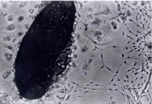
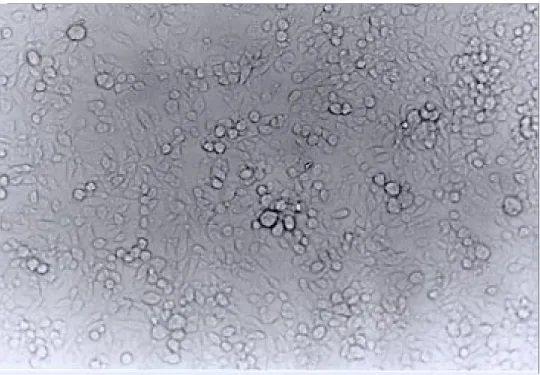
Establishing the cell line - The original Lulo culture was initiated on the 12thof July 1996 from embryonated eggs that had been incubated for six days. Explanting was carried out after ten days; some cells then started to adhere to the surface of the culture flask. These started to slowly increase in number and gave origin to cells having differ-ent morphology. The third week after the explant had been carried out, the culture was fed weekly with 2 ml of fresh culture medium. The first sub-culture was carried out on the 45th day, using a rubber policeman to remove the cells from the ture bottle surface and putting them into fresh ture medium in a 1:2 proportion. The Lulo cul-tures were initially composed of a heterogeneous cell population, being consistently elongated, spherical, irregular, small and presenting vesicles constituted by epithelioid cells. Some monolayers having fibroblastoid cells predominated in the first subcultures, while epithelioid cells were more nu-merous in others. In higher passages, there was greater cell morphology uniformity, epithelioid types being predominant (Figs 1, 2, 3). In some larval explants it was observed that cells began to appear towards the edges of the fragments but these
106 106 106 106
106 New Continuous Cell Line from Lu. longipalpis Gloria J Rey et al.
Fig. 2: different size vesicles formed by epithelioid cell aggregation around an empty space. These vesicles were produced in early Lulo subcultures.
107 107107 107107 Mem Inst Oswaldo Cruz, Rio de Janeiro, Vol. 95(1), Jan./Feb. 2000
were incapable of adapting to the medium and sub-sequently degenerated. After embryonic tissue ex-planting, the cells have been kept in MM/VP12 medium, with variations in FBS concentration; 20% FBS during the first 12 subcultures, then the concentration was gradually reduced until 10% was reached after 20 passages had been made. The op-timum pH range for cell growth in MM/VP12 medium was 6.7 to 6.9 and the average incubation temperature was 28oC. The cells did not show bac-terial, fungal, mycoplasmic nor viral contamina-tion. The viability of frozen cells was shown at two weeks and at six months after being frozen. Thawing was carried out quickly, removing a vial from liquid nitrogen and plunging it immediately into a serological bath at 37oC. The Lulo growth curve between passages 38 to 40 showed that there was one generation every 25 h.
Karyotype characteristics - The metaphases obtained from cells corresponded to early subcul-tures and to the cell line, exhibiting a diploid num-ber of eight chromosomes (2n = 8), registering haploid, aneuploid and tetraploid presence, each one having a 5% frequency, while 85% were dip-loid. Chromosome pair one was classified as be-ing subtelocentric, while pairs 2, 3 and 4 were metacentric.
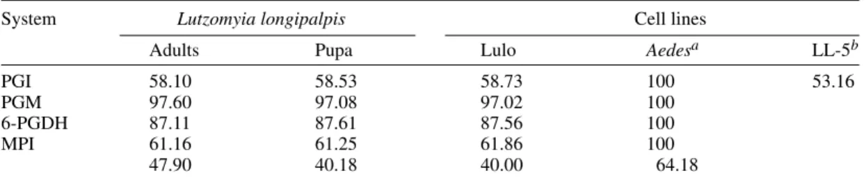
Isoenzyme profiles - The isoenzymatic pheno-types of Lulo, corresponding to the four analyzed
systems, were a band for PGI, PGM and 6-PGDH and two bands for MPI. These results also corre-sponded to the pupa and adult samples coming from the same species and colony. In Table II the aver-age values for electrophoretic mobility relating to each one of the isoenzymatic systems of Lulo are shown, taking an Ae. taeniorynchus cell line as a pattern and also comparing the PGI system with the Lu. longipalpis (LL-5) cell line.
Arbovirus susceptibility - Lulo was sensitive to infection by three of the five inoculated arbovi-ruses, representing the Togaviridae, Reoviridae and Rhabdoviridae families. The best replication ca-pability was obtained using the Vesicular Stomati-tis virus in the cell substrates which were used, while the Ilheus and Punta Toro viruses did not replicate in Lulo. None of the viruses which were examined produced cytopathic effects in the cul-tures (Table I).
Lulo interaction with Leishmania promas-tigotes - The parasites multiplied themselves in Lulo but in less quantity than that observed in Schneider’s medium, however, they conserved motility and roseate formation characteristics. By contrast, after the fifth day of being in contact with parasites, the cells started to become round and to break away from the substrate, until cell death was finally produced towards the tenth day of incuba-tion.
TABLE I
Arbovirus growth in insect cells
Family Genus Virus Cell line
AA C6/36 Lulo
Togaviridae Alfavirus Mayaro - + (10-3)a
Flaviviridae Flavivirus Ileus + (10-2)
-Reoviridae Orbivirus Changuinola - + (10-2)
Bunyaviridae Phlebovirus Punta Toro -
-Rhabdoviridae Vesiculovirus VSV + (10-3) + (10-4)
a: greater virus dilution showing greater infection capacity.
TABLE II
Relative electrophoretic mobility for the four isoenzymes used in the study
System Lutzomyia longipalpis Cell lines
Adults Pupa Lulo Aedesa LL-5b
PGI 58.10 58.53 58.73 100 53.16
PGM 97.60 97.08 97.02 100
6-PGDH 87.11 87.61 87.56 100
MPI 61.16 61.25 61.86 100
47.90 40.18 40.00 64.18
108 108 108 108
108 New Continuous Cell Line from Lu. longipalpis Gloria J Rey et al.
DISCUSSION
The MM/VP12 medium with 20% FBS, at an incubation temperature of 28oC and a pH of 6.7 to 6.9, provided the necessary environmental and nutritional conditions for explant survival and for new cell growth. Due to this, subcultures contin-ued to be carried out in MM/VP12 medium, re-ducing the FBS concentration in greater passages to 5%. Although the MM/VP12 medium was also used in the initiation of the LL-5 cell line (Tesh & Modi 1983) the FBS concentration was kept at 15% and the culture initiated with eggs laid 3-5 days before. Furthermore, differences were observed in the primary culture, while in LL-5 the primary ad-herent cells became evident 14 days after initiat-ing explant and the first subculture was made on the 107th day, a weekly pass being made thereaf-ter. The first adherent cells were obtained in Lulo 10 days after explant and their first subculture was carried out in less time (45 days), slowly dimin-ishing the time required to establish pass frequency to a week, after the twentieth passage. The forego-ing could be due to the particular characteristics of the Lu. longipalpis populations used; this being different to other subcultures obtained from Culex theileri (Oelofsen et al. 1990), Toxorhynchites amboinensis (Tesh 1980) and Anopheles gambiae
(Marhoul & Pudney 1972) mosquitoes which lasted for less time in similar conditions. Another differ-ence between the two phlebotomine established cell lines was in the morphological characteristics of the culture; the Lulo cell line was composed of epi-thelioid cells, while the presence of two types of cells was reported in LL-5: some small epithelioids and some other bigger fibroblastoids.
The Lulo karyotype, whose diploid number was eight chromosomes, coincided with that reported for this species and was found to be within the observed range of 2n = 6 to 2n = 10 for phlebotomines from the Old and the New Worlds (Carvalho et al. 1962, Kreutzer et al. 1987, 1988). The morphological char-acteristics of the Lulo chromosomes showed pair one to be subtelocentric, while pairs 2, 3 and 4 were metacentric, there being little difference in size be-tween pairs 3 and 4. None of the four chromosome pairs showed heteromorphism. A low percentage of cells in metaphase was seen with 85% of the cells diploid and the remainder heteroploid, aneuploid and haploid. Lulo can thus be considered as a diploid cell line.
Isoenzymatic profiles coincided with pupa and adult samples in four different systems from the same species and the same colony, showing the origin of Lulo from the embryonic tissue of the insect. The comparison of this cell line in the PGI system with LL-5 from Lu. longipalpis (originat-ing from a colony established with material
com-ing from Belo Horizonte, Brazil) gave different results. This suggests that, in cell cultures, the spe-cies (derived from two South American popula-tions) could also present genetic polymorphism, sustaining the idea that it is a complex of species (Ward et al. 1983, Lanzaro et al. 1993, 1998).
Other interesting data provided by the study revealed that alphavirus (Mayaro) infected Lulo cells, while no evidence of infection was found in AA C6/36, in spite of the fact that positivity has been reported by Mezencio et al. (1989) in their experiments involving this cell clone; which could probably be due to a modification of the specific clone C6/36 cell employed in the present study. The results obtained with Punta Toro virus (phlebovirus) were unexpected, due to the fact that it did not produce infection in the Lulo cells nor in the AA C6/36 cell line, the same negative results having been described in LL-5 by Tesh and Modi in 1983. The explanation could lie in the fact that the sand-fly is composed of different cell popula-tions, while the established cell line generally con-tains a single cell population. For example, Igarashi et al. (1973) showed differences in the Japanese encephalitis virus’ ability to replicate in Ae. aegypti
cells; Igarashi also showed growth variations in chikungunya and dengue viruses in selected Ae. albopictus clones’ cells. These observations sug-gest that for arbovirus growth, the type of predomi-nant cell in the cell culture must be as important a factor as the species providing the origin of cul-ture. Two of the three arbovirus isolated from phlebotomie sand flies (Changuinola and Vesicu-lar Stomatitis), and used in cell culture infection demonstrated the ability to replicate in Lulo, sug-gesting that this cell line could be used for future indigenous virus isolation, associated with sand flies and, extensively, with other arthropod vec-tors, to be considered from the perspective of also supporting viral characterization studies at molecu-lar level.
In the Lulo interaction with Leishmania
promastigote assays, the fact that the parasites con-served their viability and could multiply them-selves, was not due to the MM/VP12 medium, but rather to some type of relationship which the para-site established with the cell, perhaps taking some growth factors or ions from it, which (such as iron) are fundamental substances for parasite preserva-tion (Wilson et al. 1994).
This preliminary description of the behavior of
cul-109 109109 109109 Mem Inst Oswaldo Cruz, Rio de Janeiro, Vol. 95(1), Jan./Feb. 2000
ture physical-chemical factors; this situation will be achieved only after further experiments has been carried out to determine parasite differentiation and maturity. From the in vitro co-culturing system (i.e. Lulo with the parasite) research can be developed concerning Leishmania metabolism, anti-parasitic drug evaluation and antigen production which can be used in diagnostic tests and vaccines. These activities will constitute a great support for studies of parasite which is currently being carried out in other cell substrates derived from the tissues of humans and other mammals (Kiderlen et al. 1986, Vouldoukis et al. 1986, Carvalho et al. 1989, Konecny et al. 1999, Mukhopadhyay et al. 1999, Nandan et al. 1999).
Other potential Lulo applications could be pre-sented at physiological and genetic investigation levels, in a similar way to those effected in other lines derived from insects (Leake 1997).
ACKNOWLEDGEMENTS
To Robert Tesh of the University of Texas Medical Branch, Department of Pathology, for his valuable col-laboration with the donation of the virus and antisera used in this study.
REFERENCES
Bello F, Boshell J, Rey G, Morales A, Olano V 1995. Initiation of primary cell cultures from embryos of the mosquitoes Anopheles albimanus and Aedes taeniorhynchus (Diptera: Culicidae). Mem Inst Oswaldo Cruz90: 547-551.
Brown S, Knudson D 1980. Characterization of inverte-brate cell lines. In Vitro16: 829-832.
Carvalho de H, Falcão A, Schreiber G 1962. Cariotipo dos Phlebotomus. Ciênc Cult 14: 38.
Carvalho EM, Bacellar O, Barral A, Badaró R, Johnson WD Jr 1989. Antigen specific immunosuppression in visceral leishmaniasis is cell mediated. J Clin In-vest 83: 860-864.
Corredor A, Kreutzer R, Tesh R, Boshell J, Palau M, Cáceres E, Duque S, Peláez D, Rodriguez G, Nicholls S, Hernández C, Morales A, Ferro de Carrasquilla C 1990. Distribution and etiology of leishmaniasis in Colombia. Am J Trop Med Hyg42: 206-214. Ferro C, Pardo R, Torres M, Morrison A 1995. Larval
microhabitats of Lutzomyia longipalpis (Diptera: Psychodidae) in an endemic focus of visceral leihmaniasis in Colombia. J Med Entomol34: 719-728.
Freshney R 1987. Culture of Animal Cells. A Manual of Basic Technique, 2nd ed., Wiley-Liss, New York, 397 pp.
Grace T 1966. Establishment of a line of mosquito (Aedes aegypti L.) cells grown in vitro. Nature 211: 366-367.
Grimaldi G, Tesh R 1993. Leishmaniases of the New World: current concepts and implications for future research. Clin Microb Rev6: 230-250.
Grimaldi G, Tesh R, McMahon-Pratt D 1989. A review
of the geografic distribution and epidemiology of leishmaniasis in the New World. Am J Trop MedHyg 41: 687-725.
Hink W 1979. Cell lines from invertebrates. In W Jakoby & I Pastan (eds), Methods in Enzimology, Vol. LVII, Academic Press, San Diego, USA, p. 450-466. Hsu S, Li S, Cross J 1972. A cell line derived from
ova-rian tissue of Culex tritaeniorhynchussummorosus
Dyar. J Med Entomol9: 86-91.
Hummel K, Erdman D, Heath J, Bellini W 1992. Baculovirus expression of the nucleoprotein gene of measles virus and utility of the recombinant protein in diagnostic enzyme immunoassays. J Clin Microbiol30: 2874-2880.
Igarashi A 1978. Isolation of Singh’s Aedes albopictus
cell clone sensitive to dengue and chikungunya vi-ruses. J Gen Virol40: 531-544.
Igarashi A, Sasao F, Wungkobkiat S, Fukai K 1973. Growth of Japanese encephalitis virus in established lines of mosquito cells. Biken J16: 17-23. Kiderlen AF, Baccarini M, Lohmann Matthes ML 1986.
A fast and objetive assay for cell mediated intra and extracellular killing of Leishmania promastigotes. J Immunol Methods 86: 199-203.
Konecny P, Stagg AJ, Jebbari H, English N, Davidson RN, Knight SC 1999. Murine dendritic cells inter-nalize Leishmania major promastigotes, produce IL-12 p40 and stimulate primary T cell proliferation in vitro. Env J Immunol29: 1803-1811.
Kreutzer R, Morales A, Cura E, Ferro C, Young D 1988. Brain cell karyotypes of six new world sand flies (Diptera: Psychodidae). J Am Mosq Cont Ass4: 453-455.
Kreutzer R, Modi G, Tesh R, Young D 1987. Brain cell karyotypes of six species of New and Old World sand flies (Diptera: Psychodidae). J Med Entomol 24: 609-612.
Lanzaro G, Alexander B, Mutebi J, Montoya-Lerma J, Warburg A 1998. Genetic variation among natural and laboratory colony populations of Lutzomyia longipalpis (Lutz & Neiva 1912) (Diptera: Psychod-idae) from Colombia. Mem Inst Oswaldo Cruz93: 65-69.
Lanzaro G, Ostrovska K, Herrero M, Lawyer P, Warburg A 1993. Lutzomyia longipalpis is a species complex: Genetic divergence and inter-specific hybrid steril-ity among three populations. Am J Trop Med Hyg 48: 839-847.
Leake CJ 1997. Establishing primary cell cultures from disease vectors and maintenance of continuous cell lines. In JM Crampton, CB Beard & C Louis (eds),
The Molecular Biology of Insect Disease Vectors, Chapman & Hall, London, p. 487-499.
Marhoul Z, Pudney M 1972. A mosquito cell line (Mos. 55) from Anopheles gambiae larvae. Trans R Soc Trop Med Hyg66: 183-184.
McAteer J, Davis J 1994. Basic cell culture technique and the maintenance of cell lines. In J Davis, Basic Cell Culture. A practical Approach, Oxford Univer-sity Press Inc., New York, p. 93-148.
110 110 110 110
110 New Continuous Cell Line from Lu. longipalpis Gloria J Rey et al.
albopictus cells: an electron microscopic study. Arch Virol 104: 299-308.
Mukhopadhyay S, Sen P, Bhattacharyya S, Majumdar S, Ray S 1999. Immunoprophylaxis and immuno-therapy against experimental visceral leismaniasis.
Vaccine 17: 291-300.
Nandan D, Lo R, Reiner NE 1999. Activation of phosphotyrosine phosphatase activity attenuates mitogen-activated protein kinase signaling and in-hibits c-FOS and nitric oxide synthase expression in macrophages infected with Leishmania donovani. In-fect Immun 67: 4055-4063.
Oelofsen M, Gericke A, Smith M, De K Van der Linde T 1990. Establishment and characterization of a cell line from the mosquito Culex (Culex) theileri
(Diptera: Culicidae) and its susceptibility to infec-tion with arboviruses. J Med Entomol 27: 939-944. Russell W, Newman C, Williamson D 1975. A simple cytochemical technique for demonstrations of DNA in cells infected with mycoplasmas and viruses.
Nature2: 461-462.
Schneider I 1987. Preparation and maintenance of ar-thropod cell cultures: Diptera, with emphasis on mosquitoes. In CE Yunker, Arboviruses in Arthro-pod Cells in Vitro, Vol. 1., CRC Press, Boca Ratón, Fl, p. 31.
Tesh R, Guzman H 1996. Sand flies and the agents they transmit. In BJ Beaty & WC Marquardt (eds), The Biology of Disease Vectors, University Press of Colo-rado, USA, p. 117-127.
Tesh R, Boshell J, Young D, Morales A, de Carrasquilla C, Corredor A, Modi G, Travassos da Rosa APA, McLean R, de Rodríguez C, Gaitan M 1989. Char-acterization of five new phleboviruses recently iso-lated from sand flies in tropical America. Am J Trop Med Hyg 40: 529-533.
Tesh R, Boshell J, Modi G, Morales A, Young D, Corredor A, Ferro C, de Rodriguez C, Walters L, Gaitán M 1987. Natural infection of humans, ani-mals, and phlebotomine sand flies with the Alagoas serotype of vesicular stomatitis virus in Colombia.
Am J Trop Med Hyg 36: 653-661.
Tesh R, Boshell J, Young D, Morales A, Corredor A, Modi G, Ferro C, de Rodriguez C, Gaitán M 1986. Biology of Arboledas virus, a new phlebotomus fe-ver serogroup virus (Bunyaviridae: Phlebotovirus)
isolated from sand flies in Colombia. Am J Trop Med Hyg 35: 1310-1316.
Tesh R, Modi G 1983. Development of a continuous cell line from the sand-fly Lutzomyia longipalpis (Diptera: Psychodidae), and its susceptibility to infection with arboviruses. J Med Entomol20: 199-202.
Tesh R 1980. Establishment of two cell lines from the mosquito Toxorhynchites amboinensis (Diptera: Culicidae) and their susceptibility to infection with arboviruses. J Med Entomol 17: 338-343.
Tesh R 1979. A method for the isolation and identifica-tion of dengue viruses, using mosquito cell cultures.
Am J Trop Med Hyg28: 1053-1059.
Varma M, Pudney M, Leake C, Peralta P 1976. Isola-tions in a mosquito (Aedes pseudoscutellaris) cell line (Mos 61) of yellow fever virus strains from origi-nal field material. Intervirology 6: 50-56.
Varma M, Pudney M 1969. The growth and serial pas-sage of cell lines from Aedes aegypti (L.) larvae in different media. J Med Entomol 6: 432-439. Vouldoukis I, Alfred C, Monjour L, Mazier D,
Brandicourt O, Ploton I, Tselentis Y, Nzuzi KK, Gentilini M 1986. Culture systems for production of promastigote and amastigote forms of Leishma-nia application to serological diagnosis and thera-peutic trials. Ann Parasitol Hum Comp 61:147-154. Ward R, Ribeiro A, Ready P, Murtagh A 1983. Repro-ductive isolation between different forms of
Lutzomyia longipalpis (Lutz and Neiva), (Diptera: Psychodidae), the vector of Leishmania donovani
Chagas/Cunha and Chagas and its significance to Kala-Azar distribution in South America. Mem Inst Oswaldo Cruz78: 269-280.
Wilson M, Vorhies R, Andersen K, Britigan B 1994. Acquisition of iron from transferrin and lactoferrin by the protozoan Leishmania chagasi.Infect Immun 62: 3262-3269.
Young D, Duncan M 1994. Guide to identification and geografic distribution of Lutzomyia and sand-flies in Mexico, West Indies, Central and South America (Diptera: Psychodidae). Mem Amer Entomol Inst54: 1-881.
Yuval B, Warburg A 1989. Susceptibility of adult phlebotomine sandflies (Diptera: Psychodidae) to