Positive selection, molecular recombination structure and phylogenetic
reconstruction of members of the family Tombusviridae:
Implication in virus taxonomy
Moncef Boulila
Institut de l’Olivier, Sousse Ibn-khaldoun, Tunisia.
Abstract
A detailed study of putative recombination events and their evolution frequency in the whole genome of the currently known members of the family Tombusviridae, comprising 79 accessions retrieved from the international databases, was carried out by using the RECCO and RDP version 3.31balgorithms. The first program allowed the detection of potential recombination sites in seven out of eight virus genera (Aureusvirus, Avenavirus, Carmovirus, Dianthovirus, Necrovirus, Panicovirus, and Tombusvirus), the second program provided the same results except for genus Dianthovirus. On the other hand, both methods failed to detect recombination breakpoints in the genome of mem-bers of genusMachlomovirus. Furthermore, based on Fisher’s Exact Test of Neutrality, positive selection exerted on protein-coding genes was detected in 17 accession pairs involving 15 different lineages. Except genera Machlomovirus, and Panicovirus along with unclassified Tombusviridae, all the other taxonomical genera and the unassignedTombusviridae encompassed representatives under positive selection. The evolutionary history of all members of the Tombusviridae family showed that they segregated into eight distinct groups corresponding to the eight genera which constitute this family. The inferred phylogeny reshuffled the classification currently adopted by the International Committee on Taxonomy of Viruses. A reclassification was proposed.
Key words:bioinformatics, phylogeny, recombination, positive selection, Tombusviridae, sequence, taxonomy. Received: November 22, 2010; Accepted: May 13, 2011.
Introduction
RNA recombination is one of the major factors re-sponsible for the generation of new RNA viruses and retro-viruses. The biological mechanisms of recombination dif-fer across organisms, but in broad terms recombination results in the creation of mosaic sequences where the evolu-tionary history at each site may be different. Recombina-tion, defined as the exchange of genetic information be-tween two nucleotide sequences, is an important process that influences biological evolution at many different lev-els. Recombination explains a considerable amount of ge-netic diversity in natural populations and, in general, genes located in regions of the genome with low levels of recom-bination have low levels of polymorphism (Posada and Crandall, 2001). Recombination reshuffles existing varia-tion and even creates new variants. It has been shown that RNA recombination enables the exchange of genetic mate-rial, not only between the same or similar viruses but also between distinctly different viruses (Worobey and Holmes, 1999). Sometimes, it also permits crossovers between viral and host RNA (Greene and Allison, 1994; Aaziz and
Tep-fer, 1999; Barothet al., 2000; Nagaiet al., 2003). Taking into account the structure of viral genomic molecules and the location of crossover sites, three basic types of RNA re-combination were distinguished: homologous, aberrant ho-mologous and non-hoho-mologous (Lai, 1992; Alejskaet al., 2001). The former two occur between two identical or simi-lar RNAs (or between molecules displaying local homo-logy), while the latter involves two different molecules. Most of the collected data suggest that RNA recombinants are formed according to a copy choice model (Alejskaet al., 2001). A viral replication complex starts nascent RNA strand synthesis on one template, called RNA donor, and then switches to another template, called RNA acceptor. Accordingly, two main factors are thought to affect RNA recombination: the structure of recombining molecules and the ability of the viral replicase to switch templates. Through generations, viral populations evolve under vari-ous selective forces at different regions and sites that dis-play different functional constraints. A stringent and robust criterion for detecting adaptive evolution in a protein-coding gene is an accelerated nonsynonymous (dN, amino acid replacing) rate relative to the synonymous (dS, silent) rate of substitutions, with the rate ratiow=dN/dS> 1. As si-lent mutations do not change the amino acid whereas re-Genetics and Molecular Biology, 34, 4, 647-660 (2011)
Copyright © 2011, Sociedade Brasileira de Genética. Printed in Brazil www.sbg.org.br
Send correspondence to Moncef Boulila. Institut de l’Olivier, B.P. 14, 4061 Sousse Ibn-Khaldoun, Tunisia. E-mail: [email protected].
placement mutations do, the difference in their fixation rates provides a measure of selective pressure on the pro-tein.
Amongst positive-strand plant RNA viruses, the fam-ily Tombusviridae encompasses several viruses with an im-portant economical impact. According to the 8th ICTV (International Committee on Taxonomy of Viruses) report (Fauquetet al., 2005), the family Tombusviridae includes the following genera: Tombusvirus, Carmovirus,
Necrovirus, Dianthovirus, Machlomovirus, Avenavirus,
AureusvirusandPanicovirus. According to the Baltimore classification, the viruses in this family are classified as Type IV viruses, and are part of the luteovirus supergroup (Habili and Symons, 1989). The RNA is contained in an icosahedral (T = 3) capsid, composed of 180 units of a sin-gle coat protein 27-42 kDa in size; the virion measures 28-35 nm in diameter, and is not enveloped. All Tombusviridae have a positive- sense, single-stranded lin-ear genome, with the exception of dianthoviruses, whose genome is bipartite. The genome is approximately 4-5.4 kb in length, depending on the genus. The 3’ terminus is not polyadenylated. The 5’ terminus is capped only in Carna-tion mottle carmovirus, Red clover necrotic mosaic dianthovirusand Maize chlorotic mottle machlomovirus. The genome encodes 4-6 ORFs. The polymerase ORF en-codes an amber stop codon that is the site of a readthrough event within ORF 1 (except in dianthoviruses, where readthrough occursviaa frameshift), producing two prod-ucts necessary for replication. There is no helicase encoded by the virus. The replication process of members of family Tombusviridae comprises the following steps: (i) the virus penetrates into the host cell, (ii) the viral genomic RNA is uncoated and released into the cytoplasm, (iii) the viral RNA is translated to produce the two proteins necessary for RNA synthesis (replication and transcription), (iv) a nega-tive-sense complementary ssRNA is synthesized using the genome RNA as a template, (v) a new genomic RNA is synthesized using the negative-sense RNA as a template, (vi) the RNA-dependant RNA polymerase (RdRp) recog-nizes internal subgenomic promoters on the negative-sense RNA, to transcribe the 3’ co-terminal subgenomic RNAs that will generate the capsid and movement protein, (vii) new virus particles are formed (White and Nagy, 2004).
The main objective of this work was to determine and characterize virus evolution mechanisms of the Tombusviridae based on the occurrence of putative recom-bination events and positive selection in their full-length genome. This was achieved by the analysis of 79 accessions obtained from GenBank. As a result, we propose a reclassi-fication according to their predicted evolutionary history.
Material and Methods
The sequences of the entire genome of 79 accessions cataloged in GenBank were used in this study (Table 1).
The nucleotide sequences were aligned using pro-grams CLUSTALW 2.0.9 and CLUSTALX 2.0.9 (Larkin
et al., 2007) with default configuration. Their phylogenetic relationships were determined with the Maxi-mum-likelihood (ML) algorithm incorporated in the MEGA version 5 program (Tamuraet al., 2011) under as-sumption of the substitution models proposed by Jukes and Cantor (1969) (JC), Hasegawaet al.(1985) (HKY85), and Tamura and Nei (1993) (TN93). Bootstrap analyses with 500 replicates were performed to assess the robustness of the branches.
Using the MEGA4.1bprogram (Kumaret al., 2008), positive selection was inferred by the counting method de-scribed by Nei and Gojobori (1986) and, later on, by Suzuki and Gojobori (1999). According to this method, the phylo-genetic tree of sequences analyzed was used. For the parsi-mony method, the total numbers of synonymous (cS) and nonsynonymous (cN) substitutions as well as the average numbers of synonymous (sS) and nonsynonymous (sN) sites per codon over the phylogenetic tree for each codon site were computed according to the maximum parsimony prin-ciple (Fitch, 1971; Hartigan, 1973). The null hypothesis of selective neutrality (rS=rNorw= 1) was tested for each site by computing the probability (p) of obtaining the observed or more biased values forcSandcN, which were assumed to follow a binomial distribution with the probabilities of oc-currence of synonymous and nonsynonymous substitutions given bysS/(sS+sN) andsN/(sS+sN), respectively. Positive selection is inferred when p < 0.05 andcN/sN>cS/sS (Su-zuki, 2006).
Potential recombination events between diverged nu-cleotide sequences were explored using two programs: RDP v3.31b(Martinet al., 2005b) and RECCO (Maydt and Lengauer, 2006). RDP incorporates several published re-combination detection methods into a single suite of tools: RDP (Martin and Rybicki, 2000), GENECONV (Padidam
et al., 1999), BOOTSCAN (Martin et al., 2005a), MAXCHI (Smith, 1992), CHIMAERA (Posada and Cran-dall, 2001), SISCAN (Gibbset al., 2000), and 3SEQ (Boni
et al., 2007). In all cases, default parameters were used. Only events predicted by more than half of the methods are considered as significant. The algorithm developed and de-scribed by Maydt and Lengauer (2006) as being a fast, sim-ple and sensitive method for detecting recombination in a set of sequences and locating putative recombination breakpoints is based on cost minimization. This method has only two tunable parameters, recombination and mutation cost. In practice the only parameter considered isa, repre-senting the cost of mutation relative to recombination. Whenachanges from 0 to 1, the cost of mutation weighted byaincreases, and the cost for recombination weighted by 1 -adecreases. In other words, parameteracontrols the ambiguity between mutation and recombination.
Tombusviridae evolution 649
Table 1- Current taxonomic status of the members of the family Tombusviridae included in the study and their accession numbers.
Genus (in bold), unclassified and unassigned viruses
Virus/Isolate GenBank accession number
Aureusvirus Cucumber leaf spot virus (CLSV) NC_007816
Cucumber leaf spot virus/Canada (CLSV/Canada) EU127904
Pothos latent virus/Pigeonpea (PoLV/Pigeonpea) NC_000939 Johnsongrass chlorotic stripe mosaic virus/Iran (JCSMV/Iran) NC_005287 Maize white line mosaic virus/USA (MaWLMV/USA) NC_009533
Avenavirus Oat chlorotic stunt virus (OCSV) NC_003633
Carmovirus Cardamine chlorotic fleck virus (CCFV) NC_001600
Carnation mottle virus/China (CarMoV/China) NC_001265
Carnation mottle virus (CarMoV) X02986
Carnation mottle virus/Indian (CarMoV/Indian) AJ811998
Cowpea mottle virus (CPMoV) NC_003535
Hibiscus chlorotic ringspot virus (HCRSV) NC_003608 Hibiscus chlorotic ringspot virus.Tw (HCRSV.Tw) DQ392986
Japanese iris necrotic ring virus (JINRV) NC_002187 Melon necrotic spot virus/Yamaguchi (MeNSV/Yamaguchi) AB250687
Melon necrotic spot virus/Nagasaki (MeNSV/Nagasaki) AB250686 Melon necrotic spot virus/Kochi (MeNSV/Kochi) AB250685 Melon necrotic spot virus/Chiba (MeNSV/Chiba) AB250684
Melon necrotic spot virus/Tottori (MeNSV/Tottori) AB232925 Melon necrotic spot virus/Kochi2 (MeNSV/Kochi2) AB232926
Melon necrotic spot virus (MeNSV) NC_001504 Melon necrotic spot virus/MNSV-ISR (MeNSV/MNSV-ISR) DQ922807 Melon necrotic spot virus/MNSV-Al (MeNSV/MNSV-Al) DQ339157
Melon necrotic spot virus/MNSV264 (MeNSV/MNSV264) AY330700 Melon necrotic spot virus/nK (MeNSV/nK) AB044292
Melon necrotic spot virus/NH (MeNSV/NH) AB044291 Melon necrotic spot virus/Malfa5 (MeNSV/Malfa5) AY122286 Pea stem necrosis virus/Japan (PSNV/Japan) NC_004995
Pelargonium flower break virus/MZ10 (PFBV/MZ10) NC_005286 Pelargonium flower break virus/SP18 (PFBV/SP18) DQ256073
Saguaro cactus virus (SCV) NC_001780
Turnip crinkle virus (TCV) NC_003821
Turnip crinkle virus/UK (TCV/UK) AY312063
Unclassified Angelonia flower break virus/Florida (AFBV/Florida) NC_007733
Carmoviruses Soybean yellow mottle mosaic virus/MS1-USA (SYMoMV/MS1-USA) FJ707484
Soybean yellow mottle mosaic virus/South Korea (SYMoMV/s.Korea) NC_011643
Dianthovirus Carnation ringspot virus RNA 1 (CarRSV-RNA 1) NC_003530
Carnation ringspot virus RNA 2 (CarRSV-RNA 2) NC_003531
Red clover necrotic mosaic virus RNA 1 (RCNMV-RNA 1) NC_003756 Red clover necrotic mosaic virus RNA 2 (RCNMV-RNA 2) NC_003775
Red clover necrotic mosaic virus RNA 1/Can (RCNMV-RNA 1/Can) AB034916 Red clover necrotic mosaic virus RNA 2/Can (RCNMV-RNA 2/Can) AB034917 Sweet clover necrotic mosaic virus RNA 1/59 (SCNMV-RNA 1/59) NC_003806
Results
Recombination events during Tombusviridae evolution
Examination of the RECCO program output regard-ing the occurrence of recombination events in the complete genome of the Tombusviridae family, revealed that three out of five aureusviruses were putative recombinants (PoLV.Pigeonpea, JCSMV.Iran, MaWLMV.USA). In con-trast, CLSV (unknown isolate) and CLSV.Canada did not show any recombinant signal (Table 2). Within the genus
Aureusvirus, the most frequently recombining virus was PoLV.Pigeonpea (33 putative recombination sites), whereas only 28 possible recombination signals were de-tected in the genome of viruses JCSMV.Iran and MaWLMV.USA. Similarly, the only representative of the genus Avenavirus (OCSV) was a potential recombinant with 175 putative sites. The RDP package confirmed these results for both genera. Among the carmoviruses, 14 out of 30 members were possible recombinants. According to RECCO, the most frequently recombining virus was JINRSV with 134 putative events, while MeNSV.Nagasaki
650 Boulila
Genus (in bold), unclassified and unassigned viruses
Virus/Isolate GenBank accession number
Unclassified dianthovirus Rice virus X RNA 1 (RVX-RNA 1) AB033715
Machlomovirus Maize chlorotic mottle virus (MCMoV) NC_003627
Maize chlorotic mottle virus/Nebraska (MCMoV/Nebraska) EU358605
Necrovirus Beet black scorch virus (BBSV) NC_004452
Beet black scorch virus/Val25-Iran (BBSV/Val25-Iran) EU545828 Beet black scorch virus/CO-USA (BBSV/CO-USA) EF153268
Beet black scorch virus/Xinjiang (BBSV/Xinjiang) AY626780
Leek white stripe virus (LWSV) NC_001822
Olive latent virus 1/Citrus (OLV-1/Citrus) NC_001721
Olive latent virus 1/GM6-Portugal (OLV-1/GM6-Portugal) DQ083996 Tobacco necrosis virus A/FM1B (TNV-A/FM1B) NC_001777
Tobacco necrosis virus A/C (TNV-A/C) AY546104 Tobacco necrosis virus D/Hungarian (TNV-D/Hungarian) NC_003487 Tobacco necrosis virus D/Rhotamsted (TNV-D/Rhotamsted) D00942
Unclassified necrovirus Olive mild mosaic virus/GP-POrtugal (OMMV/GP-Portugal) NC_006939
Panicovirus Panicum mosaic virus (PMV) NC_002598
Tombusvirus Artichoke mottled crinkle virus/Bari (AMoCV/Bari) NC_001339
Carnation Italian ringspot virus (CarIRSV) NC_003500 Cucumber bulgarian latent virus (CBLV) NC_004725
Cucumber necrosis virus (CNV) NC_001469
Cymbidium ringspot virus (CymRSV) NC_003532
Grapevine algerian latent virus/nipplefruit (GALV/nipplefruit) NC_011535
Pear latent virus (PeLV) NC_004723
Tomato bushy stunt virus/Statice (TBSV/Statice) AJ249740
Tomato bushy stunt virus/Nipplefruit (TBSV/Nipplefruit) AY579432 Tomato bushy stunt virus/Pepper (TBSV/Pepper) U80935
Tomato bushy stunt virus/Cherry (TBSV/Cherry) M21958 Unclassified Tombusviruses Lisianthus necrosis virus/L (LNV/L) NC_007983
Lisianthus necrosis virus/Zantedeschia (LNV/Zantedeschia) AM711119
Pelargonium necrotic spot virus (PNSV) NC_005285
UnassignedTombusviridae Maize necrotic streak virus (MaNSV)
Pelargonium line pattern virus/PV-0193 (PLPV/PV-0193)
NC_007729 NC_007017
UnclassifiedTombusviridae Nootka lupine vein clearing virus/Alaska (NLVCV/Alaska) Pelargonium chlorotic ring pattern virus/ GR 57 (PCRPV/GR 57)
Tombusviridae
evolution
651
Table 2- Determination of inferred putative recombination events and their frequency along the sequences of the entire genome of aureusviruses, one avenavirus and carmoviruses. Algorithm RDP v3.31bshowed that only events supported by more than half of the different methods are reported. Nucleotide numbering corresponds to the aligned sequences. Abbreviations: NRS: -number of recombination sites, GIRE: -genomic interval of recombination events (the span of sequences in the viral genome where recombination events were predicted).
Recombination determined by RECCO Recombination determined by RDP v3.31b
Virus.isolate Length of breakpoint Genomic position of the longest breakpoint (size in nucleotide)
Putative parental (Major x Minor)
NRS GIRE (nt) 1 residue 2 residues 3 residues > 3 residues
PoLV.Pigeonpea 33 607-5320 10 8 3 12 2148-2184 (37) OCSV x MeNSV/MNSV-264
CarRSV RNA 1 x MeNSV-Tottori CarRSV RNA 1 x MeNSV-Kochi 2
JCSMV.Iran 28 2457-5288 11 6 3 8 3790-3802 (13) CarRSV RNA 1 x MeNSV-Tottori
CarRSV RNA 1 x MeNSV-Kochi 2
MaWLMV.USA 28 3212-4043 18 3 1 6 3836-3853 (18) OCSV x MeNSV/MNSV-264
CarRSV RNA 1 x MeNSV-Tottori
OCSV 175 766-5052 65 33 23 54 1295-1306 (12)
5036-5047 (12)
TCV.UK x MaWLMV.USA
CCFV 20 894-4681 7 2 3 8 1896-1908 (13) /
CarMoV 5 1349-2124 0 0 1 4 1986-2045 (60) /
CPMoV 14 1316-3143 4 6 2 2 2850-2854 (5) /
JINRSV 134 689-5098 58 30 20 26 1736-1760 (25) /
MeNSV.Nagasaki 2 623-4892 0 0 0 2 4847-4892 (46) /
MeNSV.Kochi 4 1843-4694 1 0 0 3 3866-3907 (42) /
MeNSV.MNSV-Al 3 619-4908 0 0 0 3 4864-4908 (45) /
MeNSV.MNSV-264 5 4940-5131 3 0 2 0 4952-4954 (3)
5129-5131 (3)
/
MeNSV.NK 2 615-4908 0 1 0 1 615-874 (60) SYMoMV/MS1-USA x TBSV.Statice
MeNSV.NH 5 615-4983 1 1 0 3 2070-2151 (82) YMoMV/MS1-USA x TBSV.Statice
MeNSV.Malfa5 13 677-4831 0 0 2 11 1736-1796 (61) /
PSNV.Japan 80 645-5174 34 15 9 22 4225-4266 (42) /
SCV 61 665-4506 28 6 13 14 1887-1898 (12)
2761-2772 (12)
/
and MeNSV.NK had the lowest number of recombination sites (two putative sites). The RDP v3.31balgorithm con-firmed the occurrence of possible recombination events only for accessions MeNSV.NK and MeNSV.NH. Recom-bination investigations of genus Dianthovirus based on RECCO analysis showed that only 80% of them were pos-sible recombinants (CarRSV-RNA 1 and 2, RCNMV-RNA 1 and 2, RCNMV.Can. RNA 1 and 2, SCNMV.59.RNA 1, and RVX.RNA 1). in contrast, SCNMV.59.RNA 2 and SCNMV.38.RNA did not show any putative recombination signals. However, the RDP package did not predict recom-bination in the dianthoviruses (Table 3). Although the most frequently recombining necrovirus was RVX (166 putative sites), RCNMV.RNA 2 had only two putative sites. Based on RECCO analysis, 50% of the necroviruses (BBSV.Val25.Iran, LWSV, TNV-A.FMB, TNV-A.C, TNV-D.Hungarian, and OMMV-GP.Portugal,) were possi-ble recombinants. Conversely, BBSV, BBSV.CO.USA, BBSV.Xinjiang, OLV-1.Citrus, OLV-1.GM6.Portugal, and TNV-D.Rhotamsted were not possible recombinants. These results were congruent with those obtained with the RDP package. While the most frequently recombining vi-rus was LWSV (39 sites), TNV.A.C recombined into two sites. Regarding the sole representative of genus
Panicovirus(PMV), the results obtained by the two meth-ods (RECCO and RDP v3.31b) were incongruent. Indeed, with RECCO, 108 possible sites were detected, whereas no recombination signals were found with the RDP package. A similar situation was observed with regard to the newly proposed carmoviruses (NLVCV.Alaska, PCRPV.GR 57, PLPV.PV-0193). According to RECCO analysis, although NLVCV.Alaska was the most frequently recombining vi-rus (65 sites), PLPV.PV-0193 recombined only into 38 sites (Table 4). Regarding the members of genus
Tombusvirus, there was an agreement between the two methods indicating that 80% of the analyzed accessions were putative recombinants. While CBLV had the highest number of putative recombination signals (67 sites), TBSV.Cherry had only two recombination sites. Further-more, it is noteworthy that the two representatives of genus
Machlomovirus(MCMoV, and MCMoV.Nebraska) were not recombinants as assessed by the two methods of analy-sis used in this study. Seeking for the recombination fre-quency in the genome of the Tombusviridae, two-thirds of the aureusviruses (JCSMV.Iran, and MaWLMV.USA) showed that in most cases, their breakpoint length was a single residue. In contrast, the breakpoint length of most putative recombination sites of PoLV.Pigeonpea was be-tween three and 37 nucleotides (Table 2). Also, the break-point length of the major recombination sites of the single representative of genusAvenavirus(OCSV) consisted of a single residue. In about 50% of the members of the genus
Carmovirus, the length of their most detected recombina-tion sites was a single residue. As opposed to that, the breakpoint interval of the remaining members exceeded
three residues reaching a size as long as 82 residues (MeNSV.NH). In 62% of the investigated dianthoviruses, the breakpoint length exceeded three nucleotides reaching 100 residues (CarRSV.RNA 1) (Table 3). In the necroviruses, the breakpoint interval distribution was simi-lari.e., 50% of the breakpoints consisted of a single residue, while the remaining breakpoints were between three and 77 nucleotides. For the sole member of the genusPanicovirus
(PMV), most of the recombination sites had a breakpoint length of a single residue (45) (Table 3). As for the tombusviruses, 75% showed a breakpoint length exceeding three residues up to 161 nucleotides (AMoCV.Bari) (Table 4).
Nucleotide sequence analysis
Maximum composite likelihood estimate of the nu-cleotide substitution pattern were made using the MEGA4.1b program. The results for Tombusviridae showed that the rates of different transitional substitutions varied from 3.18 to 14.61, and those of transversional sub-stitutions varied from 6.6 to 8.57. The nucleotide frequen-cies were: 0.269 (A), 0.258 (T/U), 0.207 (C), and 0.266 (G). The transition/transversion rate ratios werek1= 1.705 (pur-ines) and k2 = 0.482 (pyrimidines). The overall transi-tion/transversion bias wasR= 0.547, whereR= [AGk1+ TCk2]/[(A+G)(T+C)]. There were a total of 1218 positions in the final dataset. In all these analyses, the codon posi-tions included were first + second + third + noncoding. All positions containing gaps and missing data were excluded from the dataset (complete deletion option).
The MEGA4.1bprogram also incorporates the Taji-ma’s Neutrality Test. The purpose of this test is to indentify sequences which do not fit the neutral theory model at equi-librium between mutation and genetic drift. Tajima’s test compares a standardized measure of the total number of segregating sites (the polymorphic DNA sites) in the sam-pled DNA and the average number of mutations between pairs in the sample. Tajima’s D was determined (D= 5.280926).
Positive selection
The high genetic stability of viruses can be attributed to negative or purifying selection to maintain the functional integrity of the viral genome. The degree of negative selec-tion in genes, or the degree of funcselec-tional constraint for the maintenance of the encoded protein sequence, can be esti-mated, as mentioned above, by the ratio between the nucle-otide diversities in nonsynonymous and synonymous posi-tions (dN/dS). For most coding genes thedN/dSratio is < 1 which is consistent with negative selection against protein change. In contrast, adN/dSratio > 1 may be an indication that adaptive or positive selection is driving gene diver-gence. In this study, pairwise comparisons of all screened accessions showed that, none of the members of the genera
Machlomovirus and Panicovirus, and unclassified
Tombusviridae
evolution
653
Table 3- Determination of inferred putative recombination events and their frequency along the sequences of necroviruses, one panicovirus and dianthoviruses. Algorithm RDP v3.31bshowed that only events supported by more than half of the different methods are reported. Nucleotide numbering corresponds to the aligned sequences. Abbreviations: NRS: -number of recombination sites, GIRE: -genomic interval of re-combination events (the span of sequences in the viral genome where rere-combination events were predicted).
Recombination determined by RECCO Recombination determined by RDP v3.31b
Virus.isolate Length of breakpoint Genomic position of the longest breakpoint (size in nucleotide)
Putative parental (Major x Minor)
NRS GIRE (nt) 1 residue 2 residues 3 residues > 3 residues
CarRSV-RNA 1 19 709-4561 7 3 2 7 2581-2680 (100) /
CarRSV-RNA 2 3 1826-2811 0 0 0 3 1826-1835 (10)
2802-2811 (10)
/
RCNMV-RNA 1 8 1297-4754 0 1 2 5 1479-1533 (55) /
RCNMV-RNA 2 2 2696-2812 1 0 0 1 2803-2812 (10) /
RCNMV-RNA1.Can 7 720-4218 2 0 0 5 1556-1644 (89) /
RCNMV-RNA 2.Can 10 1730-2827 3 1 2 4 1730-1765 (36) /
SCNMV-RNA 1.59 12 858-4443 2 2 1 7 3126-3193 (68) /
RVX-RNA 1 166 439-5107 65 39 19 43 840-855 (16) /
BBSV-Val25.Iran 4 800-4881 2 0 0 2 1435-1468 (34) MeNSV.NH x LNV.Zantedeschia
MeNSV.Malfa5 x MaNSV SCV x CNV MeNSV.Yamaguchi x CNV
LWSV 39 1027-4853 17 9 5 8 4846-4853 (8) MeNSV.NH x LNV.Zantedeschia
SCV x CNV
TNV-A.FM1B 3 2419-2789 3 0 2 1 2419-2496 (77) OLV1.GM6-Portugal x BBSV.Xinjiang
TNV-A.C 2 2275-2619 0 0 0 2 2275-2619 (10) OLV1.GM6-Portugal x BBSV.Xinjiang
LWSV x TNV.D.Rhotamsted
TNV-D.Hungarian 4 2323-4287 2 0 0 2 3959-3964 (6) MeNSV.NH x LNV.Zantedeschia
MeNSV.Malfa5 x MaNSV BBSV x OMMV.GP.Portugal
SCV x CNV
OMMV-GP.Portugal 20 935-4857 6 3 4 7 2116-2145 (30) TNV.A.C x TNV.D.Hungarian
TNV.A.C x TNV.D.Rhotamsted TNV.D.Rhotamsted x TNV.A.FM1B OLV1.GM6-Portugal x TNV.D.Hungarian
654
Boulila
Table 4- Determination of inferred putative recombination events and their frequency along the sequences of the tombusviruses and tentative new members of genusCarmovirus. Algorithm RDP v3.31bshowed that only events supported by more than half of the different methods are reported. Nucleotide numbering corresponds to the aligned sequences. Abbreviations: NRS: -number of recombination sites, GIRE: -genomic interval of recombination events (the span of sequences in the viral genome where recombination events were predicted).
Recombination determined by RECCO Recombination determined by RDP v3.31b
Virus.isolate Length of breakpoint Genomic position of the longest breakpoint (size in nucleotide)
Putative parental (Major x Minor) NRS GIRE (nt) 1 residue 2 residues 3 residues > 3 residues
AMoCV.Bari 27 1074-4774 2 4 3 18 4614-4774 (161) CymRSV x CBLV
GALV.nipplefruit x MeNSV.Yamaguchi
CarIRSV 28 1998-5345 9 5 8 6 4775-4842 (68) TBSV.pepper x PNSV
TBSV.Statice x MeNSV.Tottori AMoCV.Bari x PNSV LNV.Zantedeschia x MaNSV
SCV x CBLV PNSV x TBSV.Pepper CBLV x MeNSV.MNSV-ISR LNV.Zantedeschia x TBSV.Statice
CBLV 67 343-5390 16 17 10 24 1804-1836 (33) TBSV.Pepper x JCSMV.Iran
CarRSV.RNA 1 x MeNSV.Tottori GALV.nipplefruit x MaWLMV.USA
PeLV x MaWLMV.USA LNV.Zantedeschia x MaNSV
CymRSV x MaNSV
CNV 34 417-5280 8 2 6 18 2681-2714 (34) CymRSV x MeNSV
TBSV.Pepper x PNSV CymRSV x SCV LNV.L x TBSV.Pepper
CymRSV x CBLV TBSV.Statice x LNV.L
CymRSV 44 675-5066 9 9 4 22 1719-1767 (49) PNSV x TBSV.Pepper
CarIRSV x CLSV.Israel TBSV.Statice x TCV.UK
CNV x CLSV.Canada CNV x TCV.UK MeNSV.MNSV.ISR x PeLV
GALV.nipplefruit 45 369-5299 7 4 11 23 4481-4532 (52) TBSV.Statice x CNV
TBSV.nipplefruit x MaNSV TBSV.nipplefruit x TBSV.Pepper
TBSV.Statice x PeLV CymRSV x CBLV
CBLV x PNSV CymRSV x PeLV AMoCV.Bari x TBSN.Pepper
Tombusviridae
evolution
655
Recombination determined by RECCO Recombination determined by RDP v3.31b
Virus.isolate Length of breakpoint Genomic position of the longest breakpoint (size in nucleotide)
Putative parental (Major x Minor) NRS GIRE (nt) 1 residue 2 residues 3 residues > 3 residues
TBSV.statice 11 379-2743 1 0 1 9 1902-1931 (30) GALV.nipplefruit x LNV.Zantedeschia
CymRSV x CBLV CymRSV x PeLV TBSV.nipplefruit x TBSV.Pepper TBSV.nipplefruit 7 618-1931 0 0 1 6 1550-1651 (102) GALV.nipplefruit x LNV.Zantedeschia
AMoCV.Bari x GALV.nipplefruit
TBSV.pepper 7 2167-4890 0 0 1 5 4852-4890 (39) CymRSV x CBLV
PeLV x AMoCV.Bari GALV.nipplefruit x LNV.Zantedeschia
TBSV.cherry 2 5324-5345 0 0 2 0 5324-5326 (3)
5343-5345 (3)
CymRSV x CBLV CarIRSV x AMoCV.Bari
CymRSV x PeLV
GALV.nipplefruit x LNV.Zantedeschia
MaNSV 54 547-5334 29 4 7 14 1932-1955 (24) GALV.nipplefruit x MaWLMV.USA
PeLV x MaWLMV.USA
PNSV 24 2025-5032 7 2 3 12 5009-5032 (23) TBSV.Statice x MeNSV.Tottori
CNV x CLSV.Canada AMoCV.Bari x TBSV.Cherry
TBSV.nipplefruit x PeLV LNV.Zantedeschia x MaNSV
TBSV.Statice x CNV CBLV x MeNSV.MNSV.ISR LNV.Zantedeschia x TBSV.Statice
NLVCV.alaska 65 771-5301 28 13 7 17 771-782 (12) /
PCRPV.GR 57 45 1109-5104 11 17 4 13 2307-2316 (20) /
PLPV.PV-0193 38 991-4908 20 7 2 9 2813-2821 (9) /
Tombusviridae was under positive selection. On the con-trary, the generaAureusvirus (JCMSV.Iran), Avenavirus
(OCSV), Carmovirus (CarMoV.China, CarMoV.Indian),
Dianthovirus (CarRSV-RNA 2, RCNMV-RNA 2, SCNMV-RNA 2.59, SCNMV-RNA 2.38), Necrovirus
(BBSV, BBSV.Val25.Iran), Tombuvirus (GALV.nip-plefruit, PeLV, TBSV.Statice, PNSV) along with the unas-signed Tombusviridae (PLPV.PV.0193) were under posi-tive selection (Table 5). It is worth pointing out that, in the viruses with a segmented genome, positive selection was detected only in RNA 2, suggesting that probably reassortment events occurred. All these results were ob-tained by testing neutrality in sequence pairs with Fisher’s Exact Test. The probability of rejecting the null hypothesis of strict-neutrality (dN=dS) in favor of positive selection for each sequence pair was determined. Values of p less than 0.05 were considered significant at the 5% level. The variance of the difference (dN-dS) was computed using the bootstrap method (500 replicates). All analyses were made using the Nei-Gojobori method incorporated in the MEGA program. All positions containing gaps and missing data were excluded from the dataset (complete deletion option). The final dataset comprised a total of 234 positions.
Phylogenetic relationships
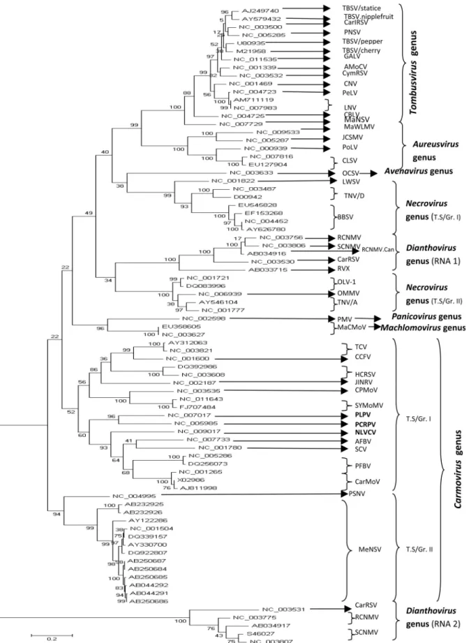
The phylogenetic relationships among members of the family Tombusviridae, based on the sequences of their complete genome, were inferred using a Maximum Likeli-hood algorithm under the assumption of three models of substitution (JC, HKY85, TN93). The topologies of the
constructed trees were identical. The inferred phylogeny showed that each taxonomical genus in the family Tombusviridae constituted a homogenous group clearly distinct from the others. However, the results obtained in this study evidenced a few differences in terms of virus spe-cies composition within each taxonomical genus compared to the current classification adopted by the ICTV. In fact, three viruses considered by the ICTV as unassigned (PLPV.PV-0193) and unclassified Tombusviridae (NLVCV.Alaska, PCRPV.GR 57) showed a close phylo-genetic relationship to known members of the genus
Carmovirus. Moreover, the viruses belonging to this genus were divided into two distinct subgroups. The first sub-group comprised viruses: TCV, CCFV, JINRV, HCRSV, PLPV, PCRPV, NLVCV, SCV, AFBV, PFBV, CPMoV, SYMoMV and CarMoV, and the second subgroup encom-passed viruses: MeNSV, and PSNV. Furthermore, it was proposed that genusNecrovirusshould be constituted by two distinct subgroups named tentative Subgroup I (BBSV, TNV.D, LWSV) and tentative subgroup II (OMMV, TNV.A, OLV-1) (Figure 1). It should be noted that here OMMV is an integral part of subgroup I rather than an un-classifiedNecrovirus. In contrast, genus Aureusvirus en-compassed members that evolved in a homogenous man-ner: CLSV, PoLV, MaWLMV, and JCSMV. Similarly, the following members of genusTombusvirusalso formed a coherent ensemble: MaNSV, CBLV, LNV.L, LNV.Zan-tedeschia, PeLV, CNV, CymRSV, AMoCV, TBSV.Stat-ice, TBSV.Nipplefruit, TBSV.Pepper, TBSV.Cherry, GALV, PNSV, and CarIRSV. Their evolutionary history
656 Boulila
Table 5- Accession pairs in family Tombusviridae under positive selection with probability determined at the 5% level, based on Fisher’s Exact Test of Neutrality and calculateddN-dS.
Accession pairs Virus.isolate pairs p value at 5% level dN-dS
NC_007017/NC_003775 PLPV/PV-0193/ RCNMV-RNA 2 0.028 0.923
AB034917/NC_003633 RCNMV-RNA 2.Can/OCSV 0.016 2.325
NC_003531/NC_003633 CarRSV-RNA 2/ OCSV 0.039 1.820
S46027/NC_003633 SCNMV-RNA 2.38/ OCSV 0.041 1.847
NC_003775/NC_003633 PLPV.PV-0193/ OCSV 0.048 1.975
NC_003807/NC_003633 SCNMV-RNA 2.59/ OCSV 0.033 1.999
AB034917/NC_001265 RCNMV-RNA 2.Can/ CarMoV.China 0.034 1.620
AB034917/AJ249740 RCNMV-RNA 2.Can/ TBSV.Statice 0.029 1.921 NC_003531/NC_005287 CarRSV-RNA 2/ JCSMV.Iran 0.037 1.790 S46027/EU545828 SCNMV-RNA 2.38/ BBSV.Val25-Iran 0.045 1.682
AB034917/AJ811998 RCNMV-RNA 2/ CarMoV.Indian 0.031 1.679 AB034917/NC_011535 RCNMV-RNA 2/ GALV.nipplefruit 0.041 1.933
NC_003775/NC_011535 PLPV/PV-0193/ GALV.nipplefruit 0.033 1.921
NC_003807/NC_005285 SCNMV-RNA 2.59/ PNSV 0.049 1.773
S46027/NC_004723 SCNMV-RNA 2.38/ PeLV 0.047 1.735
S46027/NC_004452 SCNMV-RNA 2.38/ BBSV 0.043 1.749
Tombusviridae evolution 657
reshuffled the existing classification adopted by the ICTV since 2009. In fact, according to this classification, MaNSV was considered as an unassigned Tombusviridae, whereas LNV and PNSV were included in the unclassified
Tombusvirusgroup. Concerning genusDianthoviruswhich clearly was not monophyletic, the clustering pattern showed two distinct clades representing their RNAs 1 and 2, as illustrated in Figure 1. Originally, RVX was consid-ered as an unclassified virus within genusDianthovirus.
Discussion
This study evidenced the prediction of putative re-combination events in the genome of several members of the family Tombusviridae and demonstrated that tombus-viruses and carmotombus-viruses are highly recombinant compared to viruses of the other genera. For this purpose, two meth-ods were chosen (RECCO and RDP v.3.31b), based on the fact that they are appropriate for the mosaic structure of vi-ruses as reported in previous works (Boulila, 2009; 2010). In this study, using the RECCO algorithm, it was demon-strated that the viruses belonging to the following genera contained putative recombination signals in their genome:
Aureusvirus, Avenavirus, Carmovirus, Dianthovirus,
Necrovirus,Panicovirus, andTombusvirus. These results were in good agreement with those obtained by the RDP package except for members of genusDiantovirus. By both methods, the two representatives of genusMachlomovirus
(MCMoV, MCMoV.Nebraska) were found to be non-recombinant. As revealed by RECCO, the most frequently recombining viruses were: OCSV, RVX.RNA 1, JINRSV, and PMV with 175, 166, 134, and 108 putative recombina-tion sites, respectively. All of these recombinarecombina-tion signals were constituted by a single residue. MeNSV.Nagasaki,
MeNSV.NK, RCNMV.RNA 2, TNV-A.C, and
TBSV.Cherry (2 sites), MeNSV.MNSV-Al, CarRSV-RNA 2, and TNV-A.FM1B (3 sites), MeNSV.Kochi, BBSV-Val25.Iran, and TNV-D.Hungarian (4 sites), CarMoV,
MeNSV.MNSV-264, and MeNSV.NH (5 sites),
RCNMV.RNA 1.Can, TBSV.nipplefruit, and TBSV.pep-per (7 sites), and RCNMV.RNA 1 (8 sites) showed the low-est frequency of recombination breakpoints. In contrast, most of these breakpoints had an interval exceeding three nucleotides. Furthermore, this study showed that recombi-nation may occur between viruses belonging to different genera. For example: Oat chlorotic stunt avenavirus
(OCSV) and Melon necrotic spot carmovirus (MeNSV) may give rise toPothos latent aureusvirus(PoLV). Simi-larly, OCSV itself may result from a recombination be-tweenTurnip crinkle carmovirus(TCV) andMaize white line mosaic aureusvirus (MaWLMV) (Table 2). Seem-ingly, these viruses could contain part of their sequences particularly in the coat protein-encoding gene of each other. Such an event was largely studied forCucumber necrosis
tombusvirus (CNV) andMelon necrotic spot carmovirus
(MeNSV) (Riviere and Rochon, 1990).
On the other hand, investigations of selective pres-sure acting on protein expression of virus genes led to the identification of positive selection in 17 accession pairs in-volving 15 different lineages. It is worth mentioning that numerous viruses: JCSMV.Iran, OCSV, CarRSV-RNA 2, RCNMV-RNA 2.Can, BBSV.Val25.Iran, GALV.nip-plefruit, TBSV.Statice, PNSV, and PLPV.PV.0193 evolved under both mechanisms: recombination and posi-tive selection between which synergism might be occur-ring. Such a synergism between recombination and natural selection may have played a major role in Darwinian mo-lecular evolution.
The evolutionary history of the Tombusviridae has shown that the 79 accessions split into eight clearly sepa-rated clusters representing the eight genera of the Tombus-viridae family. From the present phylogenetic study, at least two taxonomic implications can be drawn: (i) three vi-ruses (NLVCV.Alaska, PCRPV.GR 57, PLPV.PV-0193) currently considered by the ICTV as: one unassigned Tombusviridae (PLPV.PV-0193), and two unclassified Tombusviridae (NLVCV.Alaska and PCRPV.GR 57). All of them should be included in genusCarmovirus; (ii) In ad-dition to the viruses belonging to genusCarmoviruswhich have formed two separated subgroups, the members of gen-eraNecrovirus, andDianthovirusevolved separately and divided into two distinct subgroups as shown in Figure 1. In contrast, members of generaAureusvirus, andTombusvirus
formed separately a single ensemble. The evolutionary re-lationships among viruses are a reliable approach for classi-fication. As stated by Stuartet al. (2004) (who reported similar results regarding the genetic divergence of compo-nents of genusNecrovirus), the comparison of complete genomes is a more balanced approach that should provide a more precise scheme of relatedness. On the other hand, it should be pointed out that, in genusDianthovirus, the ge-netic divergence between RNAs 1 and 2 is correlated to the final products synthesized and their use by the virus to sur-vive. For example: RNA silencing is a small RNA-guided sequence- specific gene activation mechanism in eukaryotes that is involved in different biological phenom-ena (e.g.development, heterochromatin formation and de-fense against molecular parasites such as viruses). Many viruses express suppressors to counteract RNA-silencing-mediated antiviral defenses. These RNA silencing suppressors have been identified in the following genera:
Aureusvirus,Carmovirus,Tombusvirus,andDianthovirus
(Voinnete al., 1999; Quet al., 2003; Méraiet al., 2005; Takedaet al., 2005).Dianthovirususes a unique strategy to suppress RNA silencing. The dianthoviral suppressor con-sists of multiple components including P27, P88 (encoded by two ORFs in RNA 1) and viral RNA (Takeda et al., 2005). Moreover, sequence variability of the coat pro-tein-coding gene (RNA 1) may be linked to the interaction
between this structural protein and the host and vector which themselves show a major diversity among diatho-viruses. In contrast, the ORF in RNA 2 encodes the move-ment protein. All these factors can influence the divergence between the two RNAs.
Finally, to the author’s best knowledge, this is the largest study in the literature so far on recombination poten-tially occurring in the entire genome of all currently known members of the family Tombusviridae as well as positive selection operating on protein expression and their phylo-genetic reconstruction. In addition, a reclassification based on their predicted evolutionary history, is proposed.
References
Aaziz R and Tepfer M (1999) Recombination in RNA viruses and in virus-resistant transgenic plants. J Gen Virol 80:1339-1346.
Alejska M, Kurzyniska-Kokorniak A, Broda M, Kierzek R and Figlerowicz M (2001) How RNA viruses exchange their ge-netic material. Acta Bioch Polon 48:391-407.
Baroth M, Orlich M, Thiel HJ and Becher P (2000) Insertion of cellular NEDD8 coding sequences in a pestivirus. Virology 278:456-466.
Boni MF, Posada D and Feldman MW (2007) An exact non-parametric method for inferring mosaic structure in se-quence triplets. Genetics 176:1035-1047.
Boulila M (2009) Recombination structure and genetic related-ness among members of the familyBromoviridaebased on their RNAs 1 and 2 sequence analyses. Virus Genes 38:435-444.
Boulila M (2010) Putative recombination events and evolutionary history of five economically important viruses of fruit trees based on the coat protein-encoding gene sequence analysis. Biochem Genet 48:357-375.
Fauquet CM, Mayo MA, Maniloff J, Desselberger U and Ball LA (2005) Virus taxonomy: Classification and nomenclature of viruses. In: Eighth Report of the International Committee on Taxonomy of Viruses. Elsevier/Academic Press, London, 1259 pp.
Fitch WN (1971) Toward defining the course of evolution mini-mum change for a specific tree topology. Syst Zool 20:406-416.
Gibbs MJ, Armstrong JS and Gibbs AJ (2000) Sister-scanning: A Monte Carlo procedure for assessing signals in recombinant sequences. Bioinformatics 16:573-582.
Greene AE and Allison RF (1994) Recombination between viral RNA and transgenic plants transcripts. Science 263:1423-1425.
Habili N and Symons RH (1989) Evolutionary relationship be-tween luteoviruses and other RNA plant viruses based on se-quence motifs in their putative RNA polymerase and nucleic acid helicases. Nucleic Acids Res 17:9543-9555.
Hartigan JA (1973) Minimum mutation fits to a given tree. Bio-metrics 29:53-63.
Hasegawa M, Kishino H and Yano T (1985) Dating of human-ape splitting by a molecular clock of mitochondrial DNA. J Mol Evol 22:160-174.
Jukes T and Cantor C (1969) Evolution of protein molecules. In: Munro HN (ed) Mammalian Protein Metabolism. Academic Press, New York, pp 21-132.
Kumar S, Nei M, Dudley J and Tamura K (2008) MEGA: A biolo-gist-centric software for evolutionary analysis of DNA and protein sequences. Brief Bioinfor 9:299-306.
Lai MMC (1992) RNA recombination in animal and plant viruses. Microbiol Rev 56:61-79.
Larkin MA, Blackshileds G, Brown NP, Chenna R, McGettigan PA, McWilliam H, Valentin F, Wallace IM, Wilm A, Lopez R et al. (2007) Clustal W and Clustal X v. 2.0. Bioin-formatics 23:2947-2948.
Martin D and Rybicki E (2000) RDP: Detection of recombination amongst aligned sequences. Bioinformatics 16:562-563. Martin DP, Posada D, Crandall KA and Williamson C (2005a) A
modified bootscan algorithm for automated identification of recombination sequences and recombination breakpoints. AIDS Res H Retrovir 21:98-102.
Martin DP, Williamson C and Posada D (2005b) RDP2: Recom-bination detection and analysis from sequence alignments. Bioinformatics 21:260-262.
Maydt J and Lengauer T (2006) Recco: Recombination analysis using cost optimization. Bioinformatics 22:1064-1071. Mérai Z, Kerényi Z, Molnar A, Barta E, Valoczi A, Bisztray G,
Havelda Z, Burgyan J and Silhavy D (2005)AureuvirusP14 is an efficient RNA silencing suppressor that binds double-stranded RNAs without size specificity. J Virol 79:7217-7226.
Nagai M, Sakoda Y, Mori M, Hayashi M, Kida H and Akashi H (2003) Insertion of a cellular sequence and RNA recombina-tion in the structural protein coding region of cytopatho-genic bovine viral diarrhea virus. J Gen Virol 84Pt2:447-452.
Nei M and Gojobori T (1986) Simple methods for estimating the numbers of synonymous and nonsynonymous nucleotide substitutions. Mol Biol Evol 3:418-426.
Padidam M, Sawyer S and Fauquet CM (1999) Possible emer-gence of new geminiviruses by frequent recombination. Vi-rology 265:218-225.
Posada D and Crandall K (2001) Evaluation of methods for de-tecting recombination from DNA sequences. Computer sim-ulation. Proc Natl Acad Sci USA 98:13757-13762. Qu F, Ren T and Morris TJ (2003) The coat protein of Turnip
crin-kle virus suppresses post transcriptional gene silencing at an early initiation step. J Virol 77:511-522.
Riviere CJ and Rochon DM (1990) Nucleotide sequence and genomic organization of melon necrotic spot virus. J Gen Virol 71:1887-1896.
Smith JM (1992) Analyzing the mosaic structure of genes. J Mol Evol 34:126-129.
Stuart G, Moffet K and Bozarth RF (2004) A whole genome per-spective on the phylogeny of the plant virus family Tom-busviridae. Arch Virol 149:1595-1610.
Suzuki Y (2006) Statistical properties of the methods for detecting positively selected amino acid sites. Gene 365:125-129. Suzuki Y and Gojobori T (1999) A method for detecting positive
selection at single amino acid sites.Mol Biol Evol 16:1315-1328.
Tajima F (1989) Statistical-method for testing the neutral muta-tion hypothesis by DNA polymorphism. Genetics 123:585-595.
Takeda A, Tsukuda M, Mizumoto H, Okamoto K, Kaido M, Mise K and Okuno T (2005) A plant RNA virus suppresses RNA silencing through viral RNA replication EMBO J 24:3147-3157.
Tamura K and Nei M (1993) Estimation of the number of nucleo-tide substitutions in the control region of mitochondrial DNA in humans and chimpanzees. Mol Biol Evol 10:512-526.
Tamura K, Peterson D, Peterson N, Stecher G, Nei M and Kumar S (2011) MEGA5: Molecular Evolutionary Genetics Analy-sis using maximum likelihood, evolutionary distance, and maximum parsimony methods. Molecular Biology and Evo-lution. Doi 10.1093/molbev/msr121.
Voinnet O, Pinto YM and Baulcombe DC (1999) Suppression of gene silencing: A general strategy used by diverse DNA and RNA viruses of plants. Proc Natl Acad Sci USA 96:14147-14152.
White KA and Nagy PD (2004) Advances in the molecular biol-ogy of tombusviruses: Gene expression, genome replication and recombination. Prog Nucleic Acid Res Mol Biol 78:187-226.
Worobey M and Holmes EC (1999) Evolutionary aspects of re-combination in RNA viruses. J Gen Virol 80:2535-2543.
Associate Editor: Carlos F.M. Menck
License information: This is an open-access article distributed under the terms of the Creative Commons Attribution License, which permits unrestricted use, distribution, and reproduction in any medium, provided the original work is properly cited.