Simultaneous quantitative assessment of circulating cell-free mitochondrial
and nuclear DNA by multiplex real-time PCR
Peng Xia
1,2, Ramin Radpour
1, Rebecca Zachariah
1, Alex Xiu Cheng Fan
1, Corina Kohler
1, Sinuhe Hahn
1,
Wolfgang Holzgreve
3and Xiao Yan Zhong
11
Laboratory for Prenatal Medicine and Gynaecological Oncology, Women’s Hospital,
University of Basel, Switzerland.
2
Department of Oncology Surgery, First Affiliated Hospital of the Medical College,
Xi’an Jiaotong University, Xi’an, China.
3
University Medical Center Freiburg, Germany.
Abstract
Quantification of circulating nucleic acids in plasma and serum could be used as a non-invasive diagnostic tool for monitoring a wide variety of diseases and conditions. We describe here a rapid, simple and accurate multiplex real-time PCR method for direct synchronized analysis of circulating cell-free (ccf) mitochondrial (mtDNA) and nu-clear (nDNA) DNA in plasma and serum samples. The method is based on one-step multiplex real-time PCR using a FAM-labeled MGB probe and primers to amplify the mtDNA sequence of the ATP 8 gene, and a VIC-labeled MGB probe and primers to amplify the nDNA sequence of the glycerinaldehyde-3-phosphate-dehydrogenase (GAPDH) gene, in plasma and serum samples simultaneously. The efficiencies of the multiplex assays were measured in se-rial dilutions. Based on the simulation of the PCR reaction kinetics, the relative quantities of ccf mtDNA were calcu-lated using a very simple equation. Using our optimised real-time PCR conditions, close to 100% efficiency was obtained from the two assays. The two assays performed in the dilution series showed very good and reproducible correlation to each other. This optimised multiplex real-time PCR protocol can be widely used for synchronized quan-tification of mtDNA and nDNA in different samples, with a very high rate of efficiency.
Key words:circulating cell-free DNA, mitochondrial DNA, nuclear DNA, real-time PCR, quantitative PCR.
Received: June 12, 2008; Accepted: July 24, 2008.
Introduction
Eukaryotic cells have nuclear DNA (nDNA) and addi-tional cytoplasmic mitochondrial DNA (mtDNA). It has been demonstrated that cell-free nucleic acids,i.e., cell-free (ccf) nuclear DNA (cf-nDNA) and ccf mtDNA exist in cir-culation (Sozziet al., 2003). Quantification of ccf nDNA and ccf mtDNA concentrations in plasma and serum has raised great interest as a tool for non-invasive diagnosis and moni-toring of a wide variety of diseases and conditions, such as cancers (Zhong et al., 2007c), pathological pregnancies (Zhong et al., 2001), inflammatory disease (Zhong et al., 2007d) and trauma (Lamet al., 2003). It has been reported that both circulating plasma nDNA and mtDNA were in-creased after trauma (Lamet al., 2003; Lamet al., 2004). Many studies observed elevated levels of ccf nDNA in plasma or serum of various cancers (Allen et al., 2004; Gormally et al., 2004; Sozzi et al., 2003; Taback et al.,
2004). Elevated levels of mtDNA were detected in plasma of prostate cancer patients using quantitative real-time PCR amplification (Mehraet al., 2007). Recently, Ellingeret al.
(2008) observed that mtDNA in serum of patients with pros-tate cancer has a predictive value of biochemical recurrence after prostatectomy. The observations suggested that ccf DNA might be a potentially valuable prognostic marker for these patients. The similar increase of ccf mtDNA and ccf nDNA in cancers, and in patients after trauma, implies that both the nDNA and mtDNA might be released from the same tissues of origin and by similar mechanisms. Since the total amount of mtDNA per cell is unknown, determining both species in a single reaction would be the most effective and accurate method to compare relative mtDNA quantities with nDNA genome equivalents. Furthermore, the multi-plexed assay for simultaneous testing of two parameters can reduce the time consumed by the diagnostic procedures.
In our study, we developed a rapid, simple and accu-rate multiplex real-time PCR method for direct synchro-nized analysis of mtDNA and nDNA in paired plasma and serum samples. This method is based on a single-step real-time PCR, using a FAM- and a VIC-labelled probe for www.sbg.org.br
Send correspondence to Xiao Yan Zhong. Laboratory for Prenatal Medicine and Gynaecological Oncology, Women’s Hospital, De-partment of Biomedicine, University of Basel, Hebelstrasse 20, CH 4031 Basel, Switzerland. E-mail: [email protected].
determining selected mtDNA and nDNA regions of inter-est. We optimised the multiplex assays for amplifying nDNA and mtDNA simultaneously and efficiently. Since most methods for DNA extraction are established for ex-tracting and purifying nDNA, in our study we also com-pared the mtDNA and nDNA quantities by using three commercial kits for ccf DNA extraction.
Materials and Methods
Sample collection
Paired plasma and serum samples were obtained from 25 healthy blood donors, with informed consent. The study was approved by the local institutional review board.
Processing of blood samples
The 10 mL of peripheral blood samples for coagulant serum and 10 mL of peripheral blood samples for EDTA plasma were taken from blood donors. The blood samples were processed immediately by centrifugation at 1600 g for 10 min. The plasma and serum layers were transferred to new Eppendorf tubes and centrifuged again at maximum speed (16000 g) for 10 min. Plasma and serum samples were divided into aliquots of 400mL each and stored at -80 °C.
DNA extraction
Since there are no commercial kits for ccf mtDNA ex-traction from serum and plasma, we firstly compared three different DNA extraction methods for co-extracting nDNA and mtDNA using plasma and serum samples from five in-dividuals. DNA extraction from the five paired serum and plasma samples was performed using the QIAamp DNA mini kit (QIAGEN) for a first aliquot. For a second aliquot, we used the High pure PCR template preparation kit (Roche Applied Science), and for a third aliquot the auto-mated method with the MagNA Pure LC DNA Isolation Kit – large Volume (Roche Applied Science) and the MagNA Pure LC Instrument. Visually, the automated method with MagNA Pure LC DNA Isolation Kit seemed to yield larger amounts of mtDNA and nDNA, however no significant dif-ferences were observed in the quantities using the different commercial kits (Kruskal-Wallis-Test: p = 0.32 for nDNA; and p = 0.194 for mtDNA, respectively). Using the auto-mated method, ccf DNA was extracted from each 400mL plasma and serum sample, and the DNA preparations were eluted in 100mL elution buffer according to the MagNA Pure LC software.
Quantitative analysis of ccf DNA in plasma and serum samples
FivemL of DNA elution were used as template for the real-time PCR analysis. For testing nDNA, the GAPDH housekeeping gene was used with forward 5’-CCCCAC
ACACATGCACTTACC-3’ and reverse 5’-CCTAGTCCC AGGGCTTTGATT-3’ primers and 5’-MGB-TAGGAAG GACAGGCAAC – VIC-3’ as the probe. For determining mtDNA, a sequence of the MTATP 8 gene starting at locus 8446 was amplified, with forward primer 5’-AATATTAA ACACAAACTACCACCTACC-3’, reverse primer 5’-TGGTTCTCAGGGTTTGTTATAA-3’ and a 5’-6-FAM-CCTCACCAAAGCCCATA-MGB-3’ probe (Walker et al., 2005). PCR was performed using an ABI PRISM 7000 Sequence Detection System (Applied Biosystems, ABI) in a total reaction volume of 25mL, containing 5mL of DNA, 12.5mL of TaqMan® Universal PCR Master Mix, 4 prim-ers and 2 probes, using a 2 min incubation at 50 °C, fol-lowed by an initial denaturation step at 95 °C for 10 min and 40 cycles of 1 min at 60 °C and 15 s at 95 °C. For the si-multaneous multiplex TaqMan amplification of the two species, we optimised the concentration of primers and probes, which were: 0.6mM for each primer and 0.4mM for each probe.
Efficiency Measurements of the multiplex assays
The efficiency of the multiplex assay for amplifying both nDNA and mtDNA was measured with standard curves generated by dilution series. Two kinds of dilution series were used for the measurements: 1) HPLC-purified single-stranded synthetic DNA oligonucleotides (Micro-synth) specifying a 79-bp mtDNA amplicon and a 97 GAPDH amplicon with 6 concentration points ranging from 5 x 10⁷copies to 5 x 10²copies; 2) a known concentra-tion of human genomic DNA with six points ranging from 3.125 x 10⁴to 10 pg/mL (including 31250, 6250, 1250, 250, 50 and 10 pg/mL). The latter dilution series showed higher reproducible standard dilution curves than the former, and was therefore used for the further experiments.
Quantitative assessment of ccf mtDNA and nDNA
The concentrations of ccf nDNA were estimated ac-cording to the standard curves, using the known concentra-tion of human genomic DNA, and were expressed as ge-nome-equivalents (GE) per mL of plasma or serum. A conversion factor of 6.6 pg of DNA per cell was used to cal-culate the GE (Garcia Moreiraet al., 2006), as shown in our previous studies on ccf nDNA (Zhonget al., 2007a; Zhong
et al., 2007b; Zhong et al., 2007c). Fold change of ccf mtDNA could be calculated using two methods (Liu & Saint, 2002):
1) 2D 2
CT= CtnDNA-CtmtDNA
2)R R
(1+ E ) (1+ E )
0mtDNA
0nDNA
nDNA CtnDNA
mDNA CtmtDNA
=
Relative quantities of ccf mtDNA could be estimated using an equation of GEnDNAx fold-changemtDNAand
Statistical analysis
Data were analysed using the SPSS software (Statisti-cal Software Package for Windows v. 15.0). Quantities of ccf mtDNA and ccf nDNA are expressed as median, range and fold change. The Spearman Rank Test was applied to analyse the relationship between mtDNA and nDNA am-plifications. Mann-Whitney and Krurkal-Wallis tests were used to determine the statistical significance of the differ-ences between the measured concentrations of nDNA and mtDNA.
Results
Optimised experimental design and conditions for the multiplex assays
Ccf DNA extracted by two different manual methods and one automated method was amplified and compared for differences in the quantification of nDNA and mtDNA. Using the three different kits, it was possible to co-extract nDNA and mtDNA from plasma and serum samples. The automated method showed greater advantages, as it proved less time- and labour-consuming, and minimized the risk of contamination, and was therefore used for further experi-ments.
The primers and probes for amplifying GAPDH have been successfully used in our many previous studies, and the specificity of the assay has been confirmed (Lapaireet al., 2007; Zanetti-Dallenbachet al., 2007). To assess the specificity of the assay for amplifying mtDNA, ther0 cell line without mtDNA was tested. There was no false-posi-tive amplification for mtDNA in the r0 cells observed (Xiu-Cheng Fanet al., 2008).
Amplification efficiencies of the multiplex assays
We analysed 10 standard curves, using a known con-centration of human genomic DNA containing six
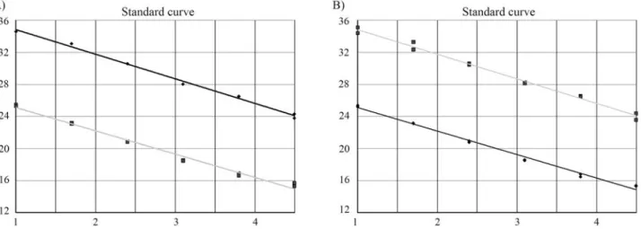
concen-tration points for both mtDNA and nDNA. The standard curves with average slopes at approximately -3.3 (~100% efficiency) were obtained using our optimised TaqMan PCR conditions. The two assays on the dilution series were very similar and showed very good correlation to each other and reproducibility. The average correlation coeffi-cient of the 10 standard curves using the Spearman Rank Test was 0.99 (range: 0.989-0.999, p < 0.001) Figure 1 shows two examples of the standard curves with a correla-tion coefficient of 0.994 and 0.997, respectively.
Quantitative assessment of ccf nDNA and ccf mtDNA in serum and plasma
The ccf nDNA equivalents were calculated according to the standardised method, using very reproducible stan-dard dilution curves, which have been described in our pre-vious studies (Lapaireet al., 2007; Zanetti-Dallenbachet al., 2007; Zhonget al., 2007a). Based on the comparative amplifications of nDNA and mtDNA with an efficiency close to 100%, the fold changes of ccf mtDNA were calcu-lated using the equation of 2CtnDNA-CtmtDNA(R0mtDNA/ R0nDNA
= (1 + EnDNA)CtnDNA / (1 + EmtDNA)CtmtDNA = (1 +
EnDNA)CtnDNA- CtmtDNA,if EnDNA= EmtDNA; (1 + EnDNA) CtnDNA-CtmtDNA
2CtnDNA-CtmtDNA, if E close to 100%).
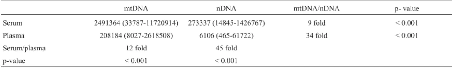
The relative equivalents of ccf mtDNA were esti-mated, and both the quantities of ccf nDNA and ccf mtDNA in the 20 paired plasma and serum samples are shown in Ta-ble 1.
Ccf mtDNA and nDNA were determined by multi-plex real-time PCR, with a mean concentration of 208,184 GE/mL and 6,106 GE/mL in the plasma samples, respec-tively, and of 2,491,364 GE/mL and 273,337 GE/mL in the serum samples, respectively. The ccf nDNA and ccf mtDNA levels in the serum samples were significantly higher than those of the plasma samples (12-45 fold). This may be a result of the release of cellular DNA artefacts dur-ing blood clottdur-ing procedures. The ccf mtDNA levels in the
plasma and serum samples were significantly higher than those of ccf nDNA (9-34 fold).
Discussion
In this study, we described the multiplex assays used to analyse ccf nDNA and ccf mtDNA simultaneously. Using the optimised PCR protocol, the amplification of nDNA and mtDNA in a single reaction tube was very simi-lar and showed a simulation of real-time PCR kinetics. With the comparative efficiencies of two assays in a single tube, we could use the nDNA level as a reference to assess the relative quantities of mtDNA. The method is simple and does not require generating standard curves, which often result in errors due to dilution inaccuracy. Using the multi-plex assays, we could rapidly and accurately determine the levels of ccf nDNA and ccf mtDNA in serum and plasma samples. The levels of ccf mtDNA in circulation are signifi-cantly higher than those of ccf nDNA, due to the fact that the number of mitochondrial genomes in a cell ranges from several hundreds to more than 10,000 copies, and each mitochondrion contains between two and 10 mtDNA mole-cules (Higuchi, 2007). The serum samples showed a signif-icantly higher concentration of ccf DNA because of cellular DNA release during the blood clotting procedures (Za-netti-Dallenbach, 2008; Zhonget al., 2007a).
Quantitative alterations of ccf nDNA and mtDNA have been observed in many conditions, especially in ag-ing, apoptosis and carcinogenesis (Goebelet al., 2005; Liu
et al., 2003; Mehraet al., 2007; Takeuchiet al., 2004). So far, the exact content of human mtDNA in different cells and tissues remains unclear. Two studies developed multi-plex assays to analyse nDNA and mtDNA simultaneously for forensic medicine (Alonsoet al., 2004; Walkeret al., 2005). In our study, we were able to use the GAPDH gene as a housekeeping gene to analyse the quantities of mtDNA. The aim of this study was to develop a rapid, sim-ple and accurate multisim-plex real-time PCR for the direct syn-chronized analysis of ccf mtDNA and ccf nDNA, which may provide a platform for further investigations leading to a better understanding of the biology of ccf mtDNA and ccf nDNA on large-scale sample sizes.
It is known that ancient mtDNA sequences, also termed as nuclear pseudogenes, are present in the human nuclear genome as multiple copies. Woischnik and Moraes (2002) found up to 612 nuclear integrations. Their homo-logy with the current mtDNA was up to 99%. An accidental
co-amplification of these nuclear copies of mitochondrial genes might bias the results (Wallace et al., 1997). We tested the specificity by using the mitochondria-negative cell liner0, and no mtDNA signals were detected in this cell line by real-time multiplex PCR.
Based on the importance of quantification of both mtDNA and nDNA in life science, we developed a rapid, accurate, simple and low-cost approach that enables the si-multaneous identification of physiological and pathogenic mtDNA and nDNA variants. Since there are no commercial kits for ccf mtDNA extraction from serum and plasma sam-ples, we, for the first time, compared three different DNA extraction methods for co-extracting nDNA and mtDNA from plasma and serum. We were also the first ones to ex-amine the efficiencies of the two assays in a single tube. Af-ter calculating the comparative efficiencies we could use the nDNA level as a reference to assess the relative quanti-ties of mtDNA, which can simplify the calculation of mtDNA content. The method is simple and does not require generating standard curves, which often result in errors due to dilution inaccuracy. This method can be considered a standard approach for widely quantifying both mtDNA and nDNA in different kinds of samples.
Acknowledgments
This work was supported in part by the Swiss Na-tional Science Foundation (320000-119722/1), Swiss Can-cer League, Krebsliga Beider Basel and the Dr Hans Altschueler Stiftung. We thank Mrs Vivian Kiefer and Mrs Nicole Chiodetti for their help. We are grateful to Mrs Regan Geissmann for proofreading the text.
References
Allen D, Butt A, Cahill D, Wheeler M, Popert R and Swaminathan R (2004) Role of cell-free plasma DNA as a diagnostic marker for prostate cancer. Ann NY Acad Sci 1022:76-80. Alonso A, Martin P, Albarran C, Garcia P, Garcia O, de Simon
LF, Garcia-Hirschfeld J, Sancho M, de La Rua C and Fer-nandez-Piqueras J (2004) Real-time PCR designs to esti-mate nuclear and mitochondrial DNA copy number in foren-sic and ancient DNA studies. Forenforen-sic Sci Int 139:141-149. Garcia Moreira V, de la Cera Martinez T, Gago Gonzalez E,
Prieto Garcia B and Alvarez Menendez FV (2006) Increase in and clearance of cell-free plasma DNA in hemodialysis quantified by real-time PCR. Clin Chem Lab Med 44:1410-1415.
Table 1- Ccf mtDNA and nDNA represented in genome equivalent (GE)/mL in serum and plasma
mtDNA nDNA mtDNA/nDNA p- value
Serum 2491364 (33787-11720914) 273337 (14845-1426767) 9 fold < 0.001
Plasma 208184 (8027-2618508) 6106 (465-61722) 34 fold < 0.001
Serum/plasma 12 fold 45 fold
Goebel G, Zitt M, Zitt M and Muller HM (2005) Circulating nu-cleic acids in plasma or serum (CNAPS) as prognostic and predictive markers in patients with solid neoplasias. Dis Markers 21:105-120.
Gormally E, Hainaut P, Caboux E, Airoldi L, Autrup H, Mala-veille C, Dunning A, Garte S, Matullo G, Overvad K,et al.
(2004) Amount of DNA in plasma and cancer risk: A pro-spective study. Int J Cancer 111:746-749.
Higuchi M (2007) Regulation of mitochondrial DNA content and cancer. Mitochondrion 7:53-57.
Lam NY, Rainer TH, Chan LY, Joynt GM and Lo YM (2003) Time course of early and late changes in plasma DNA in trauma patients. Clin Chem 49:1286-1291.
Lam NY, Rainer TH, Chiu RW, Joynt GM and Lo YM (2004) Plasma mitochondrial DNA concentrations after trauma. Clin Chem 50:213-216.
Lapaire O, Volgmann T, Huang D, Hahn S, Holzgreve W and Zhong XY (2007) Maternal smoking: Effect on circulating cell-free fetal and total DNA levels in maternal plasma from the second trimester. Obstet Gynecol 110:1358-1363. Liu CS, Tsai CS, Kuo CL, Chen HW, Lii CK, Ma YS and Wei YH
(2003) Oxidative stress-related alteration of the copy num-ber of mitochondrial DNA in human leukocytes. Free Radic Res 37:1307-1317.
Liu W and Saint DA (2002) A new quantitative method of real time reverse transcription polymerase chain reaction assay based on simulation of polymerase chain reaction kinetics. Anal Biochem 302:52-59.
Mehra N, Penning M, Maas J, van Daal N, Giles RH and Voest EE (2007) Circulating mitochondrial nucleic acids have prog-nostic value for survival in patients with advanced prostate cancer. Clin Cancer Res 13:421-426.
Sozzi G, Conte D, Leon M, Ciricione R, Roz L, Ratcliffe C, Roz E, Cirenei N, Bellomi M, Pelosi G,et al.(2003) Quantifica-tion of free circulating DNA as a diagnostic marker in lung cancer. J Clin Oncol 21:3902-3908.
Taback B, O’Day SJ and Hoon DS (2004) Quantification of circu-lating DNA in the plasma and serum of cancer patients. Ann NY Acad Sci 1022:17-24.
Takeuchi H, Fujimoto A and Hoon DS (2004) Detection of mito-chondrial DNA alterations in plasma of malignant mela-noma patients. Ann NY Acad Sci 1022:50-54.
Walker JA, Hedges DJ, Perodeau BP, Landry KE, Stoilova N, Laborde ME, Shewale J, Sinha SK and Batzer MA (2005) Multiplex polymerase chain reaction for simultaneous
quan-titation of human nuclear, mitochondrial, and male Y-chro-mosome DNA: Application in human identification. Anal Biochem 337:89-97.
Wallace DC, Stugard C, Murdock D, Schurr T and Brown MD (1997) Ancient mtDNA sequences in the human nuclear ge-nome: A potential source of errors in identifying pathogenic mutations. Proc Natl Acad Sci USA 94:14900-14905. Xiu-Cheng Fan A, Garritsen HS, Tarhouny SE, Morris M, Hahn
S, Holzgreve W and Zhong XY (2008) A rapid and accurate approach to identify single nucleotide polymorphisms of mi-tochondrial DNA using MALDI-TOF mass spectrometry. Clin Chem Lab Med 46:299-305.
Zanetti-Dallenbach RA, Schmid S, Wight E, Holzgreve W, Lade-wing A, Hahn S and Zhong XY (2007) Levels of circulating cell-free serum DNA in benign and malignant breast lesions. Int J Biol Markers 22:95-99.
Zanetti-Dallenbach RA, Wight E, Fan AXC, Lapaire O, Hahn S, Holzgreve W and Zhong XY (2008) Positive correlation of cell-free dna in plasma/serum in patients with malignant and benign breast. Disease Anticancer Research 28:921-926. Zhong XY, Hahn S, Kiefer V and Holzgreve W (2007a) Is the
quantity of circulatory cell-free DNA in human plasma and serum samples associated with gender, age and frequency of blood donations? Ann Hematol 86:139-143.
Zhong XY, Hahn S, Steinborn A and Holzgreve W (2007b) Quan-titative analysis of intact fetal cells in maternal plasma by real-time PCR. Eur J Obstet Gynecol Reprod Biol 133:20-24.
Zhong XY, Ladewig A, Schmid S, Wight E, Hahn S and Holzgre-ve W (2007c) Elevated leHolzgre-vel of cell-free plasma DNA is as-sociated with breast cancer. Arch Gynecol Obstet 276:327-331.
Zhong XY, Laivuori H, Livingston JC, Ylikorkala O, Sibai BM, Holzgreve W and Hahn S (2001) Elevation of both maternal and fetal extracellular circulating deoxyribonucleic acid concentrations in the plasma of pregnant women with preeclampsia. Am J Obstet Gynecol 184:414-419.
Zhong XY, von Muhlenen I, Li Y, Kang A, Gupta AK, Tyndall A, Holzgreve W, Hahn S and Hasler P (2007d) Increased con-centrations of antibody-bound circulatory cell-free DNA in rheumatoid arthritis. Clin Chem 53:1609-1614.
Associate Editor: Carlos F.M. Menck