I
NSIGHT INTO THE
M
ULTICOPPER
O
XIDASES
S
TABILITY
André João Tavares Fernandes
Dissertation presented to obtain the PhD degree in Biochemistry at the Instituto de Tecnologia Química e Biológica, Universidade Nova de Lisboa
Supervisor: Prof. Lígia O. Martins
Co-supervisor: Prof. Eduardo P. Melo
Oeiras, February of 2011
Supervisor: Prof. Lígia O. Martins Co-supervisor: Prof. Eduardo P. Melo President of Jury: Prof. Carlos Romão
A
CKNOWLEDGMENTSGetting to the end of this, almost, life project would not be possible without the precious help and contributions of some people to whom I would like to acknowledge all of their support.
I would also like to acknowledge Prof. Eduardo P. Melo in participating in all my work. During this period, Prof. Eduardo teached me different techniques that we further developed to specific purposes according to our work. His deep knowledge on those different techniques provided me the opportunity to further develop my scientific work and background. His collaboration was in fact fundamental to all my work since it provided, until know, insight in the obscureness of some particularities of these enzymes. His patience, without giving up gave me some windows that we further developed even if swimming against the current. For all his friendship, patience, and persistency I want to deeply thank to Prof. Eduardo, that without him certainly that this thesis would be completely different.
I only present in this thesis the work developed directly by me, but obviously, this would not be possible or at least it would be much more difficult (than what it was…) if I didn‘t have lab mates. Therefore I would like to acknowledge firstly three past members for their friendship and support, Paulo Durão, Luciana Pereira, and Carlos Covinha. I also want to express all my gratitude to the current lab members, Vânia Brissos, Zhenjia Chen, Sónia Mendes, Tânia Rosado and Rita Catarino, even if some of them did not participate directly in the work. I want to acknowledge them, because I know it is not really easy to live with me 9/10 hours a day 5 days a week… Even so it were some wonderful years or months (for some of them) to work closely with them. Their support on the bad days, the sharing of joys in the good days and more importantly their friendship was essential during all this time period.
Importantly, I also would like to thank the great contributions of Manuela Pereira, Smilja Todorovic, Isabel Bento, Catarina Silva, João M. Damas and Claúdio Soares and to Peter F. Lindley. Thank you for all the input you have given into the presented work in this dissertation.
equipment. I would also like to thank Instituto de Biologia Experimental e Biotecnológica (IBET), the European projects, Sophied and Biorenew, and off course to all FCT projects as well and again to FCT for my grant.
D
ISSERTATIONA
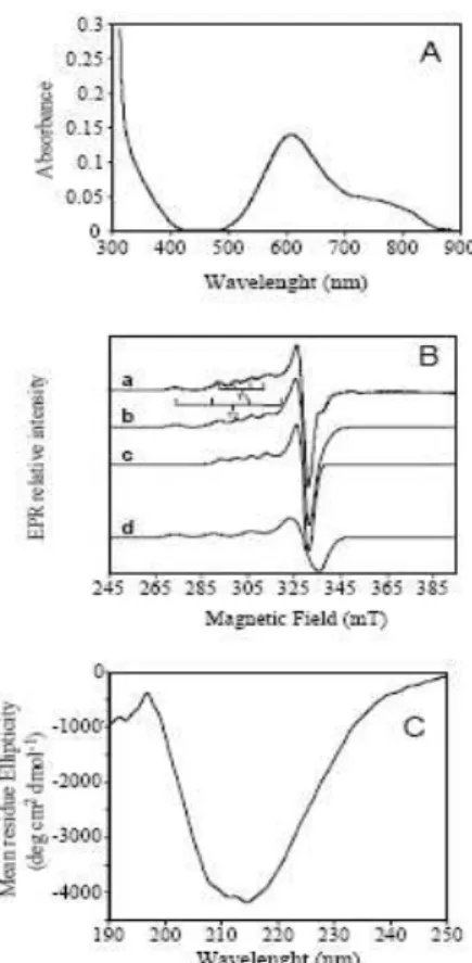
BSTRACTThis dissertation portrays recent development on the knowledge of the stability determinants and of functional characteristics of multicopper oxidases (MCO). Multicopper oxidases are a family of enzymes that includes laccases (benzenediol oxygen oxidoreductase; EC 1.10.3.2), ascorbate oxidase (L-ascorbate oxygen oxidoreductase, EC 1.10.3.3) and ceruloplasmin (Fe2+ oxygen oxidoreductase, EC 1.16.3.1). MCO are characterized by having four copper ions that are classified into three distinct types of copper sites, namely type 1 (T1), type 2 (T2) and type 3 (T3). The classical T1 copper site comprises two histidine residues and a cysteine residue arranged in a distorted trigonal geometry around the copper ion with bonding distances approx. 2.0 Å (1 Å=0.1 nm); a weaker fourth methionine ligand completes the tetrahedral geometry. The copper–cysteine linkage is characterized by an intense S(π)→Cu(dx2−y2) CT (charge transfer) absorption band at approximately 600 nm, and a narrow parallel hyperfine splitting A\\ = (43–90)×10−4 cm−1 in the electron paramagnetic resonance (EPR) spectrum. The function of the T1 copper site is to shuttle electrons from substrates to the trinuclear copper centre where molecular oxygen is reduced to two molecules of water during the complete four-electron catalytic cycle. The trinuclear center contains a T2 copper coordinated by two histidine residues and one water molecule, lacks strong absorption bands and exhibits a large parallel hyperfine splitting in the EPR spectrum (A\\ = (150– 201)×10−4 cm−1). The T2 copper site is in close proximity to two T3 copper ions, which are each coordinated by three histidine residues and typically coupled, for example, through a dioxygen molecule. The T3 or coupled binuclear copper site is characterized by an intense absorption band at 330 nm originating from the bridging ligand and by the absence of an EPR signal due to the antiferromagnetically coupling of the copper ions.
laccase highly interesting for various applications, including textile dye bleaching, pulp bleaching and bioremediation, where enzymatic catalysis could serve as a more environmentally friendly alternative than the current used chemical processes. However, using an enzyme in industrial environment requires a high stability, since most of the industrial activities are carried out at high temperatures, extreme pH values, high pressures among other physico-chemical adverse conditions. Natural enzymes are usually, far from optimally functioning under these harsh conditions. The work developed in this thesis focus precisely in the study of molecular determinants of stability in MCO. Having these in mind two different strategies were followed. We searched for hyperthermophilic MCO and simultaneously we have used site directed mutagenesis in a well characterized bacterial laccase, CotA-laccase from
Bacillus subtilis whose structure is known. Two MCO from hyperthermophilic
microorganism were isolated and characterized, one from the Bacterium Aquifex aeolicus, and the other from the Archaeon Pyrobaculum aerophilum.
Unlike most of the hyperthermophiles both A. aeolicus and P. aerophilum can
withstand the presence of oxygen, growing efficiently in microaerophilic conditions, and using it as electron acceptor in a chemiolitoauthotrophic metabolism. Aquifex aeolicus isolated from a shallow submarine hydrothermal
system at Volcano, Italy is one of the most hyperthermophilic bacteria known so far (Top 89ºC) and belongs to an ancient lineage within the bacterial domain.
Pyrobaculum aerophilum was isolated from a boiling marine water hole at
Maronti Beach in Italy as an optimal growth temperature of 100ºC. P. aerophilum is able to grow autotrophically by oxidation of molecular hydrogen,
thiosulfate or sulphur, using oxygen, as electron acceptors and is a strictly aerobic organism, a rare feature among hyperthermophiles.
The first gene to be cloned in this work was the one corresponding to the MCO from the hyperthermophilic bacterium Aquifex aeolicus and the recombinant
protein expressed in E. coli. The mcoA gene is localized in the genome as part
shows spectroscopic and biochemical characteristics typical of the well-characterized MCO. McoA presents higher specificity (kcat⁄Km) for cuprous and ferrous ions than for aromatic substrates and is therefore designated as a metallo-oxidase. Addition of copper is required for maximal catalytic efficiency. A comparative model structure of McoA has been constructed and a striking structural feature is the presence of a Met-rich region (residues 321– 363), reminiscent of those found in copper homeostasis proteins. The kinetic properties of a mutant enzyme, McoAΔP321-V363, deleted in the Met-rich region, provide evidence for the key role of this region in the modulation of the catalytic mechanism. McoA probably contributes to copper and iron homeostasis in A. aeolicus.
In order to understand the stability determinants in this family of enzymes we analyzed in more detail the thermodynamic stability of McoA. The deconvolution of the DSC traces shows that thermal unfolding is characterized by an average melting temperature of 110°C, showing that this enzyme is highly thermotolerant. Furthermore, McoA has an optimal catalytic temperature of 75 ºC and presents remarkable heat stability at 80 and 90 ºC, with activity
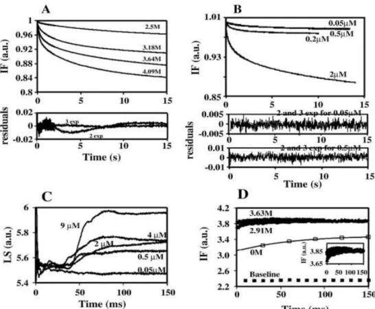
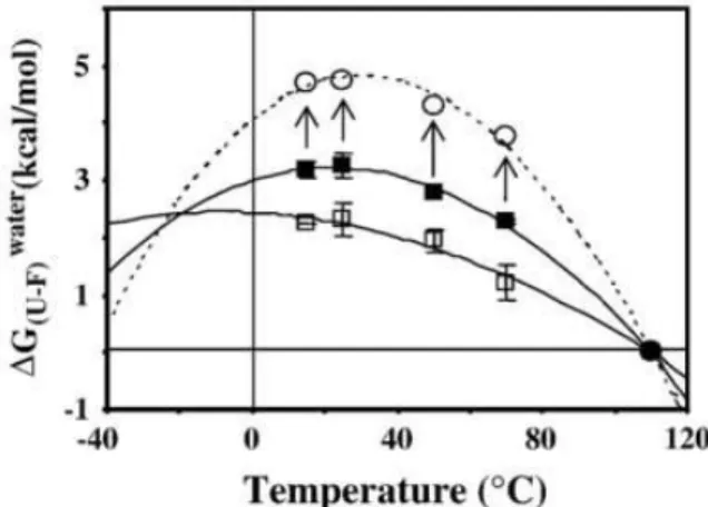
lasting for up to 9 and 5 h, respectively. However, chemical denaturation studies revealed that McoA displays, at room temperature, a very low stability (2.8 kcal/mol). Stopped-flow kinetics was performed to obtain additional information regarding the mechanism of unfolding and the role of copper in the stabilization of the protein. In McoA which is highly prone to aggregation, the kinetic partitioning between unfolding and aggregation should constitute the main factor behind the unusual low chemical stability of the enzyme. Moreover, McoA exhibits a small difference in stability between the folded and unfolded states in a broad range of temperatures. Actually, the flattening of the Gstab
this mechanism should explain, at least partially, the hyperthermophilic nature of McoA.
The second MCO (McoP) to be cloned and studied was from the hyperthermophilic archaeon Pyrobaculum aerophilum. The enzyme consists of
a single subunit with 49.6 kDa and the combined results of UV-visible, circular dichroism (CD), electron paramagnetic resonance (EPR) and resonance Raman (RR) showed the typical spectroscopic characteristics of the MCO family. McoP is an efficient metallo-oxidase that catalyzes the oxidation of Cu+ and Fe2+ metal ions with turnover rate constants of 356 and 128 min-1, respectively, at 40°C. Noteworthy, McoP that follows a ping-pong mechanism presents a 3-fold higher catalytic efficiency when using nitrous oxide (N2O) as electron acceptor as compared with dioxygen, the typical oxidizing substrate of multicopper oxidases. This finding led us to propose that McoP represents a novel archaeal nitrous oxide reductase most probably involved in the final step of the denitrification pathway of P. aerophilum. The analysis of McoP
sequence, showing similarities with bacterial MCO, allowed deriving its structure by comparative modelling methods. Mutants were designed to elucidate the substrate specificity as well as the low redox potential of the T1 center. We show that the low redox potential of this enzyme is at least in part related to a negatively charged residue (Glu296), near T1 (7.8 Å) that should stabilize the oxidized state of the copper ion, reducing the center redox potential. McoP has an optimum reaction temperature of 85°C and is a hyperthermostable enzyme, with a half-life of inactivation of around 6 h at 80°C, being one of the most hyperthermostable MCO studied so far. To gain further insight into the thermal stability of McoP we have analyzed its melting temperatures by DSC. Similar to other MCO, the endothermic peak revealed a complex and irreversible thermal transition, characterized by three independent non two-state transitions with melting temperatures ranging from 97 to 112°C.
We also evaluated the stability determinants of MCO by using site directed
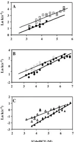
The first work performed was to evaluate the effect of the axial Met in the T1 copper center reduction potential. We have replaced the Met by a Leu or Phe residue in order to increase the redox potential of the enzyme. In fact, these mutations led to an increase of the T1 redox potential by approximately 100 mV relative to the wild-type enzyme but severely compromised the catalytic rate of the enzyme. This decrease in the catalytic efficiency was unexpected as the X-ray analysis of mutants has shown that replacement of Met ligand did not lead to major structural changes in the geometry of the T1 center or in the overall fold of the enzyme. However, these point mutations have a profound impact on the thermodynamic stability of the enzyme. The fold of the enzyme has become unstable especially with the introduction of the larger Phe residue and this instability should be related to the decrease in the catalytic efficiency. This instability of the folded state resulted in the accumulation of an intermediate state, partly unfolded, in between native and unfolded states. Quenching of tryptophan fluorescence by acrylamide has further revealed that the intermediate state is partly unfolded.
With the aim at evaluating the copper content effect on the thermal stability of CotA-laccase we have studied different forms of CotA-laccase with respect to copper content. The data shows that copper content of CotA-laccase has a profound impact in the kinetic and thermal stability as assessed by long term stability (at 80ºC) and DSC. Indeed, HoloCotA, produced in recombinant
Escherichia coli under microaerobic conditions, is a very stable enzyme,
followed by ApoCotA reconstituted in vitro with Cu+, and by ApoCotA
reconstituted with Cu2+. These results showed additionally, that copper incorporation into the protein seems to occur more effectively during the in vivo
S
UMÁRIO DA DISSERTAÇÃOEsta dissertação retrata os desenvolvimentos recentes no conhecimento dos determinantes de estabilidade e também das características funcionais de oxidases de múltiplos cobres. As oxidases de múltiplos cobres (OMCs) são uma família de enzimas que inclui lacases (oxidoreductases de benzenodiol e oxigénio, EC: 1.10.3.2,), ascorbato oxidase (oxidoreducase de L-ascorbato e oxigánio, EC. 1.10.3.3) e ceruloplasmina (oxidoreductase de Fe2+ e oxigénio, EC. 1.16.3.1). As OMCs são caracterizadas por terem 4 centros de cobre distintos, nomeadamente, tipo 1 (T1), tipo 2 (T2) e tipo 3 (T3). O centro de cobre T1 clássico é composto por dois resíduos de histidinas e um de cisteína que estão organizados numa geometria trigonal distorcida, em torno do átomo de cobre com distâncias de ligação de aproximadamente 2,0 Å (1 Å=0.1 nm); a geometria tetraédrica é completada com a presença de um quarto ligando fraco, um resíduo de metionina. A ligação entre o cobre e a cisteína é caracterizada por ter uma transferência de carga (TC) intensa entre S(π)→Cu(dx2−y2) que resulta numa banda de absorção a aproximadamente 600 nm, e uma separação do paralelo hiper-fino estreito, A\\ = (43–90)×10−4 cm−1, no espectro de
ressonância paramagnética (ERP). A função do centro de cobre T1 é transferir os electrões subtraídos aos substratos para o centro trinuclear, onde durante um ciclo completo de quatro electrões, a molécula de oxigénio é reduzida a duas moléculas de água. O centro trinuclear é composto por um centro de cobre tipo 2 (T2) coordenado por duas histidinas e uma molécula de água, não tem bandas de absorção fortes e exibe um paralelo hiper-fino largo no espectro de ressonância paramagnética (ERP) (A\\ = (150–201)×10−4 cm−1). O centro de
As laccases são enzimas que catalisam a oxidação de uma variedade de compostos fenólicos substituídos, aromáticos, aminas e, também, de alguns compostos inorgânicos, usando o oxigénio como aceitador final de electrões. A sua versatilidade de substratos faz com que as lacases sejam muito interessantes para várias aplicações biotecnológicas, incluindo descoloração de corantes têxteis, branqueamento da pasta do papel e bioremediação, onde a catálise enzimática poderá servir como uma alternativa, mais amiga do ambiente, aos processos químicos actuais. No entanto, o uso de enzimas em ambiente industrial requer que estas tenham uma elevada estabilidade, uma vez que a maioria dos processos industriais realizam-se a temperaturas elevadas, valores de pH extremos, pressões altas entre outras condições físico-químicas adversas. As enzimas naturais estão normalmente longe de funcionar optimamente sob estas condições adversas. O trabalho desenvolvido nesta tese foca precisamente o estudo dos determinantes moleculares de estabilidade das OMCs. Tendo isto em mente, foram seguidas duas estratégias. Procurámos OMCs hipertermófilas e simultaneamente usámos mutagénese dirigida numa lacase bacteriana bem caracterizada cuja estrutura tridimensional é conhecida, a CotA-lacase de
Bacillus subtilis. Duas OMCs de microrganismos hipertermófilos foram
isoladas e caracterizadas, uma da Bacteria Aquifex aeolicus e outra do Archeaum Pyrobaculum aerophilum. Ao contrário da maioria dos
hipertermófilos, ambos A. aeolicus e P. aerophilum toleram a presença de
oxigénio, crescendo de forma eficiente em condições de microaerofilia, usando, O2 como aceitador de electrões num metabolismo quimiolitoautotrófico.
Aquifex aeolicus foi isolado de um sistema hidrotermal pouco profundo em
Volcano, em Itália sendo a bactéria mais hipertermófila conhecida (Topt=89ºC) e pertence a uma linhagem antiga dentro do domínio Bacteria. O Pyrobaculum aerophilum foi isolado num buraco de água marinha fervente na praia de
Maronti, em Itália, tendo uma temperatura óptima de crescimento de 100ºC. O
P. aerophilum é capaz de crescer autotroficamente através da oxidação de
Neste trabalho a primeira OMC a ser clonada e a proteína recombinante expressa em E. coli foi a da bactéria hipertermófila Aquifex aeolicus. O gene mcoA está inserido no genoma como parte de um possível determinante
genético de resistência ao cobre. A enzima pura tem as características espectroscópicas e bioquímicas típicas de outras OCMs conhecidas. A McoA é designada como oxidase de metais uma vez que tem uma especificidade (kcat ⁄Km) elevada para o ião Cu+ e para Fe2+ quando comparado com substratos aromáticos. Para atingir a máxima eficiência catalítica é necessária a adição de cobre (Cu2+) às misturas reaccionais. Um modelo comparativo da McoA foi construído e, uma característica interessante que foi detectada, foi a presença de um segmento rico em metioninas (resíduos 321-363) reminiscente de outras enzimas envolvidas na homeostase de cobre. As propriedades cinéticas da enzima mutante McoAΔP321-V363, em que se removeu a região rica em metioninas, apontam para o papel crucial desta região na modulação do mecanismo catalítico. A McoA provavelmente contribui para a homeostase de Cu+ e Fe2+ em
A. aeolicus.
principal que determina a baixa estabilidade à temperatura ambiente. No entanto, a McoA apresenta uma baixa diferença de energia livre (Gstab) entre os estados nativo e desnaturado num intervalo largo de temperaturas. Na verdade, o alargamento e diminuição da cuva de Gstab em função da
temperatura, devido a um baixo valor de variação da capacidade calorífica (Cp), é um mecanismo usado por outras proteínas termófilas para balancear
uma temperatura de ‗unfolding‘ elevada com a necessária estabilidade termodinâmica para uma actividade catalítica óptima, sendo que este mecanismo poderá explicar, pelo menos em parte, a natureza hipertermófila da McoA.
A segunda OCM (McoP) a ser clonada e estudada foi do archaeum hipertermófilo Pyrobaculum aerophilum. A enzima é um monómero com 49.6
kDa, em que os resultados combinados de UV-Visível, dicroísmo circular, ressonância electrónica paramagnética e ressonância de Raman, mostraram que a enzima contém as características espectroscópicas típicas da família das OCM. A McoP é uma oxidase de metais eficiente com um ‗turnover‘ a 40ºC de 356 e 128 min-1 para o Cu+ e Fe2+, respectivamente. De realçar, que a McoP
segue um mecanismo tipo ‗ping-pong‘ e apresenta uma eficiência 3 vezes superior usando o óxido nitroso (N2O) como aceitador final de electrões quando
comparado com o substrato típico das OCMs, o oxigénio. Esta descoberta levou-nos a propor que a McoP representa um novo tipo de reductase de óxido nitroso envolvida no último passo da via de desnitrificação do P. aerophilum. A
catálise de 85ºC e, é uma enzima hipertermoestável com um tempo de meia vida de 6 horas a 80ºC, sendo uma das OCM‘s conhecidas mais estáveis. Com o objectivo de analisar em maior pormenor a estabilidade térmica da McoP, analisámos as temperaturas de ‗melting‘ por DSC. À semelhança do que aconteceu para outras OCM‘s, o pico endotérmico mostrou tratar-se de uma transição complexa e irreversível, caracterizada por três transições independentes, com temperaturas de ‗melting‘ de 97 a 112ºC.
Estudámos também os determinantes de estabilidade das OCM‘s usando mutagénese dirigida numa enzima bem caracterizada, a CotA-lacase de Bacillus subtilis. No primeiro estudo que desenvolvemos, avaliámos o efeito do ligando
teor em cobre na CotA-lacase, tem um efeito profundo na estabilidade cinética e térmica, tal como determinado por estudos de estabilidade cinética a 80ºC e também por varrimento diferencial de calorimetria. De facto, a holoCotA recombinante, produzida em Escherichia coli sob condições micoraeróbicas, é
uma enzima muito estável, seguido da apoCotA reconstituída in vitro com Cu+ e
pela apoCoA reconstituída com Cu2+. Estes resultados mostram, adicionalmente, que a incorporação de cobre na proteína ocorre de forma mais eficiente durante o enrolamento da enzima in vivo e que é dependente do estado
L
IST OFP
UBLICATIONSPaulo Durão, Isabel Bento, André T. Fernandes, Eduardo P. Melo, Peter F. Lindley, and Lígia O. Martins. 2006. Perturbations of the T1 copper site in the CotA-laccase from Bacillus subtilis: Structural, Biochemical, Enzymatic and
Stability Studies. J. Biol. Inorg. Chem. 11:514-526.
André T. Fernandes, Cláudio M. Soares, Manuela M. Pereira, Robert Huber, Gregor Grass, Lígia O. Martins. 2007. A robust metallo-oxidase from the hyperthermophilic bacterium Aquifex aeolicus. The FEBS Journal. 274:11
2683-2694
Paulo Durão, Zhenjia Chen, André T. Fernandes, Peter Hildebrandt, Daniel H. Murgida, Smilja Todorovic, Manuela M. Pereira, Eduardo P. Melo, Lígia O. Martins. 2008. Copper incorporation into recombinant CotA-laccase from
Bacillus subtilis: characterization of fully copper loaded enzymes. J. Biol.
Inorg. Chem. 13:183-93
André T. Fernandes, Lígia O. Martins, Eduardo P. Melo. 2009. The hyperthermophilic nature of the metallo-oxidase from Aquifex aeolicus.
Biochimica et Biophysica Acta. 1794: 75-83
André T Fernandes, João M. Damas, Smilja Todorovic, Robert Huber, M. Camilla Baratto, Rebecca Pogni, Cláudio M. Soares and Lígia O. Martins. 2010. The multicopper oxidase from the archaeon Pyrobaculum aerophilum
shows nitrous oxide reductase activity. FEBS J. 277: 3176-3189
.
T
ABLE OFC
ONTENTSD
ISSERTATIONA
BSTRACT...
IXS
UMÁRIO DA DISSERTAÇÃO...
XV1
General Introduction ... 27
1.1.
W
HITE BIOTECHNOLOGY... 2
1.1.1.
Enzymes in White Biotechnology ... 5
1.2.
M
ULTICOPPERO
XIDASES... 6
1.2.1.
Overall fold of multicopper oxidases ... 7
1.2.2.
Copper centers in multicopper oxidases ... 9
1.2.3.
reaction mechanism of Multicopper oxidases... 13
1.2.4.
Metallo-oxidases substrate specificity ... 17
1.2.5.
Distribution and role of Multicopper oxidases ... 20
1.2.6.
Biotechnological applications ... 25
1.3.
P
ROTEINS
TABILITY... 29
1.3.1.
Techniques to measure protein stability ... 30
1.3.2.
Thermal stability ... 32
1.3.3.
Protein folding ... 35
1.3.4.
Molecular determinants of stability ... 41
1.3.5.
Stability in metallo-proteins ... 44
2
A Robust Metallo-Oxidase from the Hyperthermophilic
Bacterium
Aquifex aeolicus
... 49
2.1.
I
NTRODUCTION... 51
2.2.
E
XPERIMENTALP
ROCEDURES... 53
3
The hyperthermophilic nature of the metallo-oxidase from
Aquifex aeolicus
... 73
3.1.
I
NTRODUCTION... 75
3.2.
E
XPERIMENTALP
ROCEDURES... 77
3.3.
R
ESULTS... 80
3.4.
D
ISCUSSION... 94
4
The Multicopper Oxidase from the Archaeon
Pyrobaculum
aerophilum
shows Nitrous Oxide Reductase Activity ... 101
4.1.
I
NTRODUCTION... 103
4.2.
E
XPERIMENTALP
ROCEDURES... 105
4.3.
R
ESULTS... 110
4.4.
D
ISCUSSION... 124
4.5.
S
UPPLEMENTARY MATERIAL... 127
5
Stability studies in CotA-Laccase from
Bacillus subtilis
... 129
5.1.
E
FFECT OF THE REPLACEMENT OF THE AXIAL LIGAND IN THE REDOX PROPERTIES OFC
OTA ... 132
5.1.1.
Experimental Procedures ... 134
5.1.2.
Thermodynamic stability of M502 mutants ... 136
5.1.3.
Concluding remarks ... 142
5.2.
E
FFECT OF COPPER CONTENT IN THE STABILITY OFC
OTA-LACCASE
... 143
5.3.
T
HE REMOVAL OF A DISULFIDE BRIDGE OFC
OTA
CHANGES THE SLOWER MOTION DYNAMICS INVOLVED IN COPPER BINDING BUT HAS NO EFFECT ON THE THERMODYNAMIC STABILITY... 155
5.3.1.
Abstract ... 155
5.3.2.
Introduction ... 155
5.3.3.
Experimental procedures ... 158
5.3.4.
Results and Discussion ... 161
6
General Discussion ... 175
7
References ... 193
8
Annexes ... 223
8.1.
M
COA
SUBSTRATE SPECIFICITY:
CHARACTERIZATION OF MUTANT ENZYMES... 225
1
G
ENERAL
I
NTRODUCTION
1.1.
W
HITE BIOTECHNOLOGY... 2
1.1.1.
Enzymes in White Biotechnology ... 5
1.2.
M
ULTICOPPERO
XIDASES... 6
1.2.1.
Overall fold of multicopper oxidases... 7
1.2.2.
Copper centers in multicopper oxidases ... 9
1.2.3.
reaction mechanism of Multicopper oxidases ... 13
1.2.4.
Metallo-oxidases substrate specificity ... 17
1.2.5.
Distribution and role of Multicopper oxidases ... 20
1.2.6.
Biotechnological applications ... 25
1.3.
P
ROTEINS
TABILITY... 29
1.1.
W
HITE BIOTECHNOLOGYBiotechnology can be defined as the field of applied biology involving the use of living organisms in engineering, medicine, agriculture and in products of the day to day life. The use of the term includes genetic engineering as well as cell and tissue culture technologies. This concept encompasses a wide range of procedures for modifying nature according to human purposes, going back to domestication of the living organism. Biotechnology draws on the pure biological sciences (genetics, microbiology, animal cell culture, molecular biology, biochemistry, and cell biology) and in many instances is also dependent on the knowledge and methods, outside the sphere of biology, from chemical engineering, bioprocess engineering. Biotechnology is divided in different branches and in order to better identify the terms of the biotechnology application, a code of colours was created;
The Green biotechnology is applied to agricultural processes. An example is the designing of transgenic plants to grow under specific environments in the presence or absence of chemicals, or the engineering of a plant to express a pesticide, thereby ending the need of external application of pesticides.
Red biotechnology is applied to medical processes. The designing of organisms to produce antibiotics is an example, or the engineering of genetic cures through genetic manipulation, by developing DNA or RNA vaccines.
Blue biotechnology is a term that has been used to describe the marine and aquatic applications of biotechnology, but its use is relatively rare. It involves the use of marine organisms, and their derivatives, for purposes such as increasing seafood supply and safety or controlling the proliferation of noxious water-borne organisms.
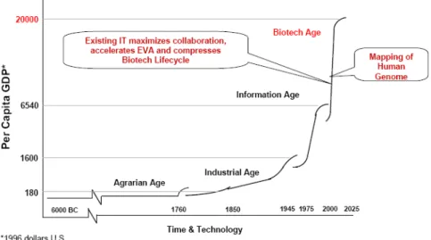
The aim is to develop cleaner processes with minimum waste generation and energy use. White biotechnology is not an end of the pipe cleaning technology; it is a clean production process that minimizes waste before it is even produced. It has substantial potential to reduce environmental impact. For example, air and water pollution can be reduced, energy use lowered, fewer raw materials needed, and waste could be diminished or substituted by biodegradable materials. White biotechnology has become more broadly applicable due to recently developed genetic techniques. Multiple enzymes variants, for example, can now be created at high speed, which are then screened to better fit the desired applications. A wide range of useful products can be produced by industrial biotechnology. These fall within the categories of fine chemicals, pharmaceuticals, food additives and supplements, colorants, vitamins, pesticides, bio-plastics, solvents, bulk chemicals and biofuels (Figure 1.1.) (EuropaBio, 2003).
Figure 1.1: The Industrial Biotechnology Value Chain.
biotechnology enables the introduction of more efficient, less energy-intensive processes. Developments are also making their first inroads into larger volume segments such as polymers, bulk chemicals and biofuels, and many other industrial sectors. Although numbers may differ, all studies agree that industrial biotechnology will play an increasingly significant role in the chemical and other manufacturing industries in the future.
Figure 1.2: Evolution of gross domestic product according to the Humanity important marks.
increase, therefore now is the time to focus attention on alternatives. It is crucial for industries to secure abundant, competitively priced and stable resources. And natural renewable resources constitute an interesting and reliable set of feedstocks.
1.1.1.
E
NZYMES INW
HITEB
IOTECHNOLOGYEnzymes are molecules present in all living organisms. The function of these biocatalysts is to catalyze chemical reactions in vivo by lowering the activation
energy of the chemical reactions. Enzymes can be considered the catalytic machinery of living systems. Industrial biotechnology aims to utilize this enzymatic capability in industrial processes. Nowadays, the industrial enzyme producers use them over a wide variety of applications. The estimated value of world market is presently about US$ 2 billion. Detergents (37%), textiles (12%), starch (11%), baking (8%) and animal feed (6%) are the main industries, which use about 75% of industrially produced enzymes. A very important field in which enzymes have proved to be of great value over the last 15-20 years is the starch industry. In 1950s, fungal amylase was used in the manufacture of specific types of syrup, which could not be produced by the conventional acid hydrolysis. The real turning point was reached early in the 1960s when the enzyme glucoamylase that completely hydrolyze starch into glucose was used for the first time. Within a few years, almost all glucose production was reorganized and enzyme hydrolysis was used instead of acid hydrolysis due to the better benefits such as greater yield, higher degree of purity and easier crystallization (James and Lee, 1997).
detergents composition was upgraded with cellulases. Cellulase is actually an enzyme complex capable of degrading crystalline cellulose to glucose. In textile, washing cellulases remove cellulose microfibrils, which are formed during washing of cotton based cloths. This can be seen as colour brightening and softening of the material. An important area of research is the investigation of enzymes that can tolerate, or even have higher activities, in hot and cold temperatures. The search for thermotolerant and cryotolerant enzymes has spanned the globe. These enzymes are especially desirable for improving laundry processes in hot water cycles and/or at low temperatures for washing colours and darks.
In contrast with hydrolases, such as lipases and proteases, some industrial processes would benefit with oxidation/reduction based enzymatic reactions. For these processes, the suitable enzymes are oxidoreductases. Most of these, usually need the addition of exogenous co-factors, electron donor/acceptors (NAD(P)H and FMNH2) which would make the process extremely expensive and not suitable for industrial application. Among the biological group of oxidoreductases, there is a ubiquitous and interesting group of enzymes that show great potential for biotechnological and environmental applications, laccases, which are a functional class among the multicopper oxidases. Laccases can oxidize a broad range of substrates, including phenolic and non-phenolic compounds. One of the main advantages of laccases is that they do not need the addition of cofactors, and use the readily available oxygen as oxidizing agent (Gianfreda, 1999).
1.2.
M
ULTICOPPERO
XIDASESfour-electron reduction to water with concomitant one-electron oxidation of the reducing substrate. These are a family that utilizes the unique redox properties of the copper ion in the catalytic centers (Giardina, et al., 2010; Nakamura and Go, 2005). MCO are characterized by having four copper ions that are classified into distinct copper centers (type 1, type 2 and type 3) (see below, section 1.2.2 for further details) (Stoj and Kosman, 2005). Two subfamilies of MCO can be distinguished based on their substrate specificity. Laccases are MCOs with higher specificity for different bulkier substrates and metallo-oxidases with higher catalytic efficiency towards low valence meal ions, such as Cu+, Fe2+ and Mn2+. The natural occurring substrates of laccases is not known, however they are involved in different processes depending on the physiological organism. Currently, laccases are a subject of intense study towards its use in biotechnological processes mainly due to their wide range of reducing substrates. Prominent metallo-oxidases, such as human ceruloplasmin, yeast ferroxidase Fet3p and CueO from Escherichia coli, are known to be critically
involved in metal homeostasis mechanisms. In aerobic metabolism, metals such as iron and copper, although essential for life, readily participate in reactions that result in the production of highly reactive oxygen species (Crichton and Pierre, 2001) which may be involved in cell damage.
1.2.1.
O
VERALL FOLD OF MULTICOPPER OXIDASESFigure 1.3: A) Cupredoxin fold from Azurin (PDB accession 1JOI). (B)Overall fold of CotA-laccase from B. subtilis (PDB accession code 1GSK) drawn with Pymol (DeLano, 2003). β
-sheets are represented in yellow, and α-helices represented in red.
1.2.2.
C
OPPER CENTERS IN MULTICOPPER OXIDASESTable 1.1: Classification of copper sites. Adapted from PROMISE database (Degtyarenko, et al., 1998).
Copper center Protein class/family
Cu(N His)2S CysR
R = S Met (azurin, plastocyanin,
laccase)
R = O Glu (phytocyanins)
R = H2O (ceruloplasmin)
Type I (blue copper proteins)
Small blue proteins
o Auracyanin o Azurin
o Phytocyanin family o Plastocyanin family o Rusticyanin
Blue oxidases
o Ascorbate oxidase o Ceruloplasmin o Laccase
Nitrite reductase
Cu(N His)mRn
L = N, O or S ligands; R = O or S ligands
m = 1 to 4; n = 0 to 3; m+n = 4 or 5
Type II
Cu, Zn superoxide dismutase Dioxygenases
Monooxygenases
o Dopamine ßhydroxylase o Methane monooxygenase o Peptidylglycine
-hydroxylating monooxygenase
o Phenylalanine hydroxylase
Nitrite reductase Non-blue oxidases
o Amine oxidase o Diamine oxidase o Galactose oxidase o Lysyl oxidase
µO2[Cu(N His)3]2
Type III
Catechol oxidase Haemocyanins Tyrosinase
Trinuclear center (type II + type III)
Blue oxidases
1.2.2.1. Type 1 copper center
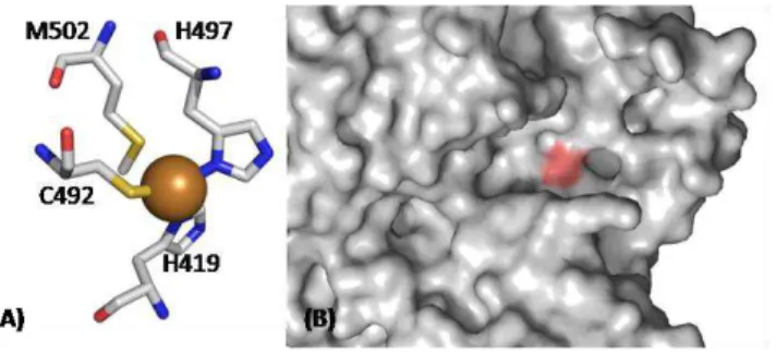
The T1 copper center, or ―blue‖ center, is usually coordinated in a distorted tetrahedral manner, with strong ligands provided by thiolate sulphur of a Cys and the imidazole nitrogens of two histidines. This active site is completed by a variable axial ligand, that commonly is a methionine residue in most of MCO (see Table 1.1 and Figure 1.4A) (Dennison, 2005; Lindley, 2001; Solomon, et al., 1996). The T1 copper center is characterized by a very strong absorption band with a molar extinction coefficient in the range from 3000 to 6000 M-1cm-1 at a wavelength of ~600 nm, associated to the typical intense blue colour. This absorption band is associated with the ligand-metal charge transfer from the sulphur atom of the cysteine to the copper atom S(π)→Cu(dx2−y2). Furthermore, the EPR spectrum of the T1 center exhibits very weak parallel hyperfine splitting lines due to the strong bond in the copper site (since the unpaired electron is considerably displaced toward the cysteine ligand, the strength of its interaction with the spin of the copper nucleus is reduced substantially (Solomon, et al., 1996; Zhukhlistova, 2008).
Figure 1.4: (A) CotA type 1 copper ligands. Two His and a Cys coordinate the T1 copper, and a Met is present as axial ligand. (B) Close-up of the active-site (T1 Cu center) region of CotA, using surface representation, highlighting (in magenta) the region of the most exposed histidine ligand of the copper, which is the residue that interacts with substrates.
and the copper ion (see Figure 1.4B) (Enguita, et al., 2003). The His2Cys equatorial ligand set is always maintained in MCO, but the residue present in the axial position variable. In related proteins different axial ligands can be present, such as in the stellacyanins where a Gln coordinates (Hart, et al., 1996; Koch, et al., 2005). The putative plantacyanins that have either a Val or Leu in the axial position (Dennison, et al., 2003; Kim, et al., 2003; Nersissian, et al., 1998; Nersissian and Shipp, 2002). The azurins the backbone carbonyl oxygen of Gly residue provides a second weak axial interaction resulting in trigonal bipyramidal geometry. In fungal laccases there is a Phe or Leu at the position of Met, which no longer coordinates T1 copper (Dennison, 2005; Ducros, et al., 1998; Kim, et al., 2003). These sites, with non-coordinating side-chain in the axial position, have trigonal active site geometries (Crowley, et al., 2001; Impagliazzo and Ubbink, 2004; Ubbink, et al., 1998). These subtle structural changes have a dramatic effect on the redox potential of the T1 center in MCO. The presence of a Leu or Phe tends to short the distance between the Cys residue and the copper atom, stabilizing the reduced state of the center originating a high redox potential. On the other hand, the presence of a Met residue as fourth ligand increases the distance between Cys and the copper atom, stabilizing the oxidized state of the center, causing a reduction of the redox potential of the center (Durao, et al., 2006; Durao, et al., 2008). The presence/absence of this fourth ligand may contribute, at least, in part to the variations of the redox potential although other elements of the protein matrix are known to affect this parameter in MCO (Kosman, 2009).
1.2.2.2. Trinuclear center
Figure 1.5: Typical trinuclear cluster containing the T2 site and the binuclear T3 site.
The T2, or ―non-blue‖, copper center is characterized by EPR signals similar to those observed for Cu2+ tetragonal complexes. The absorption spectra of these centers frequently have a low intensity. The T3 center is composed by two copper atoms bound to ligands and is called the binuclear site. The T3 coppers are paramagnetic which makes them ―invisible‖ to EPR spectroscopy, but exhibit an absorption band at ~330 nm. The trinuclear center acts in dioxygen binding and reducing it upon receiving four electrons forwarded from the mononuclear center T1. A more detailed description of the dioxygen reduction is done in the following section (section 1.2.3).
1.2.3.
REACTION MECHANISM OFM
ULTICOPPER OXIDASESwhich should occur via an outer sphere pathway. The exact mechanism of dioxygen reduction is still unknown; however several authors have been working on this issue, trying to understand the molecular basis the reduction of dioxygen to water. The different proposed mechanisms draw conclusions based on the results obtained by using different techniques such as X-ray crystal structures or spectroscopic data. We will present a more detailed description of the data obtained by Bento et al. (2005). The authors propose a reduction mechanism, by correlating the data obtained with different crystal structures of CotA-laccase. The differences in the crystal structures are in the trinuclear center were different oxygen species were modulated, a dioxygen, peroxide and a hydroxyl. The first event in this putative mechanism is the movement of molecular dioxygen to the trinuclear copper site where it binds to the type 3 copper atoms (Figure 1.6a).
Figure 1.6: Putative mechanism of the reduction of dioxygen to water, proposed by Bento et al. 2005
CotA involves substrate molecules reducing the T1 copper and then the transmission of these electrons through Cys492 and the adjacent histidines, His491 and His493 of T3 center (Figure 1.6b), which will dismember peroxide intermediate into two hydroxyl molecules (Figure 1.6c). One of these molecules will remain in a bridging position between both T3 coppers, and the other migrates to the opposite side of the T2 copper. The protons are most likely provided by the conserved acidic residues present in the access channel, like Glu498 (in the case of CotA laccase). The migration of one hydrogen peroxide to the T2 copper is difficult to explain only from the X-ray structures, once the amount of available space is limited and this phenomenon must involve a movement of the T2 copper and a local distortion of the protein geometry. However, the T2 is known to be more labile than the two T3 copper ions. It is possible that the T2 moves out the plane defined by the trinuclear cluster using the coordinating histidines as pivot points making it possible to bind a hydroxyl group and transfer it to the beginning position of the outlet solvent channel. Conformational changes in one residue coordinating the T2 copper ion were also reported for the laccase of Coprinus cinereus (Ducros, et al., 1998). After
however its catalytic ability is compromised due to the inability to reduce dioxygen, as previously reported to the laccase of Melanocarpus albomyces
(Hakulinen, et al., 2002).
A main issue that is still unsolved in the dioxygen reduction mechanism by MCO is to know the exact resting state of the enzymes. In CotA, this state seems to have a dioxygen moiety bound in the T3 center. However, contrarily to these results, the data obtained by Messerchmidt (1992) showed that the crystals of ascorbate oxidase have a hydroxyl group in the T3 binuclear center (Zaitseva, et al., 1996). Similar results were obtained in the structures of ceruloplasmin and for the laccases of Trametes versicolor (Piontek, et al.,
2002), Coprinus cinereus (Ducros, et al., 1998), and for CueO from E. coli
1.2.4.
M
ETALLO-
OXIDASES SUBSTRATE SPECIFICITYA small group of MCO, named metallo-oxidases, has a higher efficiency towards the oxidation of low valence metals ions such as, Fe2+, Cu+ and Mn2+. Their physiologic role is far better understood and characterized than in laccases, even if it only represents a fraction of MCO family. The Mn2+ oxidation by these enzymes is still not well understood, when compared to the mechanism involved in the oxidation of Cu+ and Fe2+. (Dick, et al., 2008; Francis and Tebo, 2002). For example, the CueO protein from E. coli plays a
significant role in the homeostasis of Cu+ in this microorganism (Grass and Rensing, 2001; Hall, et al., 2008; Lee, et al., 2002). CueO is a cuprous, rather than ferrous oxidase and this specific activity is due to a unique motif in the protein. It has a Met-rich region that might block the access of bulky substrates and may provide a matrix for the electron transfer between the substrate and the T1 copper center. Binding of copper to this motif has been structurally characterized and structure-function studies have demonstrated that this is the ―cuprous oxidase‖ motif, in metallo-oxidases related to bacterial copper resistance (Figure 1.7) (Kataoka, et al., 2007; Roberts, et al., 2002; Roberts, et al., 2003; Singh, et al., 2004).
Figure 1.7: View of the Met-rich segment in CueO from E. coli. Adapted from Roberts, et al.,
An equivalent Met-rich sequence is found in several bacterial PcoA homologues indicating whatever are the function of this motif is highly conserved. Although the copper binding to this region has not been demonstrated spectrally, a ‗labile‘ copper (rCu – Figure 1.7) is observed bound in CueO crystals soaked with CuCl2. The binding of the copper atom is specific, involving ligation to two Met thioether S atoms and two Asp carboxylates. One of these carboxylates appears H-bonded to the (non-coordinating) NHε2 of H443 at the T1 copper in CueO. This H-bonding network provides an attractive electronic matrix coupling pathway for electron transfer into the T1. In fact, a very recent work suggests that this binding pocket is in fact where Cu+ binds to be oxidized to Cu2+ (Djoko, et al., 2010). Two of the four ligands to this labile copper atom come from the Met-rich motif, and the other two ligands come from a subsequent strand of β-sheet. Structure-function activity studies on CueO mutants confirm that the enzyme gets ‗activated‘ most likely due to the binding of copper ions to this region. A model ferrous oxidase is the yeast Fet3p protein that contributes to the physiologic response to excess of Fe2+ in the cell (Shi, et al., 2003; Stoj and Kosman, 2003; Stoj, et al., 2007). In 2006 Stoj and co-workers presented some results that give a structural insight towards de Fe2+ specificity. The specificity of the ferric iron towards Fet3p is due to the presence of some acidic residues (Asp and Glu) in the protein surface (see Figure 1.8).
In fact, this binding site reduces the reduction potential of Fe2+ in comparison to the aqueous ferrous ion, providing a thermodynamically robust driving force for electron transfer. Furthermore, a Glu and an Asp of this binding site constitute parts of the electron transfer pathway from the bound Fe2+ to the protein T1 copper center. This electronic matrix coupling relies on H-bonds from the carboxylate of each residue to the NH group of the two His ligands of the T1 copper center. These acidic residues and its H-bond network appear to distinguish a ferroxidase from a laccase. Fet3p protein is also an essential component of the yeast high affinity iron uptake system that confers, on these opportunistic pathogens, a selective advantage in Fe-limited conditions; the AIDS related pathogen Cryptococcus neoformans illustrates this advantage in a
particularly relevant fashion (Jung and Kronstad, 2008; Jung, et al., 2008; Jung, et al., 2006). This pathway involves the coupling of Fe2+ oxidation to Fe3+ by Fet3p with the uptake by a partner ferric iron permease (Kwok, et al., 2006; Kwok, et al., 2006; Singh, et al., 2006). The coupling of ferroxidation catalyzed by a specialized MCO to a subsequent Fe3+ trafficking event, e.g. uptake or efflux, transport, or storage, is a characteristic of the mechanism by which metazoan MCO ferroxidases contribute to their organism‘s iron metabolic pathways and is well illustrated by the mammalian protein ceruloplasmin (Cp).
Both these two type of MCO, cuprous and ferrous oxidase in humans and animal models, are linked to the molecular basis of disorders in iron and copper metabolism (Hellman and Gitlin, 2002; Jeong, et al., 2009; Madsen and Gitlin, 2007; Miyajima, 2002; Petrak and Vyoral, 2005). Free living organisms tend to be highly active to relatively precipitous changes in their environment. In bacteria this adaptation is often due to the acquisition of episomally encoded enzymatic activities. In E. coli, this, resistance is due in part to the pco
metabolic pathway encoded on plasmid pRJ1004 (Brown, et al., 1995) while in
Pseudomonas syringae the homologous cop system is encoded on plasmid
is found in P. syringae. The E. coli bacterial chromosome also encodes an MCO
linked to copper resistance, CueO (previously YacK), but similar MCO have been identified in different bacteria, Campylobacter jejuni (Hall, et al., 2008), Pseudomonas aeruginosa (Huston, et al., 2002), Staphylococcus aureus
(Sitthisak, et al., 2005) Rhodobacter capsulatus (Wiethaus, et al., 2006) and Legionella pneumophila (Huston, et al., 2008). What is their physiological
activity, however, has not been directly demonstrated. The hypothesis is that these proteins act as cuprous oxidases, reducing the steady-state level of the redox active, Cu+, a known proxidant.
1.2.5.
D
ISTRIBUTION AND ROLE OFM
ULTICOPPER OXIDASESMulticopper oxidases are widely distributed in plants, fungi, bacteria and insects. Roles attributed to this enzyme include lignin synthesis by plants, pigmentation in bacteria, and morphogenesis in fungi (Baldrian, 2006; Hoegger, et al., 2006; Mayer and Staples, 2002; Nakamura and Go, 2005)
1.2.5.1. Insect Laccases
Several functions of insect laccases have been proposed. One of the first, suggested roles for an insect laccase was sclerotization of the egg case of a cockroach species, Periplaneta americana (Whitehead, et al., 1960). More
of phenoloxidases present in the insect cuticle, tyrosinase and laccase. Laccases are able to oxidize both o- and p-diphenols. Yamazaki (1969) was the first author to establish its presence in the cuticle and to correlate its activity with the process of sclerotization. During the sclerotization process, diphenols such as N-acetyldopamine and N-b-alanyldopamine are oxidized by laccase and the quinones that are generated react with cuticular proteins to form cross-links between the proteins. Of the insect genomes that have been investigated, each contains at least two putative MCO genes, and some genomes contain more (Dittmer, et al., 2004). The genome of Anopheles gambiae, a species of
mosquito, encodes five putative MCO genes (Dittmer, et al., 2004). At present, the function of just one type of insect MCO is known: cuticle tanning catalyzed by laccase 2 (Lac2) orthologs. The functions of laccase-1 type enzymes (Lac 1) and other insect MCO are still unknown. In T. castaneum, silencing of TcLac1
had no effect on cuticle tanning or viability. On the other hand, silencing TcLac2 had a profound impact in larvae (see Figure 1.9) (Arakane, et al., 2005).
Figure 1.9: The larval phenotype produced by injection of dsRNA for TcLac2. dsRNA for TcLac2 was injected into late larvae to observe the effect on larval cuticle tanning. (A) Last-instar, 1-d-old larvae, injected 3 d earlier with buffer or dsLac2. Adapted from (Arakane, et al., 2005).
The putative Lac1 ortholog in Drosophila melanogaster is expressed in
non-feeding stage larvae or pupae. This expression pattern may indicate a role in detoxification of phenolic plant compounds or in metabolizing iron or copper in the larval diet (Dittmer, et al., 2004).
1.2.5.2. Plant Laccases
Laccases are one of the oldest enzymes described and the first laccase was isolated from the lacquer tree Rhus vernicifera (Yoshida, 1883). The optional
name of urushiol oxidase for this enzyme is related to the observation that the
R. vernicifera laccase was responsible for the oxidation of urushiol, - a milky
secretion of the lacquer tree, in the presence of oxygen by a process of polymerization and cross-linking producing lacquer, a hard and strong resin that has been used in traditional oriental artefacts. When the bark of the lacquer tree is wounded, it secrets a sap, a water-in-oil emulsion composed of urushiol [60– 65% (w/w)], a catechol substituted with a long unsaturated aliphatic chain, carbohydrates (gums) [6.5–10% (w/w)], the enzyme laccase [0.1–1%(w/w)] and water [20–25% (w/w)] (Huttermann, et al., 2001). These ingredients have been used by East Asian artists and craftsman for the creation of lacquer works for more than 6,000 years. This mixture is excellently suited for plant defence against fungi, insects and phytophages. Urushiol is one of the most toxic compounds that have been discovered in the plant kingdom so far. In addition, the entire family of the Anacardiaceae, of which the lacquer tree is a member,
appears to contain laccase in the resin ducts and in the secreted resin. Reports on the presence of laccase in other plant species are more limited. Cell cultures of Acer pseudoplatanus have been shown to produce and secrete laccase
(Bligny and Douce, 1983; Tezuka, et al., 1993), and Pinus taeda tissue has been
shown to contain eight laccases, all expressed predominantly in xylem tissue (Sato, et al., 2001). Other reports are those on the presence of a laccase in leaves of Aesculus parviflora and in green shoots of tea (Gregory and Bendall,
1966). Five distinct laccases have been shown to be present in the xylem tissue of Populus americana (Ranocha, et al., 1999). Other higher plant species also
(Mayer and Staples, 2002). The laccase involvement in the plant lignification process is not a new idea, and it suffered, some turnaround along time. After the proposed role of laccases in lignification had been proposed and then discarded, in nowadays it is generally accepted that plant laccases may have a role in the early stages of lignification process in higher plants (Mayer and Staples, 2002)
1.2.5.3. Fungal Laccases
As stated above the first laccase to be discovered was from plant origin, however a few years later it was also demonstrated in fungi (Bertrand, 1896). Laccase activity is present in many fungal species and it has already been purified from dozens of species (Baldrian, 2006). However, there are many taxonomic or physiological groups of fungi that do not produce significant amounts of laccase or where laccase is only produced by a few species. Laccases are particularly widespread in ligninolytic basidiomycetes (Phanerochaete, Trametes, Pycnoporus, Nematoloma, Sporotrichum, Stropharia, etc.), and more than 125 different basidiomycetous laccase genes
have been described (Hoegger et al. 2006). It has become evident that laccases can play an important role in lignin degradation along with other extracellular enzymes such as lignin peroxidase and manganese peroxidase even though one of the strongest lignin degrading species, Phanerochaete chrysosporium, does
1.2.5.4. Bacterial Multicopper Oxidases
Protein sequences that present similarity to laccases and despite the knowledge about the widespread occurrence of prokaryotic laccases, until now, the complete purification and characterization of bacterial multicopper oxidases is much reduced when compared to the ones from fungal origin. The majority of MCO in Bacteria are in fact metallo-oxidases which are thought to be involved
in the homeostasis and detoxification of toxic metals. The first report of prokaryotic laccase is from the rhizospheric bacterium Azospirillum lipoferum
(Givaudan, et al., 1993), where laccase occurs as a multimeric enzyme composed of a catalytic subunit and one or two large chains. The enzyme plays a role in cell pigmentation and utilization of plant phenolic compounds (Faure, et al., 1994). Another laccase has been reported from a melanogenic marine bacterium Marinomonas mediterranea producing two different polyphenol
oxidases, able to oxidize substrates characteristic of both tyrosinase and laccase (Solano, et al., 1997). Laccase-like activity has also been found in other bacteria, e.g., CopA protein from Pseudomonas syringae (Mellano and
Cooksey, 1988) and PcoA protein from Escherichia coli (Brown, et al., 1995).
EpoA from Streptomyces griseus has been characterized biochemically as a
laccase or related enzyme (Endo, et al., 2003) and has been expressed as recombinant rEpoA in E. coli. It occurs as a homotrimer of 114 kDa and has
relatively narrow substrate specificity, as it does not oxidize a number of substrates including guaiacol and syringaldazine, which are known model laccase substrates. The enzyme appears to have a role in morphogenesis in
Streptomyces spp (Endo, et al., 2003). The best studied bacterial laccase is the
CotA, part of the endospore coat component of Bacillus subtilis. The cotA gene
temperature of 75ºC (Martins, et al., 2002). A laccase-like spore protein of a marine Bacillus strain SG1 has been shown to oxidize Mn2+. Mutants in genes
coding for multicopper oxidases have lost their metal-oxidizing activities. On polyacrylamide gels, laccase activity was present as complexes of enzymes from this bacterium (van Waasbergen, et al., 1996). Laccase-homologous sequences identified in thermophiles are very rare. Some of the recent reports include: laccase from Streptomyces lavendulae REN-7 (Suzuki, et al., 2003); a
laccase like protein from the hyperthermophilic archaeon, Pyrobaculum aerophilum IM2 (Fitz-Gibbon, et al., 2002); a laccase like protein from the
hyperthermophilic bacterium Aquifex aeolicus VF5 (Deckert, et al., 1998) and
laccase from Thermus thermophilus HB27 (Miyazaki, 2005).
1.2.6.
B
IOTECHNOLOGICAL APPLICATIONSLaccases present an important potential for industrial applications, in sectors such as food technology, delignification of lignocellulosics, biosensor and analytical applications. Is not, therefore, surprising that his enzyme has been studied intensively and remains a topic of intense research today.
1.2.6.1. Food Industry
stability as compared to conventional treatments by addition of ascorbic acid and sulphites. Laccases are currently of interest in baking due to its ability to cross-link biopolymers. Thus, Flander et al. (2008) showed that a laccase from the white-rot fungus Trametes hirsuta has tightened wheat dough, indicating
that the laccase treatment was beneficial to oat and oat-wheat bread. The laccase from Trametes spp. have been applied in the treatment of distillery
waste waters and olive oil wastewaters, with some promising results concerning the phenolic compounds content of these waters (Madhavi and Lele, 2009).
1.2.6.2. Delignification
Lignozym®-process (Bourbonnais, et al., 1998; Bourbonnais, et al., 1997; Carter, et al., 1997).
1.2.6.3. Decolorization of Dyes
Annually the production of dyes and pigments can reach 800 000 tons, where at least 10% of the used dyestuff enters the environment through wastes (Palmieri, et al., 2005). Most of the dye molecules are very sable to light, temperature, and microbial attack, making them recalcitrant. These industrial effluents are toxic and are characterized by high chemical and biological oxygen demands, suspended solids and intense colour. White rot fungi are the most efficient ligninolytic organisms capable of degrading various types such as azo, heterocyclic, reactive, and polymeric dyes (Novotny, et al., 2000). The decolorization of dyes by white rot fungi was first reported by Glenn and Gold (1983), who developed a method to measure the lignolytic activity of P. chrysosporium based upon the decolorization of sulphonated polymeric dyes.
The laccase enzymatic processes are considered environmental friendly since the degradation mechanisms proceeds through the release of molecular nitrogen which prohibits aromatic amines formation in contrast to anaerobic treatment that involves the activity of azoreductases, another class of dye degrading enzymes (Chivukula and Renganathan, 1995). Although there are several reports in the literature that shows the application of a few fungal laccases in the biotransformation of dyes, the first report of the use of a bacterial laccase towards the biotransformation of dyes is from our group. Pereira and co-workers (2009) had used CotA-laccase from B. subtilis to transform the azo dye
within 7 h by using 1U mL−1 of CotA-laccase at 37 ºC. Seven biotransformation products were identified using high-performance liquid chromatography and mass spectrometry and a mechanistic pathway for the azo dye conversion by CotA-laccase was proposed. The enzymatic oxidation of the Sudan Orange G resulted in the production of oligomers and, possibly polymers, through radical coupling reactions. A bioassay based on inhibitory effects over the growth of
Saccharomyces cerevisiae showed that the enzymatic bioremediation process
reduced 3-fold the toxicity of Sudan Orange G (Pereira, et al., 2009). CotA-laccase was also used to transform the commercial available anthraquinonic dye Acid Blue 62, and different spectroscopic and mass spec techniques were used to identify the intermediary and final products of the biotransformation. The main final product that was identified is the same to an intermediary product obtained when using crude fungal preparations. Some reaction products were found to be similar to the ones generated by other laccase, LAC3 from Trametes sp. C30, and is proposed that the intermediates formed undergo trough coupling
reactions. It was also shown that CotA-laccase was successful in reducing the toxicity of solutions containing Acid Blue 62 dye (Pereira, 2009).
1.2.6.4. Biosensors
important influence on the biosensor sensitivity (Freire, et al., 2001). Micropatterning is an efficient method for the immobilization of laccases on a solid surface in order to develop a multi-functional biosensor (Martele, et al., 2003). Also, Roy et al. (2005) found that cross-linked enzyme crystals (CLEC) of laccase from Trametes versicolor could be used in biosensor applications
with great advantage over the soluble enzyme. More recently, Cabrita et al. (2005) have immobilized laccase from Coriolus versicolor on
N-Hydroxysuccinimide-terminated self-assembled monolayers on gold. This procedure could be useful for the further development of biosensors. In addition, an enzyme electrode based on the co-immobilization of an osmium redox polymer and a laccase from T. versicolor on glassy carbon electrodes has
been applied to ultrasensitive amperometric detection of the catecholamine neurotransmitters dopamine, epinephrine and norepinephrine, attaining nanomolar detection limits (Ferry and Leech, 2005).


![Figure 1.10: Upper panel, shows a typical denaturation curve where the transition can be monitored by an adequate spectroscopic measurement, as a function of denaturant concentration [D]](https://thumb-eu.123doks.com/thumbv2/123dok_br/15769846.641166/57.774.171.638.552.852/denaturation-transition-monitored-adequate-spectroscopic-measurement-denaturant-concentration.webp)
![Figure 1.15: The free energy landscape for protein folding. Folding occurs through the progressive organization of ensembles of structures [shown here for the src-SH3 domain (left)] on a funnel-shaped free energy landscape (right)](https://thumb-eu.123doks.com/thumbv2/123dok_br/15769846.641166/66.774.179.575.392.660/figure-landscape-folding-progressive-organization-ensembles-structures-landscape.webp)