Comparative analysis of the genes UL1 through UL7 of the duck enteritis
virus and other herpesviruses of the subfamily
Alphaherpesvirinae
Huixin Li*, Shengwang Liu*, Zongxi Han, Yuhao Shao, Shuhong Chen and Xiangang Kong
Division of Avian Infectious Diseases, National Key Laboratory of Veterinary Biotechnology, Harbin
Veterinary Research Institute, Chinese Academy of Agricultural Sciences, Harbin, People’s Republic of China.
Abstract
The nucleotide sequences of eight open reading frames (ORFs) located at the 5’ end of the unique long region of the duck enteritis virus (DEV) Clone-03 strain were determined. The genes identified were designatedUL1, UL2, UL3, UL4, UL5, UL6 and UL7 homologues of the herpes simplex virus 1 (HSV-1). The DEV UL3.5 located between UL3 andUL4 had no homologue in the HSV-1. The arrangement and transcription orientation of the eight genes were col-linear with their homologues in the HSV-1. Phylogenetic trees were constructed based on the alignments of the de-duced amino acids of eight proteins with their homologues in 12 alpha-herpesviruses. In the UL1, UL3, UL3.5, UL5 and UL7 proteins trees, the branches were more closely related to the genusMardivirus. However, the UL2, UL4, and UL6 proteins phylogenetic trees indicated a large distance fromMardivirus, indicating that the DEV evolved dif-ferently from other viruses in the subfamilyAlphaherpesvirinae and formed a single branch within this subfamily.
Key words:duck enteritis virus, UL1-UL7 genes, phylogenetic analysis.
Received: February 7, 2008; Accepted: July 14, 2008.
Introduction
The duck enteritis virus (DEV), also known as anatid herpesvirus 1 (AnHV-1) or duck plague virus, infects birds of the orderAnseriformes(ducks, geese and swans) causing a severe epidemic of duck viral enteritis (DVE), also known as duck plague (Kaleta, 1990). This disease is acute, conta-gious and can result in host death (Davisonet al., 1993). Cases of the disease were recorded in domestic ducks in Holland as early as 1923 (Baudet, 1923). In China, the first outbreak of DVE was in 1957 (Huang, 1959). The disease affects waterfowl of all ages. Like other herpesviruses, the DEV may show latency after primary infection and survi-vors may act as carriers spreading the virus at regular inter-vals and causing epidemics. Several studies indicated that survivors of DVE may become carriers of the virus for up to four years (Burgesset al., 1979).
DEV is a member of the familyHerpesviridaewith a
linear, double stranded genomic DNA. The G+C content of the DEV genome is 64.3%, which is the highest reported
for avian herpesviruses of the subfamily
Alphaherpes-virinae(Gardneret al., 1993). Although some genes have been characterized (Plummeret al., 1998; Hansenet al.,
1999; Liet al., 2006; Liuet al., 2007), most of the genomic sequences and organization of the DEV remains unclear. DEV was classified as an unassigned virus of the family
Herpesviridaeby the Eighth International Committee on
Taxonomy of Viruses (ICTV) (Fauquetet al., 2005),
al-though it was previously considered a member of the
subfamilyAlphaherpesvirinae(Kaleta, 1990; Shawky and
Schat, 2002). The lack of the complete genome sequence and of knowledge on the genomic organization may be one of the reasons that hinders DEV classification.
To elucidate the genomic organization of the DEV, we amplified sequences of unknown regions of the viral ge-nome by ‘targeted gene walking polymerase chain reaction (PCR)’. Herein we present the molecular characteristics of eight genes located at the 5’ end of the unique long segment of the DEV genome and compare them with their
homo-logues in other herpesviruses of the subfamily
Alphaherpesvirinae. These results expand the information on the DEV genome and broaden our understanding of the evolutionary relationships of this virus within the family
Herpesviridae.
Material and Methods
Virus and cells
DEV Clone-03, a commercially available strain of the DEV vaccine, was purified from primary chicken embryo fibroblasts (CEF) using the previously published plaque as-www.sbg.org.br
Send correspondence to Xiangang Kong. Division of Avian Infec-tious Disease, National Key Laboratory of Veterinary Biotechnol-ogy, Harbin Veterinary Research Institute, Chinese Academy of Agricultural Sciences n.427 Maduan Street, Harbin 150001, P.R. China. E-mail: xgkong@hvri.ac.cn.
*These two authors contributed equally to this work.
say (Liet al., 2006; Liuet al., 2007). The virus was propa-gated in CEF grown in Dulbecco’s Modified Eagle’s Medium (DMEM, Gibco-BRL). Viral particles were har-vested when the cytopathic effect (CPE) reached 80%. The cell lysate containing DEV was subjected to three freeze-thaw cycles and stored at -70 °C.
Viral DNA extraction, PCR amplification and sequencing
The cell lysate containing the DEV was centrifugated at 10,000 rpm for 10 min in a F2402H Beckman rotor. Viral DNA was extracted from the supernatant using a Viral DNA Extraction Kit (Invitrogen) according to the manufac-turer’s instructions. Extracts of a non-infected CEF lysate were prepared as negative controls.
A modified ‘targeted gene walking PCR’ (Parkeret
al., 1991) was used to amplify unknown genes. The PCR
strategy used is shown in Figure 1. Primers P1 and P2 were
designed according to the sequence of the DEVUL6gene
(GenBank accession number AF04370). A 516-bp frag-ment amplified with this set of primers was used as the starting point for amplifying the unknown genes. Four non-specific walking primers (N1, N2, N3 and N4) were de-signed based on the conserved region observed after the alignment of the UL6 homologues of herpesviruses. These primers proved useful for ‘walking’ the genome of the DEV (Liuet al., 2007). The specific primer P2 and each of the four walking primers were used in the direction of the
UL5gene to obtain unknown gene fragments. The
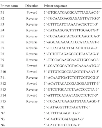
newly-obtained fragment was sequenced and used as the second starting point for PCR amplification. The same strategy was used to amplify unknown genes in the direction of the UL7 gene with primer P1 and the walking primers. After assembling the gene fragments, three sets of specific prim-ers (P10 and P11, P12 and P13, and P14 and P15) were de-signed according to the sequences obtained and used to amplify, sequence and confirm the genomic sequences. The primers used in this study are presented in Table 1.
PCR reactions were performed as previously de-scribed (Liet al., 2006). The PCR products were analyzed
on a 1.0% agarose gel and were sequenced either directly or after cloning into the pMD18-T vector (TaKaRa, Japan). Each region was sequenced at least three times from differ-ent PCR products.
ORF determination
To search for open reading frames (ORFs), the full-length assembled sequence was analyzed using the soft-ware Gene Runner (version 3.00, Hasting Softsoft-ware, Inc.). The predicted ORFs were confirmed by tblastx analysis for herpesviruses homologues. Deduced amino acid sequences identified from the DEV ORFs were compared with homo-logues of alpha-herpesviruses using the MegAlign program (DNAStar, version 7.0).
Analysis of promoter and polyadenylation signal locations
The assembled sequence of the DEV Clone-03 was submitted to the Berkeley Drosophila Genome project’s Neural Network Promoter Prediction, a eukaryotic core
promoter search engine
(http://www.fruitfly.org/seq_tools/promoter.html). The
initial search was performed at a high stringency (cutoff score of 0.85 out of 1.00). The program returned high-scoring core promoters (50-bp-long fragment) along with predicted transcription start sites (TSS). The core promot-ers found in this search were examined for the presence of Table 1- Primers used for PCR amplification.
Primer name Direction Primer sequence
P1 Foward 5’-GTGCATGAGGCATTTAGAAC-3’
P2 Reverse 5’-TGCAACGAGGAGAGTTATTG-3’
P3 Reverse 5’-GTTTCATCTAAATACGCTCT-3’
P4 Reverse 5’-TATAAGGGCTGTTTGGAGTG-3’
P5 Reverse 5’-TGCAAAGTACGGTCAAGTGA-3’
P6 Reverse 5’-AGGAGAAACATCCATAGAGT-3’
P7 Reverse 5’-TTTATAACTTACACTCTGGG-3’
P8 Reverse 5’-TCTCTTAGAGGCGTCAATAG-3’
P9 Reverse 5’-TTCCACAAGGAAGTTGCCAG-3’
U1 Foward 5’-CCATCGGATGTACAAAAATG-3’
P10 Foward 5’-GTTGTCGCCGAGGTGTAAAT-3’
P11 Reverse 5’-ACAAGTGATCTGTTCGTGCG-3’
P12 Foward 5’-ACATTACACGGAGGGAGTTT-3’
P13 Reverse 5’-GTCGTGCATCTAACCCCCTA-3’
P14 Foward 5’-ATTTCCATAATAGCCTCTCT-3’
P15 Reverse 5’-TGCAATGAAGATGTAGAAGC-3’
N1 5’-TATAGGTTT(C/A)TGTT-3’
N2 5’-CTTTTGGAGCTG-3’
N3 5’-GAATGTGA(A/g)AA-3’
N4 5’-CATGTCTGCCGA-3’
TATA box sequences using the TRANSFACFind search engine (http://motif.genome.jp/).
POLYADQ, a eukaryotic polyadenylation (poly A) signal search engine (Cold Spring Harbor Laboratory, http://rulai.cshl.org/tools/polyadq/polyadq_form.html) was used to predict transcription termination signals. All cutoff parameters were initially set at zero to return the lo-cation of all AATAAA and ATTAAA consensus signals, along with an associated score between 0 and 1. Signal pep-tide search (SignalP 3.0) and transmembrane prediction (ConPred II) of the deduced proteins were performed on-line.
The level of DNA identity between the Kozak’s con-sensus sequence (GCCGCCRCCATGG, R = A/g) (Kozak, 1986) and the flanking 13-nucleotides around the initiator AUG for each ORF was measured.
Phylogenetic analysis
Alignment was performed by MegAlign in
LASERGENE (DNAStar) with the ClustalV method. After a multiple alignment was completed, a neighbor-joining method was employed to reconstruct the phylogeny for the putative alignment of the DEV with 12 alpha-herpesvi-ruses. Reference strains and associated GenBank accession numbers were as follows: Herpes simplex virus 1 (HSV-1), X14112; Herpes simplex virus 2 (HSV-2), NC_001798; Varicella-zoster virus (VZV), NC_001348; Bovine herpes-virus 1 (BoHV-1), NC_001847; Bovine herpesherpes-virus 2 (BoHV-2), D00537; Equid herpesvirus 1 (EHV-1), AY464052; Equid herpesvirus 4 (EHV-4), NC_001844; Pseudorabies virus (PRV), NC_006151; Marek’s disease virus type 1 (MDV-1), NC_002229; Marek’s disease virus type 2 (MDV-2), NC_002577; Turkey herpesvirus (HVT), AF291866; Infectious laryngotracheitis virus (ILTV), NC_006623.
Nucleotide sequence accession number
The DNA sequences for theUL1throughUL7genes
of the DEV Clone-03 were submitted to the GenBank data-base and assigned the accession number EF449516.
Results
ORF determination, gene arrangement and predicted transcriptional elements
The full-length assembled sequence amplified by ‘targeted gene walking PCR’ is 10,374 bp long and
in-cludes the UL1, UL2,UL3,UL3.5, UL4,UL5,UL6 and
UL7genes. The arrangement of the eight genes and the
transcription orientation are shown in Figure 2. The gene
UL3.5, located betweenUL3andUL4, had no homologue
in the HSV-1 genome. TheUL1gene had a 140-nucleotide
overlap with theUL2 gene in the tail to head direction,
while theUL6gene had a 269-nucleotide overlap with the
UL7gene in a tail to head orientation.
No poly A sequences were found for theUL1,UL2
andUL3genes, but a common poly A signal for these three genes was found immediately downstream of the stop codon of theUL3.5gene. Although theUL1,UL2,UL3and
UL3.5genes shared the same transcription termination site, each gene had its own promoter for transcription initiation.
UL6andUL7shared the same poly A signal sequence
lo-cated downstream of theUL7gene. The promoters, TATA
box, poly A signal sequence and the transcription start sites of the eight genes were predicted (Table 2). The Kozak’s consensus sequence for each gene was used as a basis for
determining the start codon. The context of AUG inUL1,
UL3,UL5andUL7possessed the feature A (-3) and G (+4), but this feature could not be observed in the AUG context of other four genes.
Molecular characteristics of the genesUL1through
UL7in the DEV genome
We predicted that theUL1gene would encode a
pro-tein with 236 amino acids (aa). Amino acid alignment of the DEV UL1 protein with the same protein in 12 alpha-herpesviruses was performed. Various amino acids were conserved, including: Gly110, Val111, Phe112, His116, Cys117, Glu121, Leu124, Trp125, Ala132, Trp134, Asn136and Pro137.One transmembrane region located from aa 40 through 60 (N-terminus in) of the UL1 protein was predicted. Although the UL1 homologue in the HSV-1 was designated glyco-protein L (gL), no N-linked glycosylation site was pre-dicted in the DEV UL1 protein. Signal peptide analysis showed that the UL1 protein was a signal anchor protein.
TheUL2gene encoded 157 aa protein and overlapped
the 3’-terminus of theUL1 gene. The DEV UL2 protein
was the shortest among the homologues of the 12 reference alpha-herpesviruses. Blast analysis of the amino acid se-quence of the UL2 protein showed that it was similar to the Figure 2- Comparison of the gene content and organization of UL1 through UL7 between the DEV and species of four genera in the subfamily
DNA glycosylase of herpesviruses, but no uracil-DNA glycosylase signature consensus was found in the DEV UL2 protein. Eight conserved regions (Figure S1a), mainly located at the C-terminus of the protein, were identi-fied.
UL3 was a conserved protein with 239 aa. Eight ca-sein kinase II phosphorylation sites (Thr22, Thr27, Ser51, Thr80, Ser90, Thr94, Ser110, and Ser111) were predicted. The nuclear localization signal (NLS) at the C-terminus of the UL3 protein (193-RKPRK-197) was highly conserved among alpha-herpesviruses, in addition to other six con-served regions (Figure S1b).
UL3.5 encoded a 120 aa protein with a molecular weight of 13.4 kDa. The location of this gene was similar to
that of theUL3.5 gene in the EHV-1, EHV-4, BoHV-1,
PRV and MDV-2. Amino acid alignments of the UL3.5 protein with these six virus strains showed weak homology.
Blast analyses showed that the DEVUL3.5gene was not
homologous to a herpesvirus gene. The putative amino acid sequence of theUL3.5gene had a higher identity level with the transmembrane 7 superfamily member 3.
TheUL4gene encoded a 237 aa protein and had little homology with other alpha-herpesviruses homologues (data not shown). Two N-linked glycosylation sites were predicted at amino acids 45 and 223. A 19-aa signal peptide at the N-terminus of the UL4 protein with a cleavage site between the 18th and 19th aa was predicted.
The product of theUL5gene in the DEV encoded a
855 aa protein and was highly conserved compared with those in other alpha-herpesviruses. Six conserved helicase motifs were predicted in the UL5 protein. Amino acid alignments showed extensive conserved regions (Figure S1c).
TheUL6gene encoded a 790 aa protein which shared
a high degree of homology with homologues of 11 al-pha-herpesviruses. A leucine zipper motif with the consen-sus sequence L-X6-L-X6-L-X6-L (where X is an arbitrary amino acid) was found in the UL6 protein (463-LESYVNNLFKTIEGLKETNGEL-484), which was con-sistent with the homologous proteins of other alpha-herpes-viruses (Figure S1d). In addition, one transmembrane region containing 21 residues was predicted from aa 606 through 626 (N-terminus out).
The UL7 protein was composed of 321 aa. While no common motifs were shared with the UL7 homologues of 12 alpha-herpesviruses, amino acids 218-LNT-220 and 235-VLP-237 were completely identical among the 12 al-pha-herpesviruses. These two sites have unknown func-tions.
Phylogenetic analysis of DEV
Phylogenetic trees were constructed based on the alignments of eight DEV proteins with their homologues in 12 alpha-herpesviruses (Figure 3). In all trees, the genera
Simplexvirus, Varicellovirus, Mardivirus and Iltovirus
were distinguished as different clusters. In general, the
DEV was most closely related toMardivirus, although it
formed a distinct branch. In the UL1, UL3, UL3.5, UL5,
and UL7 trees, DEV was closer toMardivirus. The
phylo-genetic tree of the UL2 protein showed a closer relationship to the cluster ofSimplexvirus, and the branches of the UL4 and UL6 proteins were very distant fromMardivirus.
Discussion
In this paper we reported partial DNA sequences of the unique long region of the DEV genome containing the
UL1,UL2,UL3,UL3.5,UL4,UL5,UL6andUL7genes. The gene arrangement was collinear to their homologues in
the HSV-1, except for the UL3.5 gene being absent in
HSV-1. We analyzed the gene content and gene
organiza-tion ofUL1throughUL7from the typical species of four
genera ofAlphaherpesvirinae (Figure 2). Gene
transcrip-tion orientatranscrip-tion ofUL1throughUL7was identical among
five herpesviruses. A common feature was the overlapping
ofUL1andUL2, and ofUL6andUL7.There were overlaps
of theUL5andUL6genes in HSV-1, VZV and ILTV, but
not in MDV-1 and in DEV. In general, gene organization of DEV was more similar to that of MDV-1. To elucidate the taxonomic position of DEV, we performed a comparative analysis of the DEV UL1, UL2, UL3, UL3.5, UL4, UL5, UL6 and UL7 proteins and their counterparts in other virus subfamilies. Our initial analysis was within the family
Herpesviridae, which showed that DEV was grouped in the
subfamilyAlphaherpesvirinae(data not shown). A second
phylogenetic analysis focused on members of the
sub-familyAlphaherpesvirinaewas performed in order to
fur-ther identify the genus of DEV. Phylogenetic trees of the eight proteins of DEV showed a closer relationship to
Mardivirus, but formed a single branch within the sub-familyAlphaherpesivrinae.
In general, most HSV genes studied to date were not spliced and were preceded by separate promoter elements including a TATA box. Many HSV ORFs are followed by a poly A signal, but families of HSV genes have also been shown to share one poly A site (McGeogh, 1991). TheUL1,
UL2andUL3genes of HSV-1 and HSV-2 are each
pre-ceded by a TATA box, and there are poly A signals
down-stream ofUL2andUL3(McGeoghet al., 1988; Worrad and
Caradonna, 1988). In the DEV genome, theUL1,UL2,UL3
and UL3.5 genes shared the same poly A signal down-stream ofUL3.5, and theUL6andUL7genes shared a poly
A sequence downstream ofUL7.
In a eukaryotic cell, the core of the Kozak’s consen-sus is R-3CCAUGG+4(R = A/g). R-3is considered the most important flanking residue, followed by G+4(Kozak, 1987).
The flanking sequence of AUG inUL1,UL3,UL5andUL7
possessed the important feature of A-3and G+4that are criti-cal for recognition of AUG by the 40S ribosomal subunit,
while the DEV UL2, UL3.5, UL4 and UL6 contexts of
AUG did not fit the Kozak rule. We compared the homolo-gous genes in MDV-1 and PRV and not all genes were in good AUG context with Kozak’s rule. Although the nu-cleotides adjacent to AUG (CCRCCAUGG, purine at posi-tion -3 and G at posiposi-tion +4) are known to be more impor-tant for efficient translation than the rest of the Kozak’s consensus, the secondary structure of the RNA can affect the efficiency of translation (Pelletier and Sonenberg, 1987). Further experiments are needed to confirm the true translation start codon.
DEV UL1 did not show significant sequence homo-logy with the HSV-1 UL1 protein, but the gene location and
the orientation of UL1were equivalent for both viruses.
The product of the HSV-1UL1gene is the essential glyco-protein gL (Hutchinsonet al., 1992; Roopet al., 1993).
Ini-tial analysis of the DEV UL1 protein did not indicate that an N-glycosylation site was present, however further experi-mental evidence is needed to determine if the UL1 protein is a glycoprotein. HSV-1 gL has been shown to complex with the viral glycoprotein gH and is required for the
pro-cessing and cell surface expression of gH (Hutchinsonet
al., 1992). The molecular characteristics of DEV gH have been reported (Liet al., 2006) and additional experiments will be required to determine whether the product of DEV UL1 interacts with gH.
DEV UL2 is a homologue of the HSV-1 UL2 protein, which encodes a non-essential enzyme with uracil-DNA glycosylase activity (Worrad and Caradonna, 1988; Mulla-neyet al., 1989). The UL2 protein of DEV had no uracil-DNA glycosylase signature sequence. In HSV-1, interrup-tion of theUL2ORF by lacZ insertion mutagenesis resulted in a virus which grew normally in tissue culture yet was negative for uracil-DNA glycosylase activity (Mullaneyet al., 1989).
DEV UL3 had significant homology with other 12 al-pha-herpesviruses. The UL3 protein had eight casein ki-nase II phosphorylation sites and one NLS (RKPRK) at the C-terminus. We predict that the DEV UL3 protein may be a nuclear phosphoprotein similar to the UL3 protein in
HSV-2 (Worrad and Caradonna, 1993). TheUL3gene is
dispensable for viral replication in cell culture in PRV and
HSV-1 (Baines and Roizman, 1991; Klupp et al., 2004)
which provides a reason for studying the role of theUL3
gene in DEV.
The deduced amino acid sequence of DEVUL3.5had
no detectable homology to that of herpesviruses, while it showed high identity with the transmembrane 7 super-family member 3. Nevertheless, a similar gene location was found for this gene in PRV, BoHV-1, EHV-1, EHV-4 and MDV-2. In PRV, UL3.5 has been shown to be involved in virus egress (Fuchset al., 1996), but we could not speculate on the function of DEV UL3.5 because of their low homo-logy.
DEV UL4 was predicted to be a glycoprotein with
three transmembrane regions. TheUL4gene in HSV-1 and
in PRV is dispensable for virus replication (Fuchset al., 2006), but the role of theUL4gene in the DEV life-cycle remains to be determined.
DNA unwinding (Biswas and Weller, 2001). We thus pre-dict that DEV UL5 is a helicase-primase protein.
As a minor capsid protein in HSV-1, UL6 forms the portal for entry of DNA into the capsid (Newcombet al.,
2001). TheUL6gene of DEV has been used as a target
se-quence to inhibit virus replication (Mallannaet al., 2006), although the function of its product in the DEV life-cycle is still unknown.
Our homology analysis suggested that the product of
the UL7gene was not a highly conserved protein in
al-pha-herpesviruses. The UL7 protein in PRV and in HSV-2 proved to be a structural protein which affected virion for-mation and virus egress, but the UL7 gene was not consid-ered essential for virus replication (Fuchset al., 2005).
Despite the large genome of herpesviruses, not all genes are essential for virus replication in cell culture. Until
now, theUL3,UL4andUL7genes of HSV-1 and of PRV
have been reported to be dispensable for virus replication in
cell culture (Baines and Roizman, 1991; Fuchs et al.,
2005). Determining if these genes are essential for DEV replication will be important to decide on the use of DEV as a vaccine vector.
References
Baines J and Roizman B (1991) The open reading frames UL3, UL4, UL10, and UL16 are dispensable for the replication of herpes simplex virus 1 in cell culture. J Virol 65:938-944. Baudet AERF (1923) Mortality in ducks in the Netherlands
caused by a filtrable virus; fowl plague. Tijdschr Dier-geneeskd 50:455-459.
Biswas N and Weller SK (2001) The UL5 and UL52 subunits of the herpes simplex virus type 1 helicase-primase subcom-plex exhibit a comsubcom-plex interdependence for DNA binding. J Biol Chem 276:17610-17619.
Burgess EC, Ossa J and Yuill M (1979) Duck plague: A carrier state in waterfowl. Avian Dis 23:940-949.
Davison S, Converse KA, Hamir AN and Eckroade RJ (1993) Duck viral enteritis in Muscovy ducks in Pennsylvania. Avian Dis 37:1142-1146.
Fauquet CM, Mayo MA, Maniloff J, Desselberger U and Ball LA (2005) Virus taxonomy: Eighth report of the international committee on taxonomy of viruses. Elsevier Academic Press, California, 208 pp.
Fuchs W, Klupp BG, Granzow H, Rziha HJ and Mettenleiter TC (1996) Identification and characterization of the pseudo-rabies virus UL3.5 protein, which is involved in virus egress. J Virol 70:3517-3527.
Fuchs W, Granzow H, Klopfleisch R, Klupp BG, Rosenkranz D and Mettenleiter TC (2005) The UL7 gene of pseudorabies virus encodes a nonessential structural protein which is in-volved in virion formation and egress. J Virol 79:11291-11299.
Fuchs W, Granzow H, Klopfleisch R, Klupp BG and Mettenleiter TC (2006) The UL4 gene of pseudorabies virus encodes a minor infected-cell protein that is dispensable for virus rep-lication. J Gen Virol 87:2517-2525.
Gardner R, Wilkerson J and Johnson JC (1993) Molecular charac-terization of the DNA of anatid herpesvirus 1. Intervirology 36:99-112.
Hansen WR, Brown SE, Nashold SW and Knudson DL (1999) Identification of duck plague virus by polymerase chain re-action. Avian Dis 43:106-115.
Huang YX (1959) Study on duck plague-like disease. Hua Nan Nong Ye Da Xue Xue Bao 1:1-12.
Hutchinson L, Browne H, Wargent V, Davis-Poynter N, Primorac S, Goldsmith K, Minson AC and Johnson DC (1992) A novel herpes simplex virus glycoprotein, gL, forms a com-plex with glycoprotein H (gH) and affects normal folding and surface expression of gH. J Virol 66:2240-2250. Kaleta EF (1990) Herpesviruses of birds - A review. Avian Pathol
19:193-211.
Klupp BG, Granzow H, Fuchs W, Mundt E and Mettenleiter TC (2004) Pseudorabies virus UL3 gene codes for a nuclear pro-tein which is dispensable for viral replication. J Virol 78:464-472.
Kozak M (1986) Point mutations define a sequence flanking the AUG initiator codon that modulates translation by euka-ryotic ribosomes. Cell 44:283-292.
Kozak M (1987) The scanning model for translation: An update. J Cell Biol 108:229-241.
Li H, Liu S and Kong X (2006) Characterization of the genes en-coding UL24, TK and gH proteins from duck enteritis virus (DEV): A proof for the classification of DEV. Virus Genes 33:221-227.
Liu S, Chen S, Li H and Kong X (2007) Molecular characteriza-tion of the herpes simplex virus 1 (HSV-1) homologues, UL25 to UL30, in duck enteritis virus (DEV). Gene 401:88-96.
Mallanna SK, Rasool TJ, Sahay B, Aleyas AG, Ram H, Mondala B, Nautiyal B, Premraj A, Sreekumar E and Yadav MP (2006) Inhibition of anatid herpes virus-1 replication by small interfering RNAs in cell culture system. Virus Res 115:192-197.
McGeogh DJ (1991) Correlation between HSV-1 DNA sequence and viral transcription maps. In: Wagner EK (eds) Herpes-virus Transcription and its Regulation. CRC Press, Boca Raton, pp 29-48.
McGeogh DJ, Dalrymple MA, Davison AJ, Dolan A, Frame MC, McNab D, Perry LJ, Scott JE and Taylor P (1988) The com-plete DNA sequence of the long unique region in the ge-nome of herpes simplex virus type 1. J Gen Virol 69:1531-1574.
Mullaney J, Moss HW and McGeogh DJ (1989) Gene UL2 of her-pes simplex virus type 1 encodes a uracil-DNA glycosylase. J Gen Virol 70:449-454.
Newcomb WW, Juhas RM, Thomsen DR, Homa FL, Burch AD, Weller SK and Brown JC (2001) The UL6 gene product forms the portal for entry of DNA into the herpes simplex vi-rus capsid. J Virol 75:10923-10932.
Parker JD, Rabinovitch PS and Burmer GC (1991) Targeted gene walking polymerase chain reaction. Nucleic Acids Res 19:3055-3060.
Plummer PJ, Alefantis T, Kaplan S, O’Connell P, Shawky S and Schat KA (1998) Detection of duck enteritis virus by poly-merase chain reaction. Avian Dis 42:554-564.
Roop C, Hutchinson L and Johnson DC (1993) A mutant herpes simplex virus type 1 unable to express glycoprotein L cannot enter cells, and its particles lack gH. J Virol. 67:2285-2297. Shawky S and Schat K (2002) Latency sites and reactivation of
duck enteritis virus. Avian Dis 46:308-313.
Worrad DM and Caradonna S (1988) Identification of the coding sequence for herpes simplex virus uracil-DNA glycosylase. J Virol 62:4774-4777.
Worrad DM and Caradonna S (1993) The herpes simplex virus type 2 UL3 open reading frame encodes a nuclear localizing phosphoprotein. Virology 195:364-367.
Zhu L and Weller SK (1992) The six conserved helicase motifs of the UL5 gene product, a component of the herpes simplex virus type 1 helicase-primase, are essential for its function. J Virol 66:469-479.
Internet Resources
ConPred II, http://bioinfo.si.hirosaki-u.ac.jp/~ConPred2/ (Octo-ber 9, 2008) A Prediction Server of Transmembrane (TM) Topology, http://bioinfo.si.hirosaki-u.ac.jp/~ConPred2/. SignalP 3.0, http://www.cbs.dtu.dk/services/SignalP/ (October 9,
2008) SingalP 3.0 Server,
http://www.cbs.dtu.dk/cgi-bin/webface?jobid=signalp,48E D788400B31BA9&opt=none.
Supplementary Material
The following online material is available for this ar-ticle:
Figure S1 - Amino acid sequence alignment of the UL2, UL3, UL5, and UL6 genes of the DEV Clone-03 with the corresponding homologues of reference strains in the
subfamilyAlphaherpesvirinae.(a) For UL2, the signature
consensus for uracil-DNA glycosylase is underlined and eight conserved regions are boxed; (b) For UL3, six con-served regions are boxed and the nuclear localization signal is underlined; (c) For UL5, six conserved helicase motifs and their locations are presented; (d) For UL6, partial amino acid alignment is shown and the leucine zipper mo-tifs are boxed. Black shades indicate alignments with con-servation between 80 and 100% and grey shades represent less than 80% conservation but more than 60% (generated by the software GenDoc).
This material is available as part of the online article from http://www.scielo.br/gmb.
Associate Editor: Luis Carlos de Souza Ferreira