861 861 861 861 861 Mem Inst O swaldo Cruz, Rio de Janeiro, Vol. 100(8): 861-867, D ecem ber 2005
Shared and non-shared antigens from three different extracts of
the metacestode of
Echinococcus granulosus
D avid Carmena/+ +, Jorge M artínez, Aitziber Benito, Jorge A Guisantes/+
Departamento de Inmunología, Microbiología y Parasitología, Facultad de Farmacia, Universidad del País Vasco, Apartado 450, 01080-Vitoria, España
Hydatid cyst fluid (HCF), somatic antigens (S-Ag) and excretory-secretory products (ES-Ag) of Echinococcus granulosus protoscoleces are used as the main antigenic sources for immunodiagnosis of human and dog echino-coccosis. In order to determine their non-shared as well as their shared antigenic components, these extracts were studied by ELISA-inhibition and immunoblot-inhibition. Assays were carried out using homologous rabbit polyclonal antisera, human sera from individuals with surgically confirmed hydatidosis, and sera from dogs naturally infected with E. granulosus. High levels of cross-reactivity were observed for all antigenic extracts, but especially for ES-Ag and S-Ag. Canine antibodies evidenced lesser avidity for their specific antigens than antibodies from human origin. The major antigenic components shared by HCF, S-Ag, and ES-Ag have apparent molecular masses of 4-6, 20-24, 52, 80, and 100-104 kDa, including doublets of 41/45, 54/57, and 65/68 kDa. Non-shared polypeptides of each anti-genic extract of E. granulosus were identified, having apparent masses of 108 and 78 kDa for HCF, of 124, 94, 83, and 75 kDa for S-Ag, and of 89, 66, 42, 39, 37, and 35 kDa for ES-Ag.
Key words: Echinococcus granulosus - hydatid cyst fluid - somatic antigens - excretory-secretory antigens - cross-reactivity
Echinococcosis caused by cestodes of the genus Echi-nococcus is one of the major zoonotic helminthiosis, caus-ing considerable socio-economic consequences in en-demic areas. Due to its world-wide distribution and its important impact in both human and animal health, E. granulosus is considered the most relevant species (Romig 2003). The adult worm lives in the small intestine of dogs and other canids, in intimate contact with the intestinal epithelium. The intermediate larval stage (metacestode) can grow in a wide range of mammal spe-cies including humans, that acquire infection through accidental ingestion of eggs. Currently, diagnosis of hy-datidosis/echinococcosis is based on a combination of imaging techniques (ultrasonography, computerized axial tomography, X-rays) and immunodiagnostic methods such as ELISA and immunoblotting (Zhang et al. 2003).
For immunodiagnosis of human hydatidosis and dog echinococcosis, hydatid cyst fluid (HCF), somatic anti-gens (S-Ag) and excretory-secretory products (ES-Ag) of E. granulosus protoscoleces and adult worms are used as main antigenic sources. The choice of the most appro-priate antigenic extract depends on the developmental stage of the worm and the host species. Thus, HCF is mainly used for the immunodiagnosis of human cystic echinococcosis, based on the detection of antigen 5 and
Financial support: FIS, Ministry of Public Health and DEMSAC, Town Council of Vitoria, Department of Public Health, Basque Government, Spain
+Corresponding author. E-mail: oipgudej@vc.ehu.es
++Present address: MRC Clinical Sciences Centre, Membrane Transport Biology Group, Hammersmith Hospital Campus, Du Cane Road, London W12 0NN, UK
Received 2 August 2005 Accepted 23 November 2005
antigen B. The subunit of 8/12 kDa from antigen B is con-sidered the most specific component of HCF in the genus
Echinococcus (Verastegui et al. 1992, Zhang et al. 2003).
S-Ag from protoscoleces have been used for the serodi-agnosis of dog echinococcosis, as protoscoleces are the infective stage of the parasite in the definitive host. How-ever, because of their variable diagnostic sensitivity and high cross-reactivity levels with antigens from other para-site species, S-Ag are unreliable for serodiagnostic pur-poses (Gasser et al. 1988, 1994, Jenkins et al. 1990, Lightowlers & Gottstein 1995). In the last few years, ES-Ag from protoscoleces has become the main antigenic source for the immunodiagnosis of dog echinococcosis, based on the detection of parasite antigens in fecal samples (coproantigens) by ELISA (Jenkins et al. 2000, Benito & Carmena 2005).
Currently, there is very little information available about the recognition of antigenic components from different extracts of E. granulosus by sera from infected
individu-als of diverse species (Auer et al. 1988, Gasser et al. 1992). In this paper we present a comparative analysis of the cross-reactive antigenic components of HCF, S-Ag, and ES-Ag from protoscoleces of E. granulosus by ELISA-inhibition and immunoblot-ELISA-inhibition assays, in order to determine the non-shared and shared antigenic compo-nents of these extracts. These data may provide valuable information for the identification and isolation of specific antigenic components from the metacestode of E. granulosus for immunodiagnostic purposes.
MATERIALS AND METHODS
862 862 862 862
862 Shared and non-shared Ag in E. granulosus • D avid Carmena et al.
HCF was centrifuged at 6500 × g for 30 min, lyophilized and stored at 4ºC. To perform this study a pool of hydatid fluid from several large cysts from a single sheep lung was used. The concentration of proteins was 736 mg g-1
dry weight.
S-Ag - S-Ag were also prepared from protoscoleces
obtained by aseptic puncture from fertile hydatid cysts of ovine origin, washed with phosphate-buffered saline (PBS) and stored at –25ºC with proteolytic enzyme inhibitors (2 mM PMSF and 5 mM EDTA). Protoscoleces were thawed and sonicated (10 cycles of 12 s at 60 Hz frequency), freeze-thawed once more and centrifuged for 35 min at 2300 × g. Supernatants were aliquotted and stored at –25ºC. To perform this study a batch of protoscoleces from a single sheep liver infected with multiple hydatid cysts was se-lected. Protoscoleces had a viability of 97.4% at the time of their extraction from the cyst. The concentration of proteins was 2.95 mg ml-1.
ES-Ag - To obtain ES-Ag, a total of 10 cultures of
protoscoleces from sheep liver were carried out. Initial viability was assessed by morphological appearance, flame cell motility and general contractile movements (Howell 1986). Protoscoleces were cultured in PBS comple-mented with 10% glucose, 100 U ml-1 penicillin and 100 µg
ml-1 streptomycin at 37ºC in 5% CO
2 (Carmena et al. 2002).
The medium was renewed every 8 h and, after 50 h of culture, concentrated using filters with a 5 kDa pore diam-eter membrane (Ultrafree 15, Millipore). EDTA (5 mM) and PMSF (2 mM) were added, and the ES products were aliquotted and stored at –25ºC. To perform this study, a batch of protoscoleces with an initial viability of 95.2% was obtained from a single liver infected with multiple cysts. The concentration of protein was 0.2 mg ml-1.
Human sera - Thirty two pre- and post-surgery sera
from 11 individuals with confirmed liver hydatidosis were assayed by ELISA (HCF as solid phase) for levels of E. granulosus specific antibodies. Sera from the five patients with the highest absorbance values in ELISA assays were pooled and used as positive control in the inhibition as-says.
Dog sera - Dog sera were collected from the Council
Animal Rescue Mission, Vitoria, Spain. Five sera from dogs naturally infected with E. granulosus were obtained. All animals were diagnosed by autopsy. Sera from the four infected dogs with the highest absorbance values in ELISA assays using S-Ag as solid phase (Benito et al. 2001) were pooled and used as positive control in the inhibition as-says.
Hyperimmune rabbit sera - Polyclonal immunosera anti-HCF, anti-S-Ag, and anti-ES-Ag were obtained ac-cording to Gallart et al. (1985). Titration of rabbit antisera was performed by ELISA, using their homologous anti-genic extracts as solid phase.
ELISA-inhibition assays - ELISA was carried out as described by Martínez et al. (1985). Briefly, polystyrene 96-well microtitre plates (Maxisorp™, Nunc, Roskilde, Denmark) were coated with 100 µl/well of the optimal an-tigen concentrations determined according to Muñoz et
al. (1986) (HCF: 10.5 µg ml-1; S-Ag: 7.5 µg ml-1; ES-Ag: 20
µg ml-1) diluted in PBS buffer and incubated for 3 h at
room temperature (human ELISA) or 15 h at 4ºC (canine ELISA). After blocking with 1% BSA-0.05% Tween 20 in PBS, wells were filled with 50 µl/well of each inhibitory antigenic extract in PBS (serial dilutions ranging from: HCF: 3 to 0 mg ml-1; S-Ag: 1.6 to 0 mg ml-1; ES-Ag: 0.2 to 0
mg ml-1). Fifty µl/well of the tested pooled sera were added
at dilutions that had previously been optimized and incu-bated for 3 h at 37ºC. Dilutions of the first antibodies were 1:800 for pooled sera from hydatid patients and 1:50 for pooled sera from dogs with E. granulosus infections. Wells were then incubated with peroxidase-conjugated rabbit anti-human IgG (Dako, 1:2500 dilution) or anti-dog IgG (Sigma, 1:1000 dilution) in 0.05% Tween 20-PBS for 15 h at 4ºC. After washing, plates were developed with MBTH-DMAB solution at room temperature for 20 min. The reaction was stopped with 50 µl/well of 2N H2SO4 and the OD was measured at 620 nm. Assays were per-formed in triplicates. OD values from tested sera versus inhibitor concentrations were graphically depicted and percentages of inhibition were calculated with the follow-ing formula:
% inhibition[x] = 100 – (OD[x] / ODmax. × 100) where OD[x] is the absorbance value corresponding to the assayed inhibitor concentration, and ODmax. is the absorbance value corresponding to an inhibitor concen-tration equal to zero. In addition, values of Ag50 (antigen concentration required to inhibit 50% of the specific sera antibodies assayed), coefficient of linear correlation, slope of the regression line, and theoretical maximum percent-age of inhibition were also calculated for each inhibition reaction (Martínez et al. 1985).
Immunoblotting and immunoblot-inhibition assays -Proteins from HCF, S-Ag, and ES-Ag were fractionated by 12.5% SDS-PAGE under reducing conditions accord-ing to Laemmli (1970) and transferred to polyvinylidene difluoride (PVDF) membranes (Immobilon-P, Millipore), according to Towbin et al. (1979). After washing and block-ing, membranes were incubated with the sera in 20 mM Tris-buffered saline (pH 7.4), 8% skimmed milk (TBS-M) for 15 h at 4ºC. Optimal sera dilutions used were: rabbit anti-HCF: 1:1000; rabbit anti-S-Ag: 1:1000; rabbit anti-ES-Ag: 1:500; pooled sera from hydatid patients: 1:800; pooled sera from dogs with E. granulosus infections: 1:100.
Per-oxidase-conjugated swine anti-rabbit total Ig (Dako, Copenhagen, Denmark), rabbit anti-human IgG (Dako) and rabbit anti-dog IgG (Sigma) were used at 1:1000 dilution in TBS-M buffer for 4 h at room temperature. Membranes were revealed by adding 4-chloro-1-napthol solution. The immunoblotting inhibition assays were performed accord-ing to Asturias et al. (1999). Sera used in these assays were previously preabsorbed with each inhibitory anti-genic extract (HCF and S-Ag: 1 mg ml-1; ES-Ag: 0.2 mg
ml-1) for 30 min at 37 ºC with gentle shaking, and then
pat-863 863 863 863 863 Mem Inst O swaldo Cruz, Rio de Janeiro, Vol. 100(8), D ecem ber 2005
terns were analyzed and apparent molecular masses were estimated by meassuring the relative mobility of each band of interest, and interpolating the data in the standard curve. Proteins which were identified by at least four of the five inhibited antisera assayed (considering each immu-noserum used in the inhibition reaction independently) were regarded as overall shared components.
RESULTS
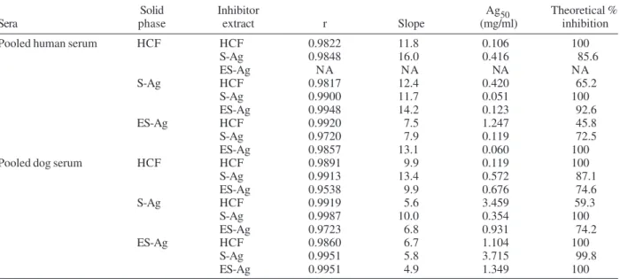
ELISA-inhibition assays - Table I summarizes the ELISA-inhibition results obtained for the pooled serum from hydatid patients and for the pooled serum from dogs infected with E. granulosus both of them being inhibited
with the antigenic extracts HCF, S-Ag, and ES-Ag. Values corresponding to correlation coefficients (r), slopes of the regression lines, Ag50, and theoretical maximum per-centages of inhibition are shown. According to the analy-sis of these data, high cross-reactivity was mainly ob-served for S-Ag and ES-Ag. Furthermore, the pool of dog sera showed considerably higher Ag50 values than the pool of human sera, when assayed under the same condi-tions.
Immunoblotting and immunoblot-inhibition assays -Fig. 1 shows the antigenic profiles of HCF, S-Ag, and ES-Ag when using the different non-inhibited and inhibited immunosera. For better comprehension, the patterns of the protein bands are also presented in a schematic fig-ure. Thirteen polypeptides ranging from 9 to 108 kDa were identified in the HCF when the non-inhibited rabbit serum against HCF was used (Fig. 1-A). Antigens of 108 and 78 kDa were specific for HCF (rabbit antibodies against these polypeptides did not bind to any S-Ag or ES-Ag compo-nents). The non-inhibited rabbit antiserum against S-Ag recognized 13 antigenic components ranging from 6 to
124 kDa when S-Ag was used as solid phase. Polypep-tides of 124, 94, 83, and 75 kDa were found to be specific for this antigenic extract (Fig. 1-B). Finally, the non-inhib-ited rabbit serum against ES-Ag reacted specifically with at least 14 components from the ES-Ag, some of which (89, 66, 42, 40, 37, and 35 kDa) are neither shared by S-Ag nor by HCF (Fig. 1-C). In addition, immunoblot-assays were carried out using each rabbit antiserum against its heterologous antigenic extracts as solid phase.
Table II summarizes the molecular masses of the major antigenic proteins shared by HCF, S-Ag, and ES-Ag, con-sidering each immunoserum used in the inhibition reac-tion independently. The antigenic components identified as shared components had apparent molecular masses of 4-6, 20-24, 52, 80, and 100-104 kDa, including doublets of 41/45, 54/57, and 65/68 kDa.
DISCUSSION
Cyst hydatid fluid and somatic antigens of pro-toscoleces are the best characterized antigenic extracts of
E. granulosus, and the main antigenic sources used for immunodiagnosis of human and dog echinococcosis (Rickard & Lightowlers 1986, Lightowlers & Gottstein 1995). During recent years, ES-Ag have acquired a role in the diagnosis of infections in the definitive host, based on the detection of these antigens in faeces by ELISA (Fraser & Craig 1997, Jenkins et al. 2000, Benito & Carmena 2005). So far, little information is available in regard to the description of ES-Ag. We have recently carried out the biochemical characterization of this antigenic extract, evaluating its potential for immunodiagnosis of human cystic echinococcosis and dog echinococcosis (Carmena et al. 2004, 2005). However, until now no studies have been performed to determine the homology degree among
TABLE I
Results of the ELISA-inhibition assays for the pool of human sera from individuals with confirmed hydatidosis and for the pool of sera from dogs naturally infected with Echinoccus granulosus
Solid Inhibitor Ag50 Theoretical %
Sera phase extract r Slope (mg/ml) inhibition
Pooled human serum HCF HCF 0.9822 11.8 0.106 100
S-Ag 0.9848 16.0 0.416 85.6
ES-Ag NA NA NA NA
S-Ag HCF 0.9817 12.4 0.420 65.2
S-Ag 0.9900 11.7 0.051 100
ES-Ag 0.9948 14.2 0.123 92.6
ES-Ag HCF 0.9920 7.5 1.247 45.8
S-Ag 0.9720 7.9 0.119 72.5
ES-Ag 0.9857 13.1 0.060 100
Pooled dog serum HCF HCF 0.9891 9.9 0.119 100
S-Ag 0.9913 13.4 0.572 87.1
ES-Ag 0.9538 9.9 0.676 74.6
S-Ag HCF 0.9919 5.6 3.459 59.3
S-Ag 0.9987 10.0 0.354 100
ES-Ag 0.9723 6.8 0.931 74.2
ES-Ag HCF 0.9860 6.7 1.104 100
S-Ag 0.9951 5.8 3.715 99.8
ES-Ag 0.9951 4.9 1.349 100
864 864 864 864
864 Shared and non-shared Ag in E. granulosus • D avid Carmena et al.
the antigenic components of HCF, S-Ag, and ES-Ag, and to find out their non-shared and shared proteins.
ELISA-inhibition results using both the pool of hu-man sera from individuals with confirmed hydatidosis and the pool of sera from dogs infected with E. granulosus
have shown a great resemblance with regard to the slopes of the regression lines corresponding to each antigenic extract used as solid phase. This fact indicates that there are antigens shared by HCF, S-Ag, and ES-Ag. The high-est cross-reactivity was observed between S-Ag and ES-Ag. These results are supported by Ag50 values, since inhibition of 50% of the specific serum antibodies in each
inhibition reaction test required higher concentrations of HCF antigens than of S-Ag and ES-Ag. Interestingly, the pool of dog sera showed considerably higher Ag50 values than the pool of human sera, when assayed under the same conditions. These results demonstrate that dog an-tibodies have a lower avidity for their specific antigens than antibodies of human origin. This phenomenon may be explained by the concept of antibody affinity matura-tion through the course of species evolumatura-tion. It is known that less evolved species have antibodies with lower af-finity for their specific epitopes than higher evolved spe-cies (Du Pasquier 2001, Frank 2002). We consider that this
865 865 865 865 865 Mem Inst O swaldo Cruz, Rio de Janeiro, Vol. 100(8), D ecem ber 2005
may be one of the several reasons why a lower sensitivity was observed in assays for the serodiagnosis of dog echi-nococcosis as compared to assays for the immunodiag-nosis of human cystic echinococcosis.
Immunoblotting and immunoblot-inhibition assays were carried out in order to describe the profiles of the non-shared and shared antigenic components among the different extracts of E. granulosus studied: HCF, S-Ag, and ES-Ag. Each antigenic extract was assayed against its homologous and heterologous rabbit antisera, the pool of human sera from individuals with confirmed hydatido-sis and the pooled sera from dogs infected with E. granulosus, with and without inhibition. It is necessary take into consideration that the reported molecular mass estimations can have slight inaccuracies as a consequence of the limitations of the measurement method used. Among the shared components of the three antigenic extracts, the polypeptide of 4-6 kDa corresponds very likely to the 8 kDa subunit of the AgB. This component is strongly recognized by the non-inhibited pool of sera from patients infected with cystic echinococcosis and the rabbit sera anti-HCF/S-Ag, but not by the non-inhibited pool of sera from dogs infected with E.granulosus (Fig. 1-A, B). This
fact confirms that AgB is present in the metacestode of the parasite, but not in the adult stage. Similarly, the polypeptide of 20-24 kDa may correspond to subunits of Ag 5 or Ag B, and the component of 41 kDa may match the major subunit of Ag 5 (Lightowlers et al. 1989, Ortona et al. 1995, González et al. 1996). Some of the products
identified in HCF by the pool of human sera in the present study may correspond to those previously described by other authors using the same antigenic extract. This may be the case of the protein of 34 kDa (recently identified by Poretti et al. 1999), and the protein of 110 kDa, probably related with the component of 100 kDa described by Shambesh et al. (1995), the protein of 110-120 kDa (Shapi-ro et al. 1992), and the p(Shapi-rotein of 116 kDa (Kanwar et al. 1992).
Some of the immunoblot-inhibition assays showed some bands in the negative controls, indicating that the inhibition reaction was not complete. This fact was de-tected more frequently when ES-Ag were used as inhibi-tory extract, and may probably be due to a lower concen-tration of protein used in the inhibition reaction.
Non-shared antigenic components of each extract from
E. granulosus (HCF, S-Ag, and ES-Ag) were determined
by analysing the profiles obtained by immunoblotting and comparing them with profiles that were obtained when the antisera were inhibited with the different antigenic extracts tested. HCF evidenced two non-shared compo-nents of 108 and 78 kDa, respectively. S-Ag showed four non-shared polypeptides with apparent molecular masses of 124, 94, 83, and 75 kDa. On the other hand, ES-Ag showed non-shared antigenic components of 89, 66, 42, 40, 37, and 35 kDa. The protein of 89 kDa has already demonstrated specificity for immunodiagnosis of human cystic echinococcosis and dog echinococcosis in previ-ous reports (Carmena et al. 2004, 2005). This component
TABLE II
Major antigenic components shared by hydatid cyst fluid (HCF), protoscoleces somatic antigens (S-Ag), and excretory-secretory antigens (ES-Ag) recognized by the different used antisera by immunoblotting and blotting-inhibition. Molecular masses are
expressed in kDa Rabbit sera
Anti-HCF Anti-S-Ag Anti-ES-Ag Pool of human sera Pool of dog sera
- - - 133-135 130-133
117 113 - - 115/122(D)
- - - - 104-110
100 99-100 100-104 -
-- - 93/99(D) - 97
89-91 91 - -
-84 86 86-88 -
-79 80 80/83(D) 81
-- - 76-77 - 76
- - 70-71 -
-- 65-69 67 65/68(D)
-62-65(D) - 63-65 - 62-64
- - - 59-60
-- 57/62(D) 57 56 56-57
54/57(D) 55 - - 54
52 52 52 52
-49 - - 49 48
41/45(D) 44/46(D) 41/45(D) 41/44(D) 41/43(D)
- 37 - 38 38-39
- - - 34 34
- 32 30 -
-23 21-23 24 20-23 21-22
866 866 866 866
866 Shared and non-shared Ag in E. granulosus • D avid Carmena et al.
may be responsible for the higher specificity shown by ES-Ag in comparison to S-Ag in ELISA assays used for serodiagnosis of dog echinococcosis.
In summary we can conclude that HCF, S-Ag, and ES-Ag share an important proportion of antigens, which ex-plains the high level of cross-reactivity found in ELISA-inhibition and immunoblot-ELISA-inhibition assays when these extracts were used. The identification of shared and non-shared immunogenic components of HCF, S-Ag, and ES-Ag may provide information that could prove very useful when searching for specific components or antigens with potential for the immunodiagnosis of human cystic echi-nococcosis and dog echiechi-nococcosis.
ACKNOWLEDGEMENTS
To Dr Sonja Kock and Dr David Guiliano (Department of Biological Sciences, Imperial College London, UK) for their critical revision of this manuscript.
REFERENCES
Asturias J, Gómez-Bayón N, Arilla MC, Martínez A, Palacios R, Sánchez-Gascón F, Martínez J1999. Molecular charac-terization of american cockroach tropomyosin (Periplaneta americana Allergen 7), a cross-reactive allergen. J Immunol 162: 4342-4348.
Auer H, Hermentin K, Aspöck H 1988. Demonstration of a specific Echinococcus multilocularis antigen in the super-natant of in vitro maintained protoscoleces. Zbl Bakt Mik Hyg268: 416-423.
Benito A, Carmena D 2005. Double-antibody sandwich ELISA for the detection of Echinococcus granulosus coproantigens in dogs. Acta Trop95: 9-15.
Benito A, Carmena D, Spinelli P, Postigo I, Martínez J, Estíbalez JJ, Martín de la Cuesta F, Guisantes JA 2001. The serologi-cal diagnosis of canine echinococcosis by an enzyme immu-noassay useful for epidemiological surveys. Res Rev Parasitol61: 17-23.
Carmena D, Benito A, Martínez J, Guisantes JA 2005. Prelimi-nary study of the presence of antibodies against excretory-secretory antigens from protoscoleces of Echinococcus granulosus in dogs with intestinal echinococcosis. Mem Inst Oswaldo Cruz, 100: 311-317.
Carmena D, Benito A, Postigo I, Arteaga J, Martínez J, Guisantes JA 2002. Short term culture of protoscoleces to obtain ex-cretory-secretory proteins of Echinococcus granulosus. Res Rev Parasitol62: 84-88.
Carmena D, Martínez J, Benito A, Guisantes JA 2004. Charac-terization of excretory-secretory products from protoscoleces of Echinococcus granulosus and evaluation of their potential for immunodiagnosis of human cystic echi-nococcosis. Parasitology129: 371-378.
Du Pasquier L 2001. The immune system of invertebrates and vertebrates. Comp Biochem Physiol129: 1-15.
Frank SA 2002. Hypothetical relations between immunology and phylogeny. In Immunology and Evolution of Infectious Diseases, Princeton University Press, New Jersey, p. 179-180.
Fraser A, Craig PS 1997. Detection of gastrointestinal helminth infections using coproantigen and molecular diagnostic ap-proaches. J Helminthol71: 103-107.
Gallart MT, Blade J, Martínez J, Sierra J, Rozman C, Vives J 1985. Multiple myeloma with monoclonal IgG and IgD of Lambda type exhibiting under treatment, a shift from mainly IgG to mainly IgD. Immunology55: 45-47.
Gasser RB, Jenkins DJ, Heath DD, Lawrence SB 1992. Use of Echinococcus granulosus worm antigens for immunodiag-nosis of Echinococcus granulosus infection in dogs. Vet Parasitol45: 89-100.
Gasser RB, Lightowlers MW, Obendorf DL, Jenkins DJ, Rickard MD 1988. Evaluation of a serological test system for the diagnosis of natural Echinococcus granulosus infection in dogs using E. granulosus protoscolex and oncosphere anti-gens. Australian Vet J65: 369-373.
Gasser RB, Parada L, Acuna A, Burges C, Laurenson MK, Gulland FMD, Reichel MP, Paolillo E 1994. Immunologi-cal assesment of exposure to Echinococcus granulosus in a rural dog population in Uruguay. Acta Trop58: 179-185. González G, Nieto A, Fernández C, Örn A, Wernstedt C, Hellman
U 1996. Two different 8 kDa monomers are involved in the oligomeric organization of the native Echinococcus gra-nulosus antigen B. Parasite Immunol18: 587-596.
Howell MJ 1986. Cultivation of Echinococcus species in vitro. In RCA Thompson, The Biology of Echinococcus and Hy-datid Disease, George Allen & Unwin, London, p. 143-163.
Jenkins DJ, Fraser A, Bradshaw H, Craig PS 2000. Detection of Echinococcus granulosus coproantigens in Australian canids with natural or experimental infection. J Parasitol86: 140-145.
Jenkins DJ, Gasser RB, Zeyhle E, Romig T, Macpherson CNL 1990. Assessment of a serological test for the detection of Echinococcus granulosus infection in dogs in Kenya. Acta Trop47: 245-248.
Kanwar JR, Kaushik SP, Sawhney IMS, Kamboj MS, Mehta SK, Vinayak VK 1992. Specific antibodies in serum of pa-tients with hydatidosis recognised by immunoblotting. J Med Microbiol36: 46-51.
Laemmli UK 1970. Cleavage of structural proteins during the assembly of the head of bacteriophage T4. Nature227: 680-685.
Lightowlers MW, Gottstein B 1995. Echinococcosis/hydatido-sis: antigens, immunological and molecular diagnosis. In RCA Thompson, AJ Lymbery (eds), Echinococcusand Hydatid Disease, CAB International, Oxon, p. 355-410.
Lightowlers MW, Liu D, Haralambous A, Rickard MD 1989. Subunit composition and specificity of the major cyst fluid antigens of Echinococcus granulosus. Mol Biochem Parasitol37: 171-182.
Martínez J, Nieto A, Vives J, Torres JM 1985. Application of ELISA-inhibition to Aspergillus antigen standardization for immunodiagnosis. J Med Vet Mycol23: 317-320.
Muñoz C, Nieto A, Gayá A, Martínez J, Vives J 1986. New experimental criteria for optimization of solid-phase anti-gen concentrations and stability in ELISA. J Immunol Meth-ods 94: 137-144.
867 867 867 867 867 Mem Inst O swaldo Cruz, Rio de Janeiro, Vol. 100(8), D ecem ber 2005
Poretti D, Felleisen R, Grimm F, Pfister M, Teuscher F, Zuercher C, Reichen J, Gottstein B 1999. Differential immunodiag-nosis between cystic hydatid disease and other cross-reac-tive pathologies. Am J Trop Med Hyg60: 193-198.
Rickard MD, Lightowlers MW 1986. Immunodiagnosis of hy-datid disease. In RCA Thompson, The Biology of Echino-coccus and Hydatid Disease, George Allen & Unwin, Lon-don, p. 217-249.
Romig T 2003. Epidemiology of echinococcosis. Langenbeck Arch Surg388: 209-217.
Shambesh MK, Craig PS, Gusbi AM, Echtuish EF, Wen H 1995. Immunoblot evaluation of the 100 and 130 kDa anti-gens in camel hydatid cyst fluid for the serodiagnosis of human cystic echinococcosis in Libya. Tran R Soc Trop Med Hyg89: 276-279.
Shapiro SZ, Bahr GM, Hira PR 1992. Analysis of host compo-nents in hydatid cyst fluid and immunoblot diagnosis of
human Echinococcus granulosus infection. Ann Trop Med Parasitol86: 503-509.
Towbin H, Staehelin T, Gordon J 1979. Electrophoretic trans-fer of proteins from polyacrylamide gels to nitrocellulose sheets: procedure and some applications. Proc Nat Acad Sci USA76: 4350-4354.
Varela-Díaz VM, Coltorti EA, Ricardes MI, Guisantes JA, Yarzábal LA 1974. The immunoelectrophoretic character-ization of sheep hydatid cyst fluid antigens. Am J Trop Med Hyg26: 1092-1096.
Verastegui M, Moro P, Guevara A, Rodríguez T, Miranda E, Gilman RH 1992. Enzyme-linked immunoelectrotransfer blot test for diagnosis of human hydatid disease. J Clin Microbiol30: 1557-1561.