Identi
fi
cation of IgE and IgG1 speci
fi
c antigens in
Echinococcus granulosus
cyst
fl
uid
S. Li
1,2, R. Qian
2, S. Wang
3, J. Ye
1and H. Zheng
11Department of Anesthesiology, First Af
filiated Hospital of Xinjiang Medical University, Urumqi, China 2Department of Anesthesiology, General Hospital, Xinjiang Command PLA, Urumqi, China 3Department of Immunology, Basic Medical College of Xinjiang Medical University, Urumqi, China
Abstract
Cystic echinococcosis (CE) is an anthropozoonotic disease with worldwide distribution and is caused by the cestode Echi-nococcus granulosus. Anaphylactic shock induced by CE rupture is a serious complication especially in patients with hydatid infections, as the resulting leakage of fluid contains highly toxic endogenous antigen. We aimed to isolate and identify the antigens of specific IgE and IgG1 (sIgE and sIgG1) inE. granulosuscystfluid (EgCF). Crude antigen for EgCF was prepared fromE. granulosus-infected sheep liver. Antigens were separated and identified by one-dimensional sodium dodecyl sulfate-polyacrylamide gel electrophoresis (1D SDS-PAGE), two-dimensional gel electrophoresis (2-DE), and immunoblotting. Results of 1D SDS-PAGE and immunoblotting showed that 40.5 kDa protein was the major antigen of sIgE, and 35.5 kDa protein was the major antigen of sIgG1 in EgCF. Results of 2-DE and immunoblotting showed that main antigens of sIgE in EgCF were four proteins with pI values ranging from 6.5 to 9.0 and a molecular weight of 40.5 kDa. Main antigens of sIgG1 in EgCF werefive proteins with pI values ranging from 6.5 to 9.0 and a molecular weight of 35.5 kDa. The antigens identified for sIgE and sIgG1 can provide critical insights into cellular and molecular mechanisms underlying anaphylactic shock induced by CE.
Key words: Cystic echinococcosis; Anaphylactic shock; Specific antigens; Specific antibody
Introduction
Cystic echinococcosis (CE) is a worldwide disease caused by the cestode Echinococcus granulosus. The prevalence of this disease in epidemic areas is estimated to be 1–7% (1), and the incidence rate is as high as 9% in
some localities in Xinjiang, China (2). The overt or unapparent rupture of the parasitic cyst can cause anaphylactic shock, becoming a serious problem (3–5). Patients that develop
type I hypersensitivity followed by echinococcosis-induced anaphylactic shock have specific clinical manifestations and immunological characteristics, and usually have poor responses to treatment and poor prognosis (6).
As the antigen components of theE. granulosuscyst
fluid (EgCF) are complex, researchers have been unable to identify the allergen responsible for the anaphylactic shock; hence, it is difficult to treat the complications effec-tively. Experiments on animals found that in addition to immunoglobulin E (IgE), IgG1 antibody is also involved in type I hypersensitivity (7,8). IgE-mast cells-histamine pathway has long been associated with anaphylaxis, but an alternative pathway mediated by IgG1 has been sug-gested to be more important in the elicitation of anaphy-laxis (9). Early research suggests that, besides specific IgE (sIgE), anaphylactic shock and even death induced
by CE are largely mediated by sIgG, especially sIgG1 subclass (10,11). However, sIgE and sIgG1 antigens produced in EgCF are still not clearly known. Determining the levels of sIgE and sIgG1 in EgCF can be of great significance for assessing the risk of anaphylactic shock, and will certainly provide critical insights into cellular and molecular mechanisms underlying anaphylactic shock induced by CE.
In this study, we conducted experiments to identify the main antigens of sIgE and sIgG1 in EgCF, and to better understand cellular and molecular mechanisms under-lying anaphylactic shock induced by CE
Material and Methods
Patients and disease controls
The study protocol was approved by the Ethics Com-mittee of the First Affiliated Hospital of Xinjiang Medical University. Patients were mainly from the Altay, Ili Kazakh, Aksu, and Tacheng epidemic areas, and were diagnosed with echinococcosis by surgery at the institution. All patients and healthy volunteers provided written informed consent. In total, 20 patients (T1-T20) admitted for surgery and 10
Correspondence: H. Zheng:<[email protected]>
healthy volunteers (C1-C10) who served as disease controls recruited outside the epidemic area in the Urumqi General Hospital, Lanzhou Command, PLA, China, were enrolled into this study. The diagnosis of anaphylactic shock and clas-sification standard for cysts were the same as used in previous studies (6,11). Serum obtained from patients and healthy volunteers was repackaged and stored at–80°C until
further use.
Inclusion and exclusion criteria
Inclusion criteria. Cystic echinococcosis surgical patients who provided written informed consent.
Exclusion criteria. Patients who had other infectious diseases at the same time (such as bacterial liver abscess, or hepatitis); and patients who had underlying immune system disease.
Healthy volunteer exclusion criteria. Patients with infec-tious diseases (all kinds of hepatitis); with accompanied immune system disease; and with respiratory infections.
Preparation of crude antigen of EgCF
EgCF was obtained from infected sheep liver from the local slaughterhouses in Urumqi, China. Freshly iso-lated EgCF was stored in sterile containers and pre-cipitated for 30 min to remove protoscoleces of E. granulosus and then centrifuged (15,000 g for 30 min at room temperature). The supernatant used was 3 kDa, obtained by centrifugal ultrafiltration (12,000gfor 20 min at room temperature) to obtain the crude antigen of EgCF. The quantity of crude antigen protein concentra-tion used was 12.5 mg/mL, which was repackaged and stored at–80°C until use.
Screening of serum-positive sIgE and sIgG1 through ELISA
The ELISA experiment followed the method adopted by Liu et al. (12). Ninety-six well plates were coated with 120 mL of 10mg/mL crude antigen of EgCF.
Determina-tion of sIgE concentraDetermina-tion was done with patient sera (undiluted) with horseradish peroxidase (HRP)-labeled mouse anti-human IgE antibody added (Southern Biotech, USA; Lot No.: J681-RB83L 1:1000 dilution). Determina-tion of concentraDetermina-tion of sIgG1 was done with patient sera (1:10 dilution), with HRP-labeled mouse anti-human IgG1 antibody (SouthernBiotech; Lot No.: L471-NB842 1:1000 dilution) added.
Serum from healthy volunteers was used as control. Absorbance was read at 450 nm on a microplate reader (xMarkt Microplate Spectrophotometer; Bio-Rad, USA). All ELISA experiments were performed in duplicate, and the data obtained are reported as means±SD. Mean absor-bance+three standard deviations from controls were used to establish a cutoff value. Patient values greater than the cutoff value were considered to be anti-EgCF sIgE or sIgG1 positive, and serum-positive specimens were used for further immunoblotting experiments.
One-dimensional SDS-PAGE and IgE and IgG1 immunoblotting
The basic experimental method used was described by Zheng et al. (10). Standard molecular protein markers were provided by Bio-Rad (range 10-250 kDa, Lot No. 161-0374) and Thermo Company (range 10-170 kDa, Lot No. 00102717, USA).
Electrophoresis was performed using a Mini-Protein 3 Cell (Bio-Rad), and the separated proteins were electro-transferred from gel to a nitrocellulose membrane (Hybond-C Extra RPN303E; Amersham Biosciences, Sweden) using Trans-Blot Electrophoretic Transfer Cell (Bio-Rad). The con-centrated EgCF proteins were separated using 10% SDS-PAGE.
IgE immunoblotting. Patient sera (1:5 dilution) was added with HRP-labeled mouse anti-human IgE antibody (1:3000 dilution). Blots were developed using SuperSignal West Femto Maximum Sensitivity Substrate (Lot no: 34095 Thermo USA), and luminescence was detected on an X-ray
film.
IgG1 immunoblotting. Patient sera (1:20 dilution) was added with HRP-labeled mouse human IgG1 anti-body (1:3000 dilution). Blots were visualized after staining with diaminobenzidine (DAB).
The results of immunoblotting were analyzed using Molecular ImagersGel DoctXR+Imaging System (Bio-Rad) with Image Lab 4.0.1 software.
2-DE and immunoblotting
The basic experimental method used was described by Liu et al. (12). To perform two-dimensional gel elec-trophoresis (2-DE) experiments combined with immuno-blotting, 200 mg of crude antigen of EgCF was diluted
in 120 mL of IPG (immobilized pH gradient) rehydration
buffer. Samples were actively rehydrated and put onto 7-cm pH 3-10 IPG strips (Amersham Biosciences) using Protean IEF cell (Bio-Rad) isoelectric focusing and then separated using SDS-PAGE. 2-DE was performed in triplicate for crude antigen of EgCF under the same con-ditions for IgE immunoblotting, IgG1 immunoblotting, and Coomassie blue G-250 staining. Sera of T1 patient, in whom sIgE and sIgG1 were all positive and typi-cal anaphylactic shock occurred due to the rupture of parasitic cyst during the surgery, was used. The method of 2-DE IgE immunoblotting experiment was the same as one-dimensional sodium dodecyl sulfate-polyacrylamide gel electrophoresis (1D SDS-PAGE) IgE immunoblotting.
preparation of crude antigen of EgCF labeled with Cy3 (Cy3 DIGE kit; GE, Co., Ltd., USA), 800 mg of crude
antigen of EgCF was diluted in 450mL of IPG
rehydra-tion buffer. Samples were actively rehydrated into 24-cm pH 3-10 IPG strips (Amersham Biosciences) using Protean IEF cell (Bio-Rad) isoelectric focusing and then separated using SDS-PAGE. Image of the EgCF sep-arated proteins was obtained byfluorescence scanning using a laser scanner.
Statistical analysis
Statistical analysis was performed using SPSS 15.0 software (SPSS Inc., USA). Absorbance values of the healthy volunteers and patients were compared with single factor analysis of variance (ANOVA).
Results
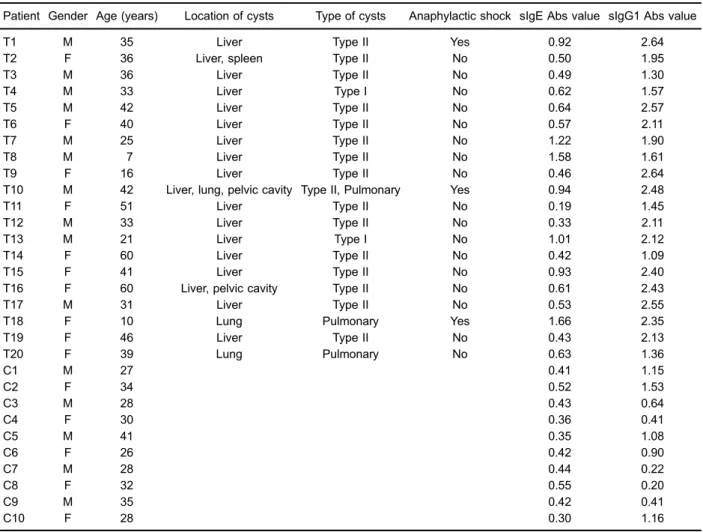
The absorbance values in patients and disease con-trols are reported in Table 1. The cutoff values of IgE and IgG1 were 0.64 and 2.15, respectively. Seven patients with higher IgE and eight patients with higher IgG1 absor-bance values than the cutoff were considered to be positive and were chosen for recognition of EgCF antigens of sIgE and sIgG1 using immunoblotting. In patients T1, T10, and T18, anaphylactic shock occurred during surgery and their sera were positive for sIgE and sIgG1.
1D SDS-PAGE and immunoblotting
The protein component in the crude antigen of EgCF was analyzed using 1D SDS-PAGE. Several protein bands
Table 1.Demographic, clinical, and antibody characteristics of patients and disease controls.
Patient Gender Age (years) Location of cysts Type of cysts Anaphylactic shock sIgE Abs value sIgG1 Abs value
T1 M 35 Liver Type II Yes 0.92 2.64
T2 F 36 Liver, spleen Type II No 0.50 1.95
T3 M 36 Liver Type II No 0.49 1.30
T4 M 33 Liver Type I No 0.62 1.57
T5 M 42 Liver Type II No 0.64 2.57
T6 F 40 Liver Type II No 0.57 2.11
T7 M 25 Liver Type II No 1.22 1.90
T8 M 7 Liver Type II No 1.58 1.61
T9 F 16 Liver Type II No 0.46 2.64
T10 M 42 Liver, lung, pelvic cavity Type II, Pulmonary Yes 0.94 2.48
T11 F 51 Liver Type II No 0.19 1.45
T12 M 33 Liver Type II No 0.33 2.11
T13 M 21 Liver Type I No 1.01 2.12
T14 F 60 Liver Type II No 0.42 1.09
T15 F 41 Liver Type II No 0.93 2.40
T16 F 60 Liver, pelvic cavity Type II No 0.61 2.43
T17 M 31 Liver Type II No 0.53 2.55
T18 F 10 Lung Pulmonary Yes 1.66 2.35
T19 F 46 Liver Type II No 0.43 2.13
T20 F 39 Lung Pulmonary No 0.63 1.36
C1 M 27 0.41 1.15
C2 F 34 0.52 1.53
C3 M 28 0.43 0.64
C4 F 30 0.36 0.41
C5 M 41 0.35 1.08
C6 F 26 0.42 0.90
C7 M 28 0.44 0.22
C8 F 32 0.55 0.20
C9 M 35 0.42 0.41
C10 F 28 0.30 1.16
ranging from 10 to 250 kDa were detected. The most abundant protein band had a molecular weight of 53.5 kDa. The differences in intensity in protein molecular weight of 51.3, 40.5, 27.0, and 24.5 kDa of IgE reaction were detected among individual sera. The 40.5-kDa
protein showed positive reaction with 7 sIgE positive sera, and was the major antigen of sIgE in EgCF. The dif-ferences in intensity in protein molecular weight of 35.5, 44.6, and 53.2 kDa of IgG1 reaction were detected among individual sera. The 35.5-kDa protein showed positive reaction with 8 sIgG1 positive sera, and was the major antigen of sIgG1 in EgCF. Sera obtained from disease con-trols showed no reaction against any of the EgCF proteins (Figures 1 and 2).
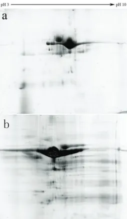
A representative 7-cm 2-DE gelfigure of crude antigen of EgCF stained with Coomassie blue shows the visuali-zation of about 50 protein spots ranging in molecular mass from 10 to 250 kDa, with pI values ranging from 3 to 10. A representative 24-cm 2-DE gelfigure of crude antigen of EgCF using laser scanner shows the visualization of about 200 protein spots ranging in molecular mass from 10 to 250 kDa, with pI values ranging from 3 to 10 (Figure 3). To identify the separated proteins, proteins of crude antigen of EgCF were transferred to a nitrocellulose mem-brane and incubated with T1 patient sera. PI values of four spots ranging from 6.5 to 9.0 comprising 40.5 kDa protein in sIgE immunoblotting and pI values offive spots ranging from
Figure 1. SDS-PAGE and IgE immunoblotting of the crude antigen ofE. granulosuscystfluid (EgCF). Crude antigen of EgCF protein extracts were incubated individually with 7 sIgE positive sera (lanes 3–9), and with disease control (lane 10); crude antigen of EgCF proteins visualized using Coomassie blue R-250
(lane 2); protein molecular weight marker (lane 1). IgE,
immuno-globulin E; SDS-PAGE, sodium dodecyl sulfate-polyacrylamide gel electrophoresis; sIgE, specific immunoglobulin E.
Figure 2. SDS-PAGE and IgG1 immunoblotting of the crude antigen of Echinococcus granulosus cyst fluid (EgCF). Crude antigens of EgCF protein extracts were incubated individually with 8 sIgG1 positive sera (lanes 2–9), and with disease control (lane 10); protein molecular weight marker (lane 1). SDS-PAGE, sodium dodecyl sulfate-polyacrylamide gel electrophoresis; sIgG1, specific immunoglobulin G1.
6.5 to 9.0 comprising 35.5 kDa protein in IgG1 immunoblot-ting showed strong reaction with T1 patient sera (Figure 4).
Discussion
Anaphylactic shock induced by CE is mediated through sIgE and sIgG1 subclass. In this study, sera from 3 patients with CE who had anaphylactic shock were col-lected and sIgE- and sIgG1-positive serum were screened using SDS-PAGE, 2-DE, and immunoblotting technique for the corresponding antigen in EgCF. The main antigens of sIgE in EgCF were identified with four protein spots with pI values ranging from 6.5 to 9.0 and a molecular weight of 40.5 kDa. The main antigens of sIgG1 in EgCF were identified with five protein spots with pI values ranging from 6.5 to 9.0 and a molecular weight of 35.5 kDa.
In this study, indirect ELISA method was used to screen sIgE- and sIgG1-positive serum in 20 patients. The absorbance values in controls showed no significant dif-ferences than in patients. As plates coated with crude anti-gen of EgCF were derived from patients with echinococcosis
containing anthropogenic IgE and IgG1, it may have pro-duced false-positive results. Hence in the former experi-ment, EgCF from sheep liver was used to prepare the crude antigen. Seven cases of sIgE-positive serum and 8 cases of sIgG1-positive serum were screened using ELISA. It is interesting to note that 3 cases with anaphy-lactic shock were caused by spillover of EgCF during the surgery; sIgE and sIgG1 were all positive. The results suggest that the content of serum sIgE and sIgG1 is not only associated with patients with hydatid infections, but also associated with normal allergic components of patients. Results of a preliminary study showed that ana-phylactic shock of patients was related to higher content of serum total IgE and IgG1 (11).
IgE immunoblotting results showed that the 40.5-kDa proteins of crude antigen of EgCF had prominent reaction with all seven sIgE-positive sera. In preliminary 1D SDS-PAGE and IgE immunoblotting experiments using DAB, TMB (tetramethylbenzidine), and SuperSignal WestPico did not show obvious immunoblotting bands. However, the use of SuperSignal West Femto Maximum Sensitivity Sub-strate showed prominent reaction in IgE immunoblotting bands. This suggests that the concentration of sIgE in the serum and sIgE antigen in EgCF was very low. Results of sIgG1 immunoblotting bands also did not distinctly show the corresponding bands or protein point on 1D SDS-PAGE and 2-DE. The results of the study showed that the sIgE and sIgG1 antigen in EgCF belongs to the low abundance pro-teins. Removing high abundance proteins extraneous to the antigen of cysticfluid protein will concentrate low abundance proteins containing purpose antigen and effectively isolate sIgE and sIgG1 of EgCF antigen.
EgCF antigen for the sensitivity and specificity of specific antibodies was the core index for screening diagnostic anti-gen. Anaphylactic shock mediated through antibody levels is closely related to the allergic constitution and immune status of the patient; therefore, allergens may not have high sensitivity and specificity of EgCF diagnostic antigens. Studies on sIgE antigen in EgCF are few, with different methods and results (13,14), and the sIgG1 subclass antigen in EgCF has not been reported. AgB is a polymeric lipoprotein with a molecular mass of 120-160 kDa, which under reducing electrophoretic conditions appears to be composed of three subunits with molecular sizes of 8, 16, and 24 kDa. AgB is considered to have high sensitivity and specificity in the diagnosis of echinococcosis (15,16), but it cannot be identified by the sIgE (10). Ag5 is a high molecular mass lipoprotein complex of 67 and 57 kDa antigen in EgCF, which under reducing conditions dissociates into 38 and 22 kDa subunits (13). Results of sIgE and sIgG1 immuno-blotting showed no obvious reaction strip in the AgB and Ag5, due to their monomer molecular weight.
Study limitation and future experimental plans
In this study, valuable serum was collected only from 3 patients with echinococcosis who experienced
Figure 4. The crude antigen of Echinococcus granulosuscyst
anaphylactic shock. Whether the recognized antigen could identify the allergen responsible for anaphylactic shock has not yet been validated. Removing high abundance proteins extraneous to the antigen of cystic
fluid protein will concentrate low abundance proteins containing target antigen, and effectively isolate sIgE and sIgG1 EgCF antigen. The purified and detected amino acid sequence was effective for the candidate antigens.
In conclusion, sIgE and sIgG1 antigens in EgCF were identified, providing critical insights into cellular and molec-ular mechanisms underlying anaphylactic shock induced by CE.
Acknowledgments
This study was supported by the National Natural Science Foundation of China (No. 81460309).
References
1. Budke CM, Carabin H, Ndimubanzi PC, Nguyen H, Rain-water E, Dickey M, et al. A systematic review of the literature on cystic echinococcosis frequency worldwide and its asso-ciated clinical manifestations.Am J Trop Med Hyg2013; 88: 1011–1027, doi: 10.4269/ajtmh.12-0692.
2. Wang G, Feng X, Chu X, ErxidingAmina, Wang Q, et al. Epidemiological study on human echinococcosis in Hobu-kesar Mongolian Autonomous County of Xinjiang. Chin J
Endemiol2009; 28: 214–217.
3. Saenz de San Pedro B, Cazana JL, Cobo J, Serrano CL, Quiralte J, Contreras J, et al. Anaphylactic shock by rupture of hydatid hepatic cyst. Follow-up by specific IgE serum antibodies.Allergy1992; 47: 568–570, doi: 10.1111/j.1398-9995.1992.tb00683.x.
4. Qi F, Xu H, Wang Y, Dou H, Wu Y, Chen K. Experience on the rescuing anaphylactic shock caused by effusion of kidney hydatidfluid.Int J Med Parasit Dis2009; 36: 85–86.
5. Vuitton DA. Echinococcosis and allergy. Clin Rev Allergy
Immunol 2004; 26: 93–104, doi:
10.1007/s12016-004-0004-2.
6. Li Y, Zheng H, Cao X, Liu Z, Chen L. Demographic and clinical characteristics of patients with anaphylactic shock after surgery for cystic echinococcosis.Am J Trop Med Hyg 2011; 85: 452–455, doi: 10.4269/ajtmh.2011.10-0448.
7. Dong N, Chen L, Xu M, Deng H, Su C, Qin C, et al. Isolation and purification of anaphylactic antibody from guinea pig and preliminary research on its role in passive cutaneous anaphylaxis test(PCA).Chin J Microbiol Immunol2010; 30: 169–173.
8. Oettgen HC, Martin TR, Wynshaw-Boris A, Deng C, Drazen JM, Leder P. Active anaphylaxis in IgE-deficient mice.
Nature1994; 37: 367–370, doi: 10.1038/370367a0.
9. Tsujimura Y, Obata K, Mukai K, Shindou H, Yoshida M, Nishikado H, et al. Basophils play a pivotal role in
immunoglobulin-G-mediated but not immunoglobulin-E-mediated systemic anaphylaxis.Immunity2008; 28: 581–589,
doi: 10.1016/j.immuni.2008.02.008.
10. Zheng H, Xu ZX, Yang GX, Wen H. [Study on the level of specific IgG, IgG1 and IgE during anaphylactic shock in sheep induced byEchinococcus granulosus].Zhongguo Ji
Sheng Chong Xue Yu Ji Sheng Chong Bing Za Zhi2003;
21: 42–45.
11. Li Y, Zheng H, Gu M, Cao X, Wen H, Liu Z, et al. Comparisons of serum total IgE, IgG, and IgG1 levels in patients with and without echinococcosis-induced anaphy-lactic shock. Am J Trop Med Hyg 2012; 87: 104–108, doi: 10.4269/ajtmh.2012.11-0694.
12. Liu R, Krishnan HB, Xue W, Liu C. Characterization of allergens isolated from the freshwaterfish blunt snout bream
(Megalobrama amblycephala).J Agric Food Chem2011; 59:
458–463, doi: 10.1021/jf103942p.
13. Khabiri AR, Bagheri F, Assmar M, Siavashi MR. Analysis of specific IgE and IgG subclass antibodies for diagnosis
of Echinococcus granulosus. Parasite Immunol2006; 28:
357–362, doi: 10.1111/j.1365-3024.2006.00837.x.
14. Ortona E, Vaccari S, Margutti P, Delunardo F, Rigano R, Profumo E, et al. Immunological characterization of
Echino-coccus granulosuscyclophilin, an allergen reactive with IgE
and IgG4 from patients with cystic echinococcosis.Clin Exp
Immunol 2002; 128: 124–130, doi: 10.1046/j.1365-2249.
2002.01807.x.
15. Gao C, Wang J, Yang Y, Shi F, Zhu H, Jiao W. Serological evaluation of five Echinococcus granulosus antigens for detecting cystic echinococcosis by ELISA.Chin J Zoonoses 2012; 28: 811–814.
16. Li Y, Meng C, Zhang Z. Diagnostic value of ELISA using antigen B in cystic echinococcosis: A meta-analysis.Chin J