311 311 311 311 311 Mem Inst O swaldo Cruz, Rio de Janeiro, Vol. 100(3): 311-317, M ay 2005
Preliminary study of the presence of antibodies against
excretory-secretory antigens from protoscoleces of
Echinococcus granulosus
in dogs with intestinal echinococcosis
D avid Carmena/+ +, Aitziber Benito, Jorge M artínez, Jorge A Guisantes/+
Departamento de Inmunología, Microbiología y Parasitología, Facultad de Farmacia, Universidad del País Vasco, Apartado 450, 01080-Vitoria, España
The aim of the present study was to analyze the antibody response against excretory-secretory antigens (ES-Ag) from Echinococcus granulosus protoscoleces, using sera from dogs infected with E. granulosus and other helminths. ES-Ag were obtained from the first 50 h maintenance of protoscoleces in vitro. Immunochemical characterization was performed by immunoblotting with sera from dogs naturally infected with E. granulosus (n = 12), sera from dogs infected with helminths other than E. granulosus (n = 30), and helminth-free dog sera (n = 20). These findings were compared to those obtained from a somatic extract of protoscoleces (S-Ag). ES-Ag only showed four cross-reacting proteins of 65, 61, 54, and 45-46 kDa. Antigens with apparent masses of 89 and 50 kDa in ES-Ag and of 130 and 67 kDa in S-Ag were identified by sera of dogs infected with E. granulosus only, whereas a protein of 41-43 kDa was recognised by the majority of the sera from dogs with non-echinococcal infection. Employing ELISA to study the same sera, S-Ag revealed higher immunoreactivity than ES-Ag, but also showed higher cross-reactivity levels when sera from dogs with non-echinococcal infection were assayed in immunoblotting.
Key words: Echinococcus granulosus - excretorysecretory antigens protoscoleces intestinal echinococcosis -parasite antigens
Cystic hydatid disease in man is a zoonosis caused by infection with the larval stage of the dog tapeworm Echi-nococcus granulosus. The adult worm lives in the small intestine of dogs and other canids, in intimate contact with the intestinal epithelium. Parasite eggs (the infective stage for the intermediate host) are excreted in faeces, and may thus contaminate soil, grass and water. Detec-tion of E. granulosus adults in dogs is very important in order to evaluate its prevalence and to develop surveil-lance and control programmes for hydatidosis/echinococ-cosis (WHO 2001). Diagnosis of taeniid cestode infec-tions in dogs can be carried out using arecoline hydro-bromide, but this technique has a highly variable sensi-tivity, is time consuming and some dogs suffer undesired side-effects (Wachira et al. 1990). Coprological exams, however, have low sensitivity since excretion of the eggs occurs sporadically. Furthermore, the eggs of taeniid ces-todes are morphologically indistinguishable by light mi-croscopy.
In an attempt to improve the sensitivity of E. gra-nulosus diagnostics, different serological tests have been developed, mainly based on the detection of specific IgG
Financial support: FIS, Ministry of Public Health; DEMSAC, Town Council of Vitoria, and Department of Health, Basque Government
+Corresponding author. E-mail: oipgudej@vc.ehu.es
++Present address: MRC Clinical Sciences Centre, Membrane
Transport Biology Group, Hammersmith Hospital Campus, Du Cane Road, London W12 0NN, UK
Received 17 February 2005 Accepted 27 April 2005
antibodies against the adult parasite. Somatic extracts of protoscoleces (S-Ag) are the most commonly used anti-genic source. However, ELISA results showed highly vari-able sensitivities, ranging from 40 to 90% (Jenkins et al. 1990, Gasser et al. 1992, 1993, 1994). These studies also demonstrated that 25-60% of the sera from dogs infected with E. granulosus did not show significant levels of spe-cific antibody (Jenkins et al. 1990, Gasser et al. 1994), and that cross-reactivity with other parasite species may oc-cur (Gasser et al. 1988).
During recent years, diagnosis of intestinal echino-coccosis has been mainly based on the detection of ex-cretory-secretory products of E. granulosus protos-coleces in dog faeces using ELISA (Allan et al. 1992, Deplazes et al. 1992, Jenkins et al. 2000). This technique considerably improves both diagnostic sensitivity and specificity, permits the detection of the parasite during the prepatent period and shows the current status of the infection (Fraser & Craig 1997).
Although characterization of somatic antigens of E. granulosus protoscoleces for diagnostic purposes has been described in several reports (Gasser et al. 1989, 1991, 1992, Rafiei & Craig 2002), very little information is avail-able about excretory-secretory products of protoscoleces (Gasser et al. 1992, Carmena et al. 2004). In this preliminary work we show the presence of antibodies that recognize excretory-secretory antigens of E. granulosus proto-scoleces in sera from dogs infected with E. granulosus
312 312 312 312
312 D og antibodies against ES-Ag of E. granulosus • D avid Carmena et al.
MATERIALS AND METHODS
Somatic antigens (S-Ag) - E. granulosus parasite material was isolated from sheep hepatic hydatid cysts collected from an abattoir in La Rioja, Spain. Somatic anti-gens were obtained from protoscoleces removed by asep-tic cyst puncture as described by Smyth and Davies (1974), washed with phosphate-buffered saline (PBS) and stored at –20ºC with proteolytic enzyme inhibitors (2 mM PMSF and 5 mM EDTA). Protoscoleces were thawed and soni-cated (10 cycles of 12 s at 60 Hz frequency), freeze-thawed once more and centrifuged for 35 min at 2300 × g. Super-natants were aliquotted and stored at –20ºC.
Excretory-secretory antigens (ES-Ag) -To obtain ex-cretory-secretory products, protoscoleces with viability higher than 90% were selected. Viability was assessed by morphological appearance, flame cell motility and general contractile movements (Smyth & Davies 1974, Howell 1986). Protoscoleces were cultured in PBS complemented with 10% glucose, 100 U ml-1 penicillin and 100 µg ml-1 streptomycin at 37ºC in 5% CO2, which promoted parasite survival for several days (Carmena et al. 2002). Every 8 h the medium was removed and replaced with fresh medium. Protein recovery from the media was achieved by using Ultrafree 15 filters with a 5 kDa pore diameter membrane (Millipore, Bedford, US). EDTA (5 mM) and PMSF (2 mM) were added, and the ES products were aliquotted and stored at –20ºC. Protein concentration was determined by the bicinchoninic acid method (Sigma-Aldrich, Dorset, UK). In total, 8 cultures of hepatic protoscoleces from sheep were carried out. Medium corresponding to the first 50 h of culture was subsequently used. The concentra-tion for proteins obtained for each preparaconcentra-tion was: ES-Ag: 0.2 mg ml-1 and S-Ag: 2.95 mg ml-1.
Enzyme linked immunosorbent assay (ELISA) - ELISA was carried out as described by Benito et al. (2001). The following optimal antigen concentrations were used: ES-Ag: 20 µg ml-1 and S-Ag: 7.5 µg ml-1. Microtitre plates (Maxisorp, Nunc, Roskilde, Denmark) were coated with antigenic extract diluted in PBS buffer (100 µl/well) and incubated overnight at 4ºC. Blocking was carried out with PBS-1% bovine serum albumin (BSA, 200 µl/well) for 1 h at 37ºC. Dog sera were tested in PBS-0.5% BSA buffer (100 µl/well) using serial dilutions ranging from 1:50 to 1:200 in duplicate. The plates were incubated for 1 h at 37ºC. Peroxidase-conjugated rabbit anti-dog IgG (Sigma) was used at 1:1000 dilution in PBS-4% SAB-0.05% Tween 20 buffer for 1 h at 37ºC. Binding was visualised with 5-aminosalicilic acid. The reaction was stopped by adding 25 µl/well NaOH 1N, and the absorbance value was mea-sured at 450 nm.
SDS-PAGE and immunoblotting - Proteins were sepa-rated by 12.5% SDS-PAGE under reducing conditions ac-cording to Laemmli (1970). For immunoblotting, proteins were transferred to polyvinylidene difluoride (PVDF) mem-branes (Immobilon-P, Millipore), according to Towbin et al. (1979).Primary sera were used at 1:50 in 20 mM Tris-buffered saline (pH 7.4), 8% skimmed milk (TBS-M), incu-bating overnight at 4ºC. Peroxidase-conjugated rabbit anti-dog IgG (Sigma) was used at 1:1000 in TBS-M buffer for 4
h at room temperature, and binding was visualised with 4-chloro-1-napthol.
Dog sera - Include: a) dog sera obtained from the Coun-cil Animal Rescue Mission of Vitoria, Spain. All animals were diagnosed by autopsy, but their previous infection history was unknown. Five sera from dogs naturally in-fected with E. granulosus were obtained, with worm bur-dens ranging from 2 to 155 helminths. Thirty sera were obtained from dogs that harboured infections with Tae-nia spp. (including T. hydatigena and T. pisiformis),
Mesocestoides spp., Dipylidium caninum, Toxocara canis, Toxascaris leonina, Trichuris vulpis, Uncinaria stenocephala, and Ancylostoma caninum. Sera from 20 helminth-free dogs that were under 2 years of age at the time of autopsy were also used; b) seven serum samples from farm dogs naturally infected with E. granulosus in the Chubut Province (Argentina). These samples were diagnosed by identifying worms following arecoline pur-gation, and were kindly provided by Dr Eduardo Fernán-dez, Laboratorio de Control de Patologías Prevalentes, Hospital Zonal de Trelew, Chubut, Argentina; c) 20 sera from 5 dogs experimentally infected with E. granulosus, kindly provided by Dr P Spinelli, Instituto de Higiene, Montevideo (Uruguay). Animals had been orally infected with 60,000 protoscoleces (Spinelli et al. 1996) and four serum samples were obtained at day 0 and days 5, 15, and 29 post-infection. A pool of sera from dogs with E. granu-losus natural infections was also used as a positive con-trol in ELISA assays, with an assigned value of 8000 arbi-trary units ml-1 (Benito et al. 2001).
Statistical methods - ELISA results were analyzed by means of variance analysis (ANOVA), using SPSS 11.5 statistical software.
RESULTS
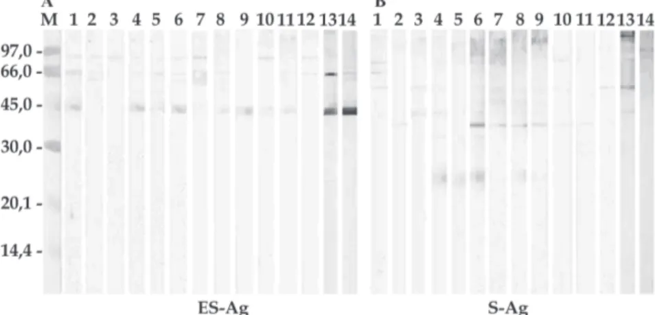
Immunoblotting - Fig. 1 shows an analysis of recogni-tion of individual components of ES-Ag and S-Ag with the 12 sera from dogs naturally infected with E. gra-nulosus. These sera identify seven major components in the ES-Ag, standing out those of 89, 46, and 50 kDa. With regard to S-Ag, the same sera recognize 15 antigenic pro-teins, the most prominent being those of 67, 55, 48, 43, 39, and 24 kDa. Figs 2 and 3 show the antigenic profiles of the ES-Ag and S-Ag identified with sera from dogs infected with other helminths, as well as with sera from helminth-free dogs. Proteins of 41-43 kDa and 85 kDa, both shared by ES-Ag and S-Ag, were recognized by many of these sera. Table I summarizes the data obtained by im-munoblotting.
313 313 313 313 313 Mem Inst O swaldo Cruz, Rio de Janeiro, Vol. 100(3), M ay 2005
Fig. 1: immunoblotting profiles using the sera from dogs naturally infected with Echinococcus granulosus (lanes 1-12). Lane 13: serum from dog infected with Taenia hydatigena (control). Lane 14: serum from helminth-free dog (negative control). M: molecular mass marker (kDa). A: excretory-secretory antigens (ES-Ag); B: somatic antigens (S-Ag)
Fig. 2: immunoblotting profiles for excretory-secretory antigens (ES-Ag) and somatic antigen (S-Ag) using sera from dogs infected with helminths other than Echinococcus granulosus. Taenia spp.: lanes 1 to 8; Mesocestoides spp.: lane 9; D. caninum: lanes 10 to 14; T. canis: lanes 15 to 19; T. leonina: lanes 20 to 22; T. vulpis: lane 23; U. stenocephala: lanes 24 to 27; A. caninum: lanes 28 to 30. M: molecular mass marker (kDa). A: ES-Ag; B: S-Ag
Fig. 3: immunoblotting profiles for excretory-secretory antigens (ES-Ag) and somatic antigens (S-Ag) using sera from helminth-free dogs. M: Molecular mass marker (kDa). A: ES-Ag; B: S-Ag
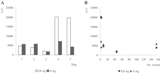
Fig. 5 summarizes the ELISA results for the 20 sera from dogs experimentally infected with E. granulosus. The highest specific antibody levels were observed for the bleeds obtained at days 15 and 29 post-infection from dogs 1 and 2. S-Ag was the most immunoreactive extract and also showed more homogeneous absorbance values among the studied sera than the ES-Ag. Regarding the sera from dogs naturally infected with E. granulosus, no
significant differences were found between the means of the absorbance values obtained for both antigenic ex-tracts (P < 0.05).
DISCUSSION
echino-314 314 314 314
314 D og antibodies against ES-Ag of E. granulosus • D avid Carmena et al.
coccosis by ELISA and/or immunoblotting (Jenkins & Rickard 1986, Gasser et al. 1992). However, information about the characterization of ES-Ag from this parasite is very rare, which can be partially due to difficulties in ob-taining enough amounts of ES-Ag. Recently, our labora-tory has identified over 20 major protein components in the ES-Ag which could be distinguished by 1-dimensional SDS-PAGE and revealed apparent masses between 9 and 300 kDa (Carmena et al. 2004). In this study, we present preliminary data about the immunochemical characteriza-tion of ES-Ag from protoscoleces of E. granulosus, which may help to identify new candidate antigens with poten-tial for immunodiagnosis of dog echinococcosis.
Secreted products of E. granulosus adults have been characterized by immunoblotting using sera from dogs
naturally infected with this cestode (Gasser et al. 1992). These authors identified three antigenic components larger than 94 kDa, two triplets of 68/94 and 39/43 kDa, and seven proteins smaller than 30 kDa. In our study, we have iden-tified seven secreted components from E. granulosus
protoscoleces ranging from 46 to 133 kDa. When S-Ag from protoscoleces was used, Gasser et al. (1992) found antigenic proteins of 94, 76, 35, 27, and others smaller than 14 kDa. In our work, we have recognised 15 anti-genic components ranging from 24 and 130 kDa in the same extract. Only three of them had similar molecular masses (76, 34, and 24 kDa) compared to the components described by Gasser et al. (1992).
We have evaluated by immunoblotting the possible cross-reactivity of S-Ag with sera from dogs infected with
TABLE I
Immunoblotting results. Components of excretory-secretory antigens and somatic antigens recognized by different dog sera. The percentage of sera which bound to each antigenic component is expressed in brackets. Molecular masses are expressed in kDa
Excretory-secretory antigens Somatic antigens
With Other With Other
E. granulosus parasitoses Helminth-free E. granulosus parasitoses Helminth-free
- 140 - - 137 137
(20%) (27%) (20%)
133 - - 130 -
-(58%) (33%)
- - - 115/122 112/120 113
(D, 83%) (D, 70%) (55%)
- - - 103 104
-(58%) (20%)
- - - - 94 98/104 (D)
(53%) (50%)
89 - - - -
-(92%)
- 85 85 85 - 85
(20%) (70%) (42%) (30%)
- - - - 82 81
(33%) (30%)
- 76 77 76 - 78
(17%) (65%) (42%) (25%)
- - 71 - 70
-(20%) (30%)
- - 68 67 -
-(60%) (25%)
65 65 - 65 65
-(67%) (10%) (17%) (17%)
61 61 61 - 61 60
(25%) (43%) (25%) (57%) (30%)
54 54 - 55 52
-(17%) (37%) (67%) (10%)
46/50 (D) 45 46 48 46
-(68%) (17%) (40%) (33%) (40%)
- 41-43 41 41/43 (D) 41
-(80%) (100%) (50%) (20%)
- - - 39 38 39
(67%) (10%) (10%)
- - - 34 36
-(25%) (17%)
- - - 24 26
-(42%) (10%)
315 315 315 315 315 Mem Inst O swaldo Cruz, Rio de Janeiro, Vol. 100(3), M ay 2005
helminths other than E. granulosus as well as with sera from helminth-free dogs. Using S-Ag, 40% of sera from dogs with non-echinococcal infections reacted specifi-cally with a 46 kDa protein, and 20% with a 41 kDa polypep-tide, which were also recognized by sera from dogs natu-rally infected with E. granulosus. Additional cross-react-ing proteins of 113, and 85 kDa were identified by 55%, and 30% of helminth-free dog sera, respectively, and to a lower degree, proteins of 78, and 39 kDa. S-Ag with mo-lecular masses of 130 and 67 kDa may have diagnostic potential, since they were only identified by sera from dogs infected with E. granulosus, but not by sera from dogs with non-echinococcal infections.
On the other hand, only 4 components from ES-Ag (polypeptides of 65, 61, 54, and 45-46 kDa) showed sig-nificant cross-reactivity levels. These proteins were rec-ognized by 10-43% of sera from dogs infected with helm-inths other than E. granulosus or by sera from helminth-free dogs. In this antigenic extract, 2 proteins of 89 and 50 kDa, respectively, were recognized by sera from dogs
in-fected with E. granulosus only. Interestingly, the compo-nent of 89 kDa was identified by 92% of sera that were tested. This fact suggests that this protein might be a promising diagnostic antigen that could be used in immu-nological tests for the detection of E. granulosus in dogs. These data demonstrate that, although S-Ag showed the highest specific antibody levels in ELISA, ES-Ag is more specific than S-Ag for immunodiagnosis of dog echino-coccosis.
It was difficult to highlight specific recognition pat-terns attributable to individual infections, except in the case of the sera from dogs infected with Toxascaris le-onina. All of them showed a specific protein of 36 kDa when using S-Ag (Fig. 2B, lanes 20 to 22).
The lack of specificity of the 41-43 kDa component from the excreted products of E. granulosus adults has been previously described by Gasser et al. (1992). In our work, we have corroborated this result, identifying this antigenic protein both in ES-Ag and S-Ag. Recently, we have also found the 41-43 kDa component in the ES-Ag
Fig. 4-A: ELISA results for different antigenic extracts using sera from dogs naturally infected with Echinococcus granulosus; B: ELISA results for different antigenic extracts considering the worm burden found at autopsy. ES-Ag: excretory-secretory antigens; S-Ag: somatic antigens; A.U.: arbitrary units
316 316 316 316
316 D og antibodies against ES-Ag of E. granulosus • D avid Carmena et al.
by immunoblotting with non-hydatidic human sera, in-cluding sera from individuals with other parasitoses, from individuals with non-parasitological pathologies and from healthy donors (Carmena et al. 2004).
ELISA results for sera from dogs infected with E. granulosus showed that highest worm burdens are not always corresponding with highest specific antibody lev-els, especially when ES-Ag is used (Gasser et al. 1992, 1993). This finding seems to demonstrate that there is no relationship between the number of worms in the intes-tine and the circulating antibody level, confirming previ-ous results of Jenkins and Rickard (1985), Gasser et al. (1988, 1992, 1994) and Jenkins et al. (1990, 1991). This fact may either be due to the sequestration of antibodies and the formation of circulating immunocomplexes (Gasser et al. 1988, 1993, Spinelli et al. 1996), or to immune evasion mechanisms of the parasite (Gasser et al. 1992, 1994), or to a low immune response of the host (Gasser et al. 1988, 1993, 1994). Host nutritional status may also have an im-pact on the antibody levels (Jenkins et al. 1991, Gasser et al. 1992). On the other hand, the broad differences of an-tibody levels found among the studied dog sera also may reflect the influence by physiological and environmental factors, like re-infection or co-infection with other para-site species.
In ELISA, sera from dogs experimentally infected with
E. granulosus showed less variability regarding the lev-els of specific antibodies than sera from dogs with natural infections (Figs 4 and 5). This may be due to the stan-dardized environmental conditions under which these animals were maintained. The highest specific antibody levels were reached using S-Ag, especially when sera from dogs experimentally infected were assayed. These results are in agreement with those obtained by Benito et al. (2001), who found higher discrimination levels between positive and negative controls using S-Ag than with ES-Ag or hydatid cyst fluid as antigenic extracts.
We conclude that ES-Ag from protoscoleces of E. granulosus contain potential diagnostic antigens to be used in the immunodiagnosis of canine echinococcosis. In particular, the protein of 89 kDa has evidenced the most promising features. However, a more comprehensive analy-sis will be necessary to determine more accurately the significance of these observations, since only a compara-tively low number of sera was available for the present study.Further studies to clone and express individual pro-tein components from secreted products of protoscoleces which display both high immunoreactivity and specific-ity. may be of great interest for the diagnosis of dog echi-nococcosis.
ACKNOWLEDGEMENTS
To Prof. Murray E Selkirk and Dr Sonja Kock (Department of Biological Sciences, Imperial College London, UK) for their critical revision of this manuscript.
REFERENCES
Allan JC, Craig PS, García Noval J, Mencos F, Liu D, Wang Y, Wen H, Zhou P, Stringer R, Rogan M, Zeyhle E 1992. Coproantigen detection for immunodiagnosis of echinococ-cosis and taeniasis in dogs and humans. Parasitology104: 347-355.
Benito A, Carmena D, Spinelli P, Postigo I, Martínez J, Estíbalez JJ, Martín de la Cuesta F, Guisantes JA 2001. The serologi-cal diagnosis of canine echinococcosis by an enzyme immu-noassay useful for epidemiological surveys. Res Rev Parasitol61: 17-23.
Carmena D, Benito A, Postigo I, Arteaga J, Martínez J, Guisantes JA 2002. Short term culture of protoscoleces to obtain ex-cretory-secretory proteins of Echinococcus granulosus.
Res Rev Parasitol62: 84-88.
Carmena D, Martínez J, Benito A, Guisantes JA 2004. Charac-terization of excretory-secretory products from pro-toscoleces of Echinococcus granulosus and evaluation of their potential for immunodiagnosis of human cystic echi-nococcosis. Parasitology129: 371-378.
Deplazes P, Gottstein B, Eckert J, Jenkins DJ, Ewald D, Jiménez-Palacios S 1992. Detection of Echinococcus
coproantigens by enzyme-linked immunosorbent assay in dogs, dingoes and foxes. Parasitol Res78: 303-308.
Fraser A, Craig PS 1997. Detection of gastrointestinal helminth infections using coproantigen and molecular diagnostic ap-proaches. J Helminthol71: 103-107.
Gasser RB, Jenkins DJ, Heath DD, Lawrence SB 1992. Use of
Echinococcus granulosus worm antigens for immunodiag-nosis of Echinococcus granulosus infection in dogs. Vet Parasitol45: 89-100.
Gasser RB, Jenkins DJ, Paolillo E, Parada L, Cabrera P, Craig PS 1993. Serum antibodies in canine echinococcosis. Int J Parasitol23: 579-586.
Gasser RB, Lightowlers MW, Obendorf DL, Jenkins DJ, Rickard MD 1988. Evaluation of a serological test system for the diagnosis of natural Echinococcus granulosus infection in dogs using E. granulosus protoscolex and oncosphere anti-gens. Australian Vet J65: 369-373.
Gasser RB, Lightowlers MW, Rickard MD 1989. Identification of protein components of Echinococcus granulosus
protoscolex antigens for specific serodiagnosis of Echino-coccus granulosus in dogs. Parasite Immunol11: 279-291.
Gasser RB, Lightowlers MW, Rickard MD 1991. Echinococ-cus granulosus: antigenic proteins in oncospheres and on the surface of protoscoleces identified by serum antibodies from infected dogs. Res Vet Sci50: 340-345.
Gasser RB, Parada L, Acuna A, Burges C, Laurenson MK, Gulland FMD, Reichel MP, Paolillo E 1994. Immunologi-cal assesment of exposure to Echinococcus granulosus in a rural dog population in Uruguay. Acta Trop58: 179-185.
Howell MJ1986. Cultivation of Echinococcus species in vitro. In RCA Thompson, The Biology of Echinococcus and Hy-datid Disease, George Allen & Unwin, London, p. 143-163.
Jenkins DJ, Rickard MD 1985. Specific antibody responses to
Taeniahydatigena, Taenia pisiformis and Echinococcus granulosus infection in dogs. Australian Vet J62: 72-78.
Jenkins DJ, Rickard MD 1986. Specific antibodies responses in dogs experimentally infected with Echinococcus granulosus. Am J Trop Med Hyg35: 345-349.
Jenkins DJ, Fraser A, Bradshaw H, Craig PS 2000. Detection of
317 317 317 317 317 Mem Inst O swaldo Cruz, Rio de Janeiro, Vol. 100(3), M ay 2005
Jenkins DJ, Gasser RB, Romig T, Zeyhle E 1991. Antibody responses against natural Taenia hydatigena infection in dogs in Kenya. Int J Parasitol21: 251-253.
Jenkins DJ, Gasser RB, Zeyhle E, Romig T, Macpherson CNL 1990. Assessment of a serological test for the detection of
Echinococcus granulosus infection in dogs in Kenya. Acta Trop47: 245-248.
Laemmli UK 1970. Cleavage of structural proteins during the assembly of the head of bacteriophage T4. Nature 227: 680-685.
Rafiei A, Craig PS 2002. The immunodiagnostic potential of protoscolex antigens in human cystic echinococcosis and the possible influence of parasite strain. Ann Trop Med Parasitol96: 383-389.
Smyth JD, Davies Z 1974. In vitro culture of the strobilar stage of Echinococcus granulosus (sheep strain): a review of ba-sic problems and results. Int J Parasitol4: 631-644.
Spinelli P, Carol H, Nieto A 1996. Niveles de anticuerpos y antígenos circulantes en perros con infección natural y ex-perimental por Echinococcus granulosus. Inmunología15: 21-29.
Towbin H, Staehelin T, Gordon J 1979. Electrophoretic trans-fer of proteins from polyacrylamide gels to nitrocellulose sheets: procedure and some applications. Proc Nat Acad Sci USA76: 4350-4354.
Wachira T, McPherson CNL, Gathuma JM 1990. Hydatid dis-ease in the Turkana District of Kenya VII. Analysis of the infection pressure on definitive and intermediate hosts of
E. granulosus. Ann Trop Med Parasitol84: 361-368.
WHO 2001. WHO/OIE Manual on Echinococcosis in Humans and Animals: a Public Health Problem of Global Concern