Cop
yright
© ABE&M t
odos os dir
eit
os r
eser
vados
.
Marrow hypoplasia: a rare
complication of untreated
Grave’s disease
Hipoplasia de medula óssea: uma rara complicação da doença de Graves não tratada
Juliana Garcia1, Bruna Silveira Rodrigues1, Larissa de França1, Mônica Wolff1, Renato Torrini1, Vivian Ellinger1, Carlos Campos1, Vera Leal1, Dayse Caldas1
SUMMARY
Atypical presentation forms of hyperthyroidism are always a challenge to the clinician. We pre sent a female patient with the typical symptoms of thyrotoxicosis, without any thionamides treatment before, associated with pancytopenia, which recovered after euthyroidism state was achieved. Although the major cases of pancytopenia in Grave’s disease are seen as a compli cation of antithyroid drugs (thioamides), in this case report the alteration in blood tests was associated with untreated hyperthyroidism. In the literature review, we found 19 case reports between 1981 to 2012, but it has been related to a hypercellular bone marrow with periferic destruction. Our case, however, is about a hypocellular bone marrow without ibrosis or fat tissue replacement, which proceeded with a periferic improvement following thyroid treatment. Although rare, pancytopenia, when present, may develop as an unusual and severe manifesta tion in untreated subjects. Arq Bras Endocrinol Metab. 2014;58(9):953-7
SUMÁRIO
Formas atípicas de apresentação do hipertireoidismo são sempre um desaio para o clínico. Apresentamos uma paciente do sexo feminino, com sintomas típicos de tireotoxicose associa do a um quadro de pancitopenia sem nenhum tratamento prévio com tionamidas. A melhora da alteração hematológica ocorreu após recuperação do eutireoidismo. Embora a maioria dos casos de pancitopenia na doença de Graves seja uma complicação das drogas antitireoidianas (tionamidas), neste caso a alteração hematológica foi associada ao quadro de hipertireoidismo não tratado. Após uma revisão na literatura, encontramos 19 relatos de caso descritos no perío do de 1981 a 2012, nos quais o quadro de pancitopenia estava relacionado à hipercelularidade medular com destruição periférica das células sanguíneas. Nosso caso, entretanto, tratase de uma pancitopenia com medula óssea hipocelular, sem iniltração por tecido adiposo ou ibrose, que evoluiu com melhora dos elementos do sangue periférico após tratamento do hipertireoi dismo. Embora rara, a pancitopenia, quando presente, pode se manifestar como uma severa manifestação se não tratada a condição desencadeadora. Arq Bras Endocrinol Metab. 2014;58(9):953-7
1 Institute of Diabetes and
Endocrinology Luiz Capriglione (IEDE), Rio de Janeiro, RJ, Brazil
Correspondence to:
Juliana Garcia
Rua Gildásio Amado, 55, sala 1901 22631-020 – Rio de Janeiro, RJ, Brazil [email protected] Received on Jan/10/2014 Accepted on Apr/7/2014
DOI: 10.1590/0004-2730000003216
INTRODUCTION
G
rave’s disease (GD) usually presents with the several well-known symptoms and signs, but in some cases there is atypical manifestations of thyrotoxi-cosis, which include hematological alterations, as ane-mia. Pancytopenia is a rare, but a serious complication that physicians may come across.Neverthe-Cop
yright
© ABE&M t
odos os dir
eit
os r
eser
vados
.
less, a good response to hyperthyroidism treatment is almost guaranteed.
CASE REPORT
A 54 year-old female was referred to our hospital com-plaining of palpitations, hand tremor and a 23 kg weight loss in the last 5 months. The physical examination re-vealed thyroid (approximately double the normal size), irregular, irm, without lumps, sinus tachycardia (120 bpm) with holosystolic murmur in the mitral focus, hy-pertension (BP 160 x 100 mmHg) as well as moist and warm skin. There was no evidence of exophthalmos or pretibial myxedema. The patient was diagnosed with GD and stated never having been submitted to any pre-vious treatment.
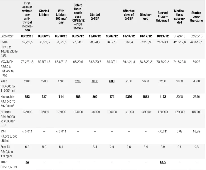
Besides the hyperthyroidism, her blood test results, at the time of the clinic visit included microcytic hypochro-mic anemia, thrombocytopenia, associated with neutro-philia (Table 1). We had her admitted for compensation of the thyroid disease and started treatment with Lithium – 900 mg/day. After 18 days of treatment with Lithium, she was submitted to radioiodine I-131 (15 mci).
An evaluation by the hematology department (HEMORIO) was asked in order to rule out other pos-sible causes of pancytopenia. They requested laboratory tests and proceeded with aspirate and biopsy of bone marrow. Due to the worsening of neutropenia (Table 1) treatment with granulocyte stimulating factor (G-CSF) was initiated.
Subsequent results of laboratory tests (HIV and vi-ral hepatitis serology, B12 vitamin, serum iron, ferritin,
Table 1. Evolution of blood tests (hematological values, thyroid hormones and TSH receptor antibodies – TRAb)
First consult without
any anti-thyroid medica-tion
Started Lithium
With Lithium 900 mg/
day
Before Thera-peutic dose (09/26/12
– I131 15mci)
Started G-CSF
After ten days of
G-CSF Dischar-ged
Started Propyl-thiouracil
Medica-tions
suspen-ded
Started Levo-thyroxine
Laboratory 08/22/12 09/06/12 09/10/12 09/24/12 10/04/12 10/07/12 10/14/12 10/17/12 10/24/12 01/24/13 02/22/13 Ht/Hb
RR:12 to 16g/dL /36 to 48%
32,2/9,5 30,6/9,5 30,8/9,5 27,6/8,5 28,9/8,7 26,3/7,8 30/9,4 32/10,3 28,9/9,1 42,3/12,8 42,0/12,1
MCV/MCH RR:80 to 96fL/27 to 33pg
72,2/21,3 69,5/21,6 68,8/21,2 68/20,9 68,6/20,7 64,3/21 69,4/21,8 68,8/22,2 70,7/22,2 74,3/22,5 80/25
WBC RR:4000 to 11000/mm3
2100 1900 1700 1200 1000 600 7100 2600 2200 3400 4600
Neutrophils RR:1640 TO 7920/mm3
882 627 714 288 260 174 5396 1872 1122 2040 2896
Platelets RR:150000 to 450000/ mm3
137000 136000 122000 103000 140000 106000 141000 149000 170000 179000 187000
TSH RR:0,3 to 5,0 µUI/mL
< 0,011 – < 0,011 – – – – – < 0,011 0,03 16,82
Free T4 RR: 0,8 to 1,9 ng/dL
6,9 5,9 5,1 – 3,4 2,9 2,6 2,4 2,9 0,6 0,3
TRAb RR:< 1,5 UI/L
34 – – – – – – – 18,5 – –
Cop
yright
© ABE&M t
odos os dir
eit
os r
eser
vados
.
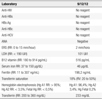
DHL, ANA, VHS, hemoglobin electrophoresis) (Table 2) and the total abdominal ultrasonography were nor-mal. The bone marrow biopsy revealed hypocellularity wi thout any iniltration or ibrous/fatty tissue replace-ment (Figures 1 and 2).
After thirty-eight days of lithium, twenty days of radioiodine I-131 (15 mci) and ten days of G-CSF as-sociated with decreased levels of thyroid hormones, im-provement of pancytopenia could be noticed (Table 1). The patient was discharged after a 42 days period of
hospitalization with prescription for lithium (900 mg/ day), and referral for our outpatient clinic.
She returned ten days after, as lab results were still abnormal (Table 1), propylthiouracil was added to the treatment with lithium. The decrease of thyroid hor-mones and complete improvement of pancytopenia i-nally occurred after two months of hospital discharge and the medications were therefore discontinued. Five months after the radioiodine treatment, she developed hypothyroidism. Levothyroxine was then initiated for the control of thyroid hormones.
DISCUSSION
Our patient presented with the classical signs and symp-toms of hyperthyroidism due to diffuse toxic goiter (DTG), with the irst lab results showing a pancyto-penia that required investigation since she have never been submitted to any previous treatment. The Ameri-can Thyroid Association as well as the AmeriAmeri-can Asso-ciation of Clinical Endocrinologists suggests that, prior to initiating antithyroid drug therapy (ATD), patients should have a complete blood count, and in case the pa-tient has an absolute neutrophil count below 500/mm3
ATD is not recommended (item 15 of reference 1). Since the main cause of hematological alteration in hyperthyroidism is known to be the treatment with thionamides, this would be a relative contraindication. Although, in a systematic review about agranulocytosis induced by nonchemotherapy drugs, it was shown that propiltiouracil rarely causes agranulocytosis, and low doses of MMI are even less likely to do so (2).
Figures 1 and 2. Marrow biopsy revealed hypocellularity without any iniltration or ibrous/fatty tissue replacement.
Table 2. Laboratory tests
Laboratory 9/12/12
Anti-HIV No reagent
Anti-HBs No reagent
HBs Ag No reagent
Anti-HBc No reagent
Anti-HCV No reagent
ANA Negative
ERS (RR: 0 to 15 mm/hour) 2 mm/hora
LDH (RR: < 190 UI/l) 101 UI/l
B12 vitamin (RR: 180 to 914 pg/mL) 516 pg/mL
Serum iron RR: 37 to 150 µg/dL) 46 µg/dL
Ferritin (RR: 11 to 307 ng/mL) 198,2 ng/mL
Transferrin saturation 18% (RV: 20 to 50%)
Hemoglobin electrophoresis (Hg A1 RR: > 95%; Hg A2 RR: < 3,5%; Fetal Hg RR: < 0,5%)
Hg A1: 96,4%; Hg A2 3,4%; Hg Fetal 0,2%
Transferrin (RR: 200 to 360 mg/dL) 233 mg/dL
Cop
yright
© ABE&M t
odos os dir
eit
os r
eser
vados
.
Another study examined clinical features of hema-topoietic disorders that occur after the administration of ATD. In a group of 50,385 patients analyzed in a Japanese hospital, only ive developed pancytopenia af-ter treatment for GD, and the majority patients who had been diagnosed with GD were treated with ATD. So, the incidence of pancytopenia in that study group can be estimated to have been minimal. In four of the patients in that study, pancytopenia was preceded by agranulocytosis. In addition, agranulocytosis and pan-cytopenia showed no manifest differences in their in-tervals of onset from the start of ATD administration. The authors suggest that these facts indicate, in this in-stance, these disorders could belong to the same disease with some probable overlap in their pathogenesis, only differing in severity. This would mean that ATD does damage hematopoietic stem cells. The pathogenic fac-tors involved in erythropoietic organ disorders caused by ATD are diverse and require further research (3).
These evidences led us to choose the use of lithium as adjunct to RAI, so we could increase the radiation dose delivered to the thyroid (4). As seen in a retro-spective cohort study that compared the eficacy of ra-dioactive iodine therapy (RAI) given with or without concomitant lithium treatment in patients with newly diagnosed Graves’ disease, patients treated with RAI plus lithium had a higher rate of recovery than those treated with RAI alone. In addition, patients treated with RAI and lithium were cured more rapidly than those treated with RAI alone. Treatment with lithium prevented serum free T4 increase after methimazole withdrawal and RAI therapy. At last, studies have shown that RAI is safe and except of transient or per-manent iatrogenic hypothyroidism no signiicants out-comes have been reported (5). In a follow up study for 20 years, Ron and cols. didn’t observe an increased mortality from cancer, including leukemia and hema-tological effects after low radioiodine therapy in GD. Cell killing, rather than mutation, should be the pre-dominant effect (6,7).
Regarding the pancytopenia, our approach was to rule out other causes such as HIV, viral hepatitis, dei-ciency of vitamin B12 or iron, collagenosis, hemoglo-binopathy and hypersplenism. With the hematological service support (HEMORIO), we got the marrow’s biopsy and aspirate, which indicated that there was a hypocellularity without any iniltration or ibrous/fatty tissue replacement. This suggested that the pancytope-nia was due to a complication of hyperthyroidism itself,
since improvement in the hematological alterations oc-curred with euthyroid state.
It is well known that GD is an autoimmune disease associated with hyperthyroidism. Both thyrotoxicosis and the underlying autoimmunity of GD affect multi-ple tissues and their functions, including hematopoesis. Anemia is common, resembles the anemia of chronic disease, and it corrects promptly with return to the eu-thyroid state following treatment (8).
The pathogenesis of DTG has been linked to genetic factors such as HLA-DR3 and cytotoxic T-lymphocyte antigen-4 (CTLA-4) and to environmental factors such as stress, female sex steroids, and certain infections (9). As already mentioned, pancytopenia is a rare but se-rious complication of thyrotoxicosis. We found 19 case reports in a literature review between 1981 to 2012, that made references to this relationship and all of them demonstrated reversible pancytopenia in response to hyperthyroidism’s improvement. Single lineage abnor-malities related to hyperthyroidism are more commonly reported than pancytopenia (5), e.g. anemia, and even in these cases it is demonstrated that after correction of hyperthyroidism an increase in hemoglobin values is detected, even in non-anemic patients (8,10).
In a very interesting case where the patient had a protracted period of pancytopenia prior to hyperthy-roidism, therapy with anti-thyroid led to a sustained improvement in his blood cell levels. The authors sug-gest that thyroid hormones may have a direct effect on hematopoiesis at a stage previous to erythropoietic stem cell differentiation, disturbing maturation and differen-tiation of the pluripotent stem cells (11). However, the exact pathogenic mechanism is still unclear, since hy-perthyroidism could affect hematopoiesis in many ways (although clinically important abnormalities are rare). Some causes are suggested: ineffective hematopoiesis caused by an excess of thyroid hormones; reduction in blood cell life span caused by hypersplenism imunologi-cal mechanisms, such as antineutrophil antibodies and antiplatelet antibodies; toxicity of thyroid hormone to bone marrow stem cells (12,13).
Cop
yright
© ABE&M t
odos os dir
eit
os r
eser
vados
.
Acknowledgment: thanks to MD Heloisa Miranda, chief hema-tology service at Hemorio, RJ, and to MD Vivian Pessoa, staff clinician at Hemorio, RJ.
Disclosure: no potential conlict of interest relevant to this article was reported.
REFERENCES
1. Bahn Chair RS, Burch HB, Cooper DS, Garber JR, Greenlee MC, Klein I, et al.; American Thyroid Association; American Asso ciation of Clinical Endocrinologists. Hyperthyroidism and other causes of thyrotoxicosis: management guidelines of the Ameri can Thyroid Association and American Association of Clinical En docrinologists. Thyroid. 2011;21(6):593646.
2. Andersohn F, Konzen C, Garbe E. Systematic review: agranulo cytosis induced by nonchemotherapy drugs. Ann Intern Med. 2007;146(9):65765.
3. Watanabe N, Narimatsu H, Noh JY, Yamaguchi T, Kobayashi K, Kami M, et al. Antithyroid druginduced hematopoietic damage: a retrospective cohort study of agranulocytosis and pancytopenia involving 50,385 patients with Graves’ disease. J Clin Endocrinol Metab. 2012;97(1):E4953.
4. Dunkelmann S, Künstner H, Nabavi E, Eberlein U, Groth P, Schümi chen C, et al. Lithium as an adjunct to radioiodine therapy in Graves’ disease for prolonging the intrathyroidal effective halflife of radioiodine. Useful or not? Nuklearmedizin. 2006;45(5):2138. 5. Aizawa Y, Yoshida K, Kaise N, Fukazawa H, Kiso Y, Sayama N, et al.
The development of transient hypothyroidism after iodine131 treat ment in hyperthyroid patients with Graves’ disease: prevalence, mechanism and prognosis. Clin Endocrinol (Oxf). 1997;46(1):15.
6. Cooper DS. Radioiodine for hyperthyroidism: where do we stand after 50 years? JAMA. 1998;280(4):3756.
7. Bogazzi F, Giovannetti C, Fessehatsion R, Tanda ML, Campomori A, Compri E, et al. Impact of lithium on eficacy of radioactive io dine therapy for Graves’ disease: a cohort study on cure rate, time to cure, and frequency of increased serum thyroxine after antithy roid drug withdrawal. J Clin Endocrinol Metab. 2010;95(1):2018. 8. Gianoukakis AG, Leigh MJ, Richards P, Christenson PD, Haki mian
A, Fu P, et al. Characterization of the anaemia associated with Graves’ disease. Clin Endocrinol (Oxf). 2009;70(5):7817. 9. Chaar BT, Kudva GC, Olsen TJ, Silverberg AB, Grossman BJ.
Thrombotic thrombocytopenic purpura and Graves disease. Am J Med Sci. 2007;334(2):1335.
10. Nightingale S, Vitek PJ, Himsworth RL. The haematology of hy perthyroidism. Q J Med. 1978;47(185):3547.
11. Shaw B, Mehta AB. Pancytopenia responding to treatment of hy perthyroidism: a clinical case and review of the literature. Clin Lab Haematol. 2002;24(6):3857.
12. Akoum R, Michel S, Waic T, Emile B, Marwan M, Khaled H, et al. Myelodysplastic syndrome and pancytopenia responding to treatment of hyperthyroidism: peripheral blood and bone mar row analysis before and after antihormonal treatment. J Cancer Res Ther. 2007;3(1):436.
13. Chen YH, Lin HJ, Chen KT. Rare presentations of hyperthyroid ism—Basedow’s paraplegia and pancytopenia. Am J Emerg Med. 2009;27(2):258.e12.
14. Lima CS, Zantut Wittmann DE, Castro V, Tambascia MA, Lorand Metze I, Saad ST, et al. Pancytpenia in untreated patients with Grave’s disease. Thyroid. 2006;16(4):4039.