Major Article
Corresponding autor: Dr. Zaida Araujo. e mail: [email protected] Received 26 September 2016 Accepted 6 March 2017
Concordance between IFN
γ
gene +874 A/T polymorphism and
interferon-
γ
expression in a TB-endemic indigenous setting
Zaida Araujo
[1],
Andrea Palacios
[1],
Rubén Biomon
[1],
Bruno Rivas-Santiago
[2],
Carmen Judith Serrano
[2],
Leonor Enciso-Moreno
[2],
Juan Ernesto López-Ramos
[2],[3],
Albina Wide
[4],
Juan Carlos Jiménez
[5],
Carlos Fernández de Larrea
[6]and José Antonio Enciso-Moreno
[2][1]. Laboratorio de Inmunología de Enfermedades Infecciosas, Instituto de Biomedicina “Dr. Jacinto Convit”, Universidad Central de Venezuela, Caracas, Venezuela. [2]. Unidad de Investigación Biomédica de Zacatecas, Instituto Mexicano del Seguro Social, Zacatecas, México. [3]. Universidad Autónoma de Aguascalientes, Aguascalientes, México. [4]. Laboratorio de Biotecnología, Facultad de Medicina, Instituto de Medicina Tropical, Universidad Central de
Venezuela, Caracas, Venezuela. [5]. Laboratorio de Bioquímica, Instituto de Inmunología, Universidad Central de Venezuela, Caracas, Venezuela.
[6]. Departamento de Hematología, Hospital Clínic, Catalonia, Barcelona, España.
Abstract
Introduction: Interferon-γ (IFN-γ) plays a crucial role in resistance to mycobacterial diseases; accordingly, variants of the
gene encoding this cytokine may be associated with elevated risk of contracting pulmonary tuberculosis (TB). Methods: Blood
samples were collected from 135 Warao indigenous individuals with newly diagnosed sputum culture-positive TB. Of these, 24 were diagnosed with active tuberculosis (ATB). The study comprised 111 participants, who were grouped as follows: 1) 14
tuberculin skin test (TST)-positive Warao indigenous individuals and 4 that were QuantiFERON-TBGold In-Tube (QFT-IT)
test-positive, collectively comprising the latent TB infection group (LTBI), n = 18), and 2) healthy controls who were QFT-IT- and TST-negative, comprising the control group (CTRL, n = 93). Detection of the IFN γ gene (IFNG) +874A/T polymorphism
was performed via PCR and quantiication of IFNG expression via qPCR. Results: Relative to indigenous and white Americans,
ATB and CTRL groups had a higher frequency of the IFNG SNP (+874A): 23 (95.8%) and 108 (97.3%), respectively. Indigenous Warao individuals homozygous for the IFNG (+874) A allele exhibited 3.59-fold increased risk of developing TB (95% conidence interval, 2.60-4.96, p =0.0001). A decreased frequency of the AT genotype was observed in individuals with pulmonary TB (4.16%) and controls (0.90%). The frequency of the TT genotype was decreased among controls (1.80%); none of the patients with TB were found to have this genotype. The differences in IFNG expression between the groups, under unstimulated and
stimulated conditions, were not statistically signiicant. Conclusions: Preliminary results demonstrate concordance between
IFNG +874 A/A genotype and low expression of IFNG.
Keywords: Warao. TB. IFNG +874 A/T polymorphism. Genotype. Allele.
INTRODUCTION
Tuberculosis (TB), caused by Mycobacterium tuberculosis,
is one of the most common infectious diseases with high morbidity and mortality worldwide. According to a report of the World Health Organization (WHO), it causes > 2 billion infections and about 2 million deaths annually, mainly in the
developing world1. The Venezuelan Health Services reported a
rate of TB infection between 24.8 and 26.1 per 100,000 among the non-indigenous population, and a rate of between 129.4 and 155.6 per 100,000 inhabitants among the indigenous population between 1997 and 20012,3,4.
One-third of the population is infected with M. tuberculosis;
however, it is estimated that only 5-10% will develop clinical disease5. Interferon-γ (IFN-γ) is a regulatory cytokine that
participates in the immune response by acting as a pro-inlammatory mediator; it has been shown that IFN-γ levels inluence susceptibility towards, and the outcome of, infectious such as TB5. IFN-γ is the most important cytokine in resistance
to mycobacterial diseases. Macrophage activation by IFN-γ, which secreted by CD40 ligand-expressing Th1 cells, is central to the host response to pathogens, such as M. tuberculosis, that
proliferate in macrophage vesicles5,6. Several polymorphisms
in the promoter region of the IFN-γ gene (IFNG), in particular those at position +874, are considered to inluence the levels of expression of this cytokine. Several studies have demonstrated the association of this single nucleotide polymorphism (SNP)
with susceptibility towards, and severity of, this disease7,8.
Active tuberculosis (ATB) is characterized by lower levels of IFN-γ production by peripheral blood mononuclear cells (PBMCs) relative to latent infection. In this context, an earlier study carried out in patients with pulmonary TB revealed that the combination of the IFNG +874 polymorphism and allele A homozygosity was associated with signiicantly lower levels of IFNG compared to those in individuals carrying one or two copies of the T allele7, 8.
In Venezuela, there are 28 different ethnic groups; among
these, the Warao peoples are of lowsocio-economic status and
lack access to healthcare compared with the Creole peoples from
urban areas9. We previously investigated cytokine secretion by
PBMCs and puriied protein derivative (PPD)-induced responses in Warao and Creole individuals with TB. Our indings revealed that PBMCs from Creole patients exhibited a tendency for secretion of TNF-α, IL-12, and IFN-γ, while those of Warao individuals exhibited a higher tendency for IL-5 and IL-4 secretion. Therefore, among Warao patients, a tendency for production of lower IFN-γ and higher IL-4 levels, relative to Creole patients, was observed10.
The aim of the present work was to examine the incidence of the IFNG (+874 A/T) polymorphism in the Warao indigenous
population and elucidate its association with M. tuberculosis
active infection relative to white American individuals. We also aimed to identify concordance between the IFNG +874A/T polymorphism present and IFNG expression in this population.
METHODS
Study design and inclusion and non-inclusion criteria
A transversal study was carried out among individuals of both sexes from the Warao indigenous population living in the Orinoco Delta area. A total of 135 Warao indigenous patients and controls were enrolled and classiied according a previously reported scheme, which took into account clinical, nutritional, and epidemiological factors such as tuberculin skin test (TST)-positive and -negative status (TST+ and TST-). Participants were grouped as follows: A) indigenous individuals with active TB (ATB, n = 24), according to clinical evaluation of smear and sputum culture-positive tuberculosis, and B) healthy volunteers (n = 111). Whole peripheral blood samples from all
Characteristics ATB LTBI CTRL
Ages in years (Mean±SD) 32.0±17.5 42.3±19.8 36.6±14.3
Number of Male/Female 10/14 10/08 28/65
Smear+ (%) 64.0(a) 0(b) ND
Culture+ (%) 100.0(c) 0(d) ND
QFT-IT + (%) 25.0(e) 22.0(f) 0.0(g)
TST+ (%) ND 77.7(h) 0.0(i)
TABLE 1
Age and gender characteristics and immunological and bacteriological markers of the study population.
healthy indigenous individuals without TB symptoms or disease
activity were drawn for QuantiFERON–TB Gold In-Tube
(QFT-IT) testing (Commercial test QuantiFERON–TB Gold
In-Tube, Cellestis, Victoria, Australia). These individuals were additionally subjected to TST; results were used as the basis for classiication as the TB latent infection group (LTBI, n = 18), which consisted of Warao indigenous TST+ individuals, (n = 14) and QFT-IT-positive (QFT-IT+), (n = 4) individuals. The healthy control group, (CTRL, n = 93) was composed of Warao indigenous QFT-IT- and TST- individuals (Table 1).
Inclusion and non-inclusion criteria for individuals with or without evidence of clinical symptoms of pulmonary TB. Individuals with evidence of clinical symptoms suggesting pulmonary TB infection were diagnosed as having pulmonary TB, using at least one of the following previously applied criteria: X-ray suggestive of TB and positive sputum smear or
positive sputum culture11. For conirmatory diagnosis of TB,
sputum was collected for investigation of alcohol/acid-fast bacilli among all individuals presenting respiratory symptoms. Smears were stained using the Ziehl-Neelsen direct method. For each sputum sample, 2 tubes of modiied Ogawa egg medium and Lowėnstein-Jensen were inoculated using the swab
method of Kudoh and Kudoh12. Patients with coinfection with
the human immunodeiciency virus were excluded from the study. Tuberculosis medication was initiated in all cases of TB identiied via X-rays, bacilloscopy, or culture as recommended by the Venezuelan National TB Control Program.
Inclusion criteria for healthy controls comprised lack of evidence of clinical symptoms suggesting pulmonary TB infection, in which HIV and active TB were ruled out by blood tests, microbiological assays, and X-rays, respectively. Individual prescribed immunosuppressive drugs (i.e., corticosteroids, azathioprine, and cyclophosphamide) were excluded. Participants who did not sign an informed consent agreement were additionally excluded.
Ethical considerations
The present study complied with the Helsinki Declaration. This study was approved by the Ethical Committee of the Biomedicine Institute. (Protocol number
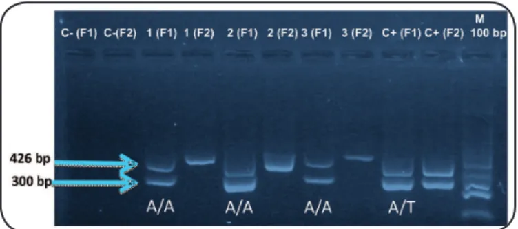
FIGURE 1 -Genotypes of Warao indigenous individuals according to IFNG polymorphism status. The genotype of each individual (+874A/T) was analyzed using the ampliication-refractory mutation system-PCR technique. A pair of primers based on the nucleotide sequence of the human growth hormone (GH1f) was added to amplify a 426-bp product (Control +) to assess the success of PCR; negative control (C-), A allele (F1), T allele (F2), Base pair marker (100bp) (M).
UCV-8007/2011-FONACIT-2013002319/2013-UNU-BIOLAC/2016). Voluntary informed consent forms were signed by individuals.
Quantiication of IFN-γ
Blood samples were transported and processed soon after collection. Briely, tubes were incubated for 24h at 37°C; then, plasma was separated from blood by centrifugation. Measurement of IFN-γ levels in plasma was performed by enzyme-linked immunosorbent assay (ELISA) using the QFT-IT test kit, following the manufacturer’s instructions. The cut-off value for a positive result was ≥ 0.35 International Units (IU)/ mL of IFN-γ in the sample after stimulation witha mixture of the TB7.7 antigen and two synthetic peptides of;the early secreted antigen-6 (ESAT-6), the culture iltrate protein-10 (CFP10), and TB7.7, regardless of the result obtained for the positive control. In accordance with the manufacturer’s instructions, the test result was considered indeterminate if the value of the antigen-stimulated sample was negative or if the value of the positive control was < 0.5IU/mL after subtraction of the value of the nil control, and/or if the negative control was > 8.0IU/mL.
Tuberculin skin test
Tuberculin skin test (TST) was carried out after performing
the QuantiFERON–TB test on Warao indigenous individuals
without TB symptoms or disease activity. The TST was administered according to the Mantoux method, by injecting intradermally 2 tuberculin units; 0.1 mL of puriied protein derivate (RT23 PPD; Statens Serum Institute, Copenhagen, Denmark) as previously described9, 10. Reading was performed by trained professionals between 48 and 72 hours after administration. The criterion for positive test reactivity was based on measurements of the transversal diameter of the indurations on the volar surface of the forearm; (value ≥ 10mm), according to national guidelines2. Due to ethical considerations and norms of
the Venezuelan National TB Control Program, the TST test was not performed on Warao indigenous diagnosed with active TB.
Genotyping of subjects
The two alleles of the polymorphic region of the irst intron of IFNG (+874 A/T), corresponding to each individual were analyzed in two different reaction samples using ampliication refractory mutation system polymerase chain reaction (PCR). Briely, a blood sample of 3-5 mL was collected in a Vacutainer tube with EDTA as anticoagulant. Total DNA was extracted from peripheral blood cells using the Total DNA Isolation System kit (Promega Corporation, WI, US), following the instructions of the supplier. Two sense primers; 5'-TTCTTACAACACAAAATCAAATCA-3' (primer A) and 5'-TTCTTACAACACAAAATCAAATCT-3' (primer T), and one antisense primer 5'-TCAACAAAGCTGATACTCCA-3' were used to amplify the IFNG +874 A/T polymorphism. Each reaction employed a generic antisense primer and one of the two allele-speciic sense primers. In every reaction sample, a pair of primers based on the nucleotide sequence of the human growth hormone (Gh1f); 5′-GCCTTCCAACCATTCCCTTA-3′ (sense) and 5′-TCACGGATTTCTGTTGTGTTTC-3′ (antisense) was included as internal control. The PCR protocol comprised 10 cycles:
95°C for 1 min, 95°C for 15 s, 62°C for 50 s, and 72°C for 40 s, followed by 20 cycles; 95°C for 20 s, 56°C for 50 s, and 72°C for 50 s. The PCR assay was performed using 2µl of DNA (30 ng/µl), 5µl of the master-mix SYBER Green (Promega Corporation, WI, US), 1.2µl of sterile and nuclease-free water and 0.6 µl of each primer (5 mM) added in a inal volume of 10 µl. The ampliied products were separated by electrophoresis on a 2% agarose gel stained with SYBR Green (Sigma-Aldrich Co, St. Louis MO, US) and visualized by a Benchtop UV transilluminator,
MultiDoc-It Digital Imaging System camera combination (Figure 1).
IFNG ampliication
Quantitative PCR (qPCR) was performed on 50 blood samples under non-stimulation and stimulation; under stimulation, 3-5ml of peripheral blood sample was collected in a Vacutainer tube with EDTA as anticoagulant and stimulated
withantigens (ESAT-6, CFP10, and TB7.7) for 24h at 37°C,
after incubation, both non-stimulated and stimulated blood cells were obtained by centrifugation. Total RNA was extracted from blood cells using the Total RNA Isolation System kit (Promega Corporation, WI, US) following the instructions of the supplier, and the RNA content was measured using a spectrophotometer at 260nm. cDNA was generated from 5µg of RNA using the Reverse Transcription System kit (Promega Corporation, WI US), following the manufacturer’s protocol. Briely, RNA was incubated with a 1µl of oligo dT primer (50µM); the reaction was made up to 12µl with sterile and RNaseOut-free water, and incubated at 70ºC for 10 min, after which it was quickly cooled on ice. A total of 2µl of 10 × irst-strand buffer (100mM Tris-HCl, pH 8.8 at 25ºC; 500 mM KCl; 1% Triton X-100),
2µl MgCl2 (25mM), 2µl deoxynucleoside trisphosphate mix
Primers for IFNG selected gene were was designed using the GENBANK database; GTTTTGGGTTCTCTTGGCTGTTA (sense), and AAAAGAGTTCCATTATCCGCTACATC (antisense), (IDT®, USA, NM-000619.2 amplicon size: 112 (bp)) were used to amplify IFNG, data were normalized to those
for the reference gene5. We analyzed expression levels of the
frequently used reference genes; ACTβ, HPRT, and GAPDH to identify the most suitable ones for our study. ACTβ and HPRT were excluded as their ampliication rates were too low. GAPDH was used as the reference gene. AGCCACATCGCTCAGACAC (sense), and GCCCAATACGACCAAATCC (antisense), (IDT®, USA, NM-002046.3 amplicon size: 66 (bp) were used as primers for GAPDH. All primers were designed to anneal at 60°C, and primer speciicities and assay eficiencies were tested on control cDNA.
Quantitative PCR was performed as follows: 2µl of cDNA (50ng) was incubated with 8µl of reaction mix, which was composed of 5µl of SsoFast™ EvaGreen® 2x (BioRad, USA), 2.5µl of sterile and nuclease-free water, and 0.25 µl of IFNG or GAPDH forward and reverse primers (20 μM) each. The reaction mix was placed in a Light Cycler 480® (Roche Life Sciences, USA). Cycling parameters were 1 cycle for enzyme activation at 95°C for 30 s, 45 cycles of ampliication at 95°C for 5 s and 60°C for 10 s, 1 cycle for melting curve analysis at 60°C and 90C for 15 s, and 1 cycle for cooling at 40°C for 30 s. Assay eficiencies were tested on positive and negative cDNA control samples prepared by pooling cDNA from several individuals, as well as on blank control without cDNA; these controls were included in every assay.
Quantiication of gene expression
Acceptance criteria for specific amplification of each targeted cDNA were set as follows: 1) Crossing point (Cp) value for speciic ampliication of control cDNA to be below 40; 2) Single dominant peak in the derivative of the melting curve; 3) No ampliication of non-template controls.
The mRNA relative expression of each gene was calculated based on the values of Cp. Normalization of Cp was performed against corresponding values for the constitutive gene GAPDH, using the following equation: 2-ΔΔct 13.
ER=2-((Cp problem-Cp GAPDH)-(Cp control- Cp GAPDH control))
Data analysis
Genotype frequencies were compared by the Fisher’s exact test, and the relative risk and odds ratio for disease susceptibility or clinical course were calculated. Genotype frequencies in patients and control subjects were not signiicantly different from those predicted under Hardy-Weinberg equilibrium. Results were analyzed by the Mann-Whitney rank-sum test. A multiple logistic regression model employing the likelihood ratio was used to examine the inluence of different genotypes
and ex vivo IFNG expression on susceptibility to tuberculosis.
For comparison of IFNG expression between groups, Shapiro-Wilks for normality, Kruskal-Wallis, and Dunn’s multiple comparison tests were performed using GraphPadPrism, 6.0-Windows (San Diego, Ca. USA).
RESULTS
Characteristics of study participants
A total of 135 participants were enrolled into this study, 24 (17.8%) of who were found to have culture-positive pulmonary TB or ATB. Of the 135 study participants, 111 (82.2%) were classiied as follows: 18 (16.2%) with LTBI (QFT-IT+ or/and TST+) and 93 (83.8%) were healthy controls (CTRL) with no reactivity to QFT-IT (QFT-IT-) or TST (TST-). The age results are shown as value X ± SD years. The average age was 32.0 ± 17.5 years, 42.3 ± 19.8 years, and 36.6 ± 14.3 years for ATB, LTBI, and CTRL groups (Table 1). There were no statistically signiicance differences between males and females in the ATB group (10/14), the LTBI group (10/8), and the CTRL group (28/65), respectively (Table 1). Demographic and clinical data
for the participants are shown in Table 1.
Bacteriological, QFT-IT, and TST
Table 1 shows results for the bacteriological, QFT-IT, and
TST. Smears or smear plus culture were performed for all indigenous individuals with TB symptoms or disease activity (Table 1). Bacteriological assays revealed a statistically
signiicant differencebetween ATB and LTBI groups, both
for smears as well as cultures; p < 0.0001 (Table 1). Chest
radiograph studies conirmed radiographic characteristics with lesions suggestive of active TB (data not shown). QFT-IT
results showed a statistically signiicant differencebetween
indigenous QFT-IT+ and QFT-IT- in ATB (6/24) and LTBI (4/18) versus CTRL (0/93) groups; p < 0.0001and p < 0 .0005,
respectively (Table 1). The skin reactivity test was carried
out in all indigenous individuals without TB symptoms or disease activity in order to study delayed-type hypersensitivity (DTH); reactions for which the transversal diameter of indurations ≥ 10mm were considered positive. There was a statistically signiicant difference between indigenous TST+ and TST- in LTBI (14/18) and CTRL (0/93) groups, p < 0.0001 (Table 1).
Cytokine genotype and allele frequencies of Warao individuals
Analysis of genotype and allele frequencies for IFNG +874 A/T in indigenous patients with TB as well as controls revealed
that the AA genotype for IFNG was the most common (Table
2). The AA genotype was present in 108 (0.973) controls
and 23 (0.958) patients, respectively, p < 0.0001 (Table 2);
Analysis of allele frequency for the IFNG +874 A/T genotype showed that the A allele was markedly overrepresented among Warao indigenous individuals compared with the T allele. A signiicant difference in allele frequencies was observed when comparing A allele between controls [217 (0.977)] and patients with TB [47 (0.979)]. Using the approximation of Katz, a RR of [0.21 (95% CI; 0.1457-0.3113, p < 0.0001) was found (Table 2); however, there was no signiicant difference between
indigenous controls [5 (0.022)] and patients with TB [1 (0.020)] with respect to the T allele, RR of [0.20 (95% CI; 0.032-1.241, p < 0.080)], (Table 2).
Cytokine phenotype frequencies for Warao indigenous and white American individuals
Genotype and allele frequencies for IFNG +874 A/T among the Warao indigenous and white American individuals
are presented in Table 3. Compared with the white American
population, Warao indigenous individuals exhibited a higher frequency of the IFNG +874 allele SNP, whose phenotypic expression is associated with decreased production of IFN-γ. Results showed that the AA genotype for IFNG +874 A/T differed signiicantly between Warao indigenous and white Americans: the frequency of this genotype was higher in Warao indigenous individuals [131(0.97)] compared with white American individuals 27 (0.27) (Table 3). However,
AT and TT genotypes were less common among Warao indigenous individuals [2 (0.014) for both genotypes] compared with white American individuals [53 (0.53) and 20 (0.20)],
respectively, (Table 3). Using the approximation of Katz, a
signiicant difference in genotype frequencies was observed when comparing AA genotype between indigenous and white Americans; AA-homozygous individuals had a RR of [3.59 (95% CI; 2.600-4.968, p < 0.0001)], (Table 3). Additionally,
a signiicant difference was found between indigenous with the AT and TT genotypes and white Americans RR of [0.02 (95% CI; 0.0069-0.1120, p < 0.0001)] and RR of [0.07 (95% CI; 0.0177-0.3098, p < 0.0001)], respectively, Table 3. In relation to allele frequency for IFNG +874A/T, results showed that the A allele was markedly overrepresented among Warao indigenous individuals compared with the T allele. Using the approximation of Katz, a signiicant difference for allele frequencies was obtained when comparing A allele between Warao indigenous [264 (0.98)] and white American individuals [107 (0.54)] RR of [1.82 (95% CI; 1.604-2.082, p < 0.0001)], (Table 3). Additionally, there was a signiicant difference
between indigenous [6 (0.02)] individuals and white Americans [93(0.47)] with respect to the T allele RR of [0.04 (95% CI; 0.0213-0.1069, p < 0.0001)], (Table 3).
Analysis of IFNG expression by quantitative assay Figure 2 shows the medians of mRNA relative expression
between groups studied. A total of 50 individuals composing the quantitative PCR set were studied; these were grouped into three diagnostic categories: 4 indigenous patients with ATB, 18 indigenous patients with LTBI, and 28 healthy indigenous controls (CTRL). The indings are shown as medians and inter-quartile range (IQR) of expression relative of the ATB, LTBI,
Genotypes/Alleles Healthy Controls (n = 111)
Patients with TB (n = 24)
RR CI
(95%)
A:A T:A T:T
108( 0.973)(a) 1(0.009) 2(0.018)
23(0.958)(b) 1(0.041) 0.984.62
-0.9011-1.0077 0.2995-71.423
-Fischer’ s exact Test (p value)
p < 0.0001*
A Allele/T Allele 217(0.977)(c)
5(0.022) 47(0.979) (d) 1(0.020) 0.210.20
0.1457-0.3113 0.0320-1.2410
Fischer’ s exact Test (p value)
p < 0.0001** TABLE 2
Genotype distribution and allele ratio for the Warao indigenous population.
There were statistically signiicant differences between controls and patients with TB with AA genotype, (a) & (b) p < 0.0001; and controls and patients with
TB cases with the A allele, (c) & (d) p < 0.0001. TB: tuberculosis; RR: relative risk; CI: conidence interval.
and CTRL for non-stimulated and stimulated conditions. There were no statistically signiicant differences between groups (Figure 2).
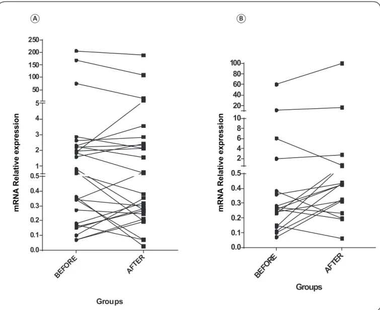
Eighteen blood samples from indigenous patients in the LTBI group, under non-stimulated conditions, showed a decreased IFNG level (4.83 ± 14.51 of mRNA relative expression under non-stimulated conditions, while under stimulated conditions was 6.94 ± 23.53 of mRNA relative expression); however, there was no statistically signiicant difference between the two conditions, p < 0.15 (Figure 3).
Analysis of the 28 blood samples from CTRL indigenous individuals under non-stimulated conditions showed that the median IFNG levels were higher in this group: 17.50 ±51.15 of mRNA relative expression compared with that under stimulated conditions; 14.45 ± 41.31 of mRNA relative expression; there were no statistically signiicant differences between groups under each condition, p < 0.96 (Figure 3). Analysis of 4
blood samples from indigenous individuals with ATB without stimulation revealed that the median IFNG levels in response to
M. tuberculosis-speciic antigens at baseline evaluation was 0.56
± 0.59 of mRNA relative expression. In comparison, the median IFNG levels under stimulated conditions were 1.54 ± 2.62 of mRNA relative expression; there were no statistically signiicant differences under the two conditions (p < 0.49) (data not shown).
DISCUSSION
The Venezuelan Warao indigenous group consists of 25,125 individuals, comprising 17% of the total American
Indian population of Venezuela2. Although the Warao have
The genetic component of TB susceptibility in this population likely involves interaction between alleles located on different genes, as well as chromosomes; this higher susceptibility is considered to arise as a result of the different genetic backgrounds of Warao indigenous individuals and white Americans. Immunological studies performed in the Warao population conirmed inheritance and segregation of DW, an antigen of class II Human Leukocyte Antigen (HLA), HLA haplotypes, linkage disequilibrium deined only in homozygous typing cells of Warao origin, and DR/DQ associations not
seen previously in other human populations14,15. The role
of immunogenicity factors in inluencing the incidence of pulmonary TB in a population is well known14, 15. In the present
work, we aimed to examine a polymorphism in IFNG, which is the most important cytokine in resistance to mycobacterial diseases, and its association with elevated susceptibility to TB in the Warao indigenous population. Present indings showed the highest frequency of the AA genotype of IFNG among Warao CTRL or indigenous controls and Warao patients with ATB. Further, a low frequency of heterozygous AT (AT genotype) among these indigenous groups: Warao CTRL and ATB, and a low frequency of T homozygous (TT genotype) within the Warao CTRL group, were observed. A signiicant difference in genotype frequencies was obtained when comparing the AA genotype between CTRL and ATB groups; AA homozygous indigenous individuals showed a relative risk of 0.98. The IFN-γ cytokine is the archetypical readout for cell-mediated
immune response (CMI) assays16, and has been recognized as
the deining cytokine of Th1 cells. IFN-γ–mediated immune
activation appears to play an importantrole in immunity to
intracellular pathogens, both in mice andhumans17-19. Early
studies demonstrated that mice with disruptionof IFNG are
more susceptible to infection by M. tuberculosis thanwild-type
strains20. Zembruzuski et al. have investigated the Xavante
Genotypes/Alleles
Warao Indigenous (n = 135)
White American
(n = 100)
RR
CI (95%)
A:A T:A T:T
131(0.970)(a) 2 (0.014)(c) 2 (0.014)(e)
27(0.270)(b) 53(0.530)(d) 20(0.200)(f)
3.59 0.02 0.07
2.600-4.968 0.006-0.112 0.017-0.309
Fischer’ s
exact Test (p value) p < 0.0001p < 0.0001 p < 0.0001
A Allele/ T Allele
264(0.977)(g)
6(0.022)(i) 107(0.535) (h)
93(0.465)(j) 1.820.04 1.604-2.0820.021-0.106
Fischer’ s
exact Test (p value) p < 0.0001p < 0.0001 TABLE 3
IFN-γ genotype frequencies in Warao indigenous and white American individuals.
AA genotype and AT + TT genotype frequencies were signiicantly different between Warao indigenous and white American individuals, (a) & (b), (c) & (d) and (e)
& (f) p < 0.0001, Additionally, statistically signiicant differences in A and T allele frequencies were observed between Warao indigenous and white American individuals, (g) & (h), (i) & (j) p < 0.0001; Relative risk (RR) and Conidence Interval (CI).
CTR L N
S
TBL NS
TBA NS
CTR L S
TBL S
TBA S
0.0 0.5 1.0 1.5 2 4 6 8 10 50 100 150 200 250
Groups
mRNA
Relative expression
Groups
BE
FORE AFTER 0.0
0.1 0.2 0.3 0.4 0.5 2 4 6 8 10 20 40 60 80 100
Groups
BEF
ORE
AFT
ER 0.0
0.1 0.2 0.3 0.4 0.5 1 2 3 4 5 50 100 150 200 250
m
RN
A
Relative expression
mRNA
Relative expression
FIGURE 3 - Relative expression ofIFNG measured by quantitative PCR before and after the stimulated condition for CTRL and LTBI groups; (A) and (B), respectively. The differences between the relative expression values in each group before and after stimulation were not signiicant by Mann Whitney test.
A B
population, who live in Brazil and exhibit high TB prevalence, and revealed that the absence of TST response (anergy) may be associated with a predominantly Th2 cytokine pattern, which may increase susceptibility to TB21.
The present work describes the relative expression of IFNG in Warao individuals with TB. The expression of this cytokine relects the antigen-speciic response of cells stimulated ex vivo
in whole blood samples from ATB, LTBI, and CTRL indigenous groups by speciic-M. tuberculosis T-cell antigens for 24h. There were no statistically signiicant differences between non-stimulated and non-stimulated conditions between groups; IFNG was found to be expressed at low levels, especially in the ATB and LTBI groups (0.56 ± 0.59 and 4.83 ± 14.51 of mRNA relative expression, respectively under non-stimulation, and 1.54 ± 2.62 and 6.94 ± 23.53 of mRNA relative expression, respectively, under stimulation). There were no statistically significant differences between either condition for the CTRL group,
demonstrating the existence of concordance between the highest frequency of the AA genotype among Warao healthy controls and patients with TB, and IFNG expression. IFN-γ cytokine exhibits poor potential as a biomarker in the Warao indigenous population owing to its failure to discriminate between ATB and LTBI indigenous individuals. The low IFNG expression levels detected may be correlated with the present indings in relation to QFT-IT assays; among the LTBI indigenous individuals tested, only 4 of were QFT-IT+. In this context, the value of the QFT-IT as an adjunct in diagnosing latent infection in Warao individuals may be limited: in Warao children for whom both TST and QFT-IT results are available, the proportion of children with a positive TST was high compared with those with a positive QFT-IT22. The latter correlates with the present results;
therefore, replacement of the TST by QFT-IT for detection of
M. tuberculosis infection is not recommended in this indigenous
It has been reported that the clinical symptoms in patients
with TB correlate with M. tuberculosis–stimulated IFN-γ
production by PBMCs, which is lower in patients with TB than in healthy tuberculin reactors, and even lower in patients with extensive disease10,23. Differences in the distribution of the IFNG
genotype may explain the lower production of this cytokine in patients with TB; these patients have a higher frequency of the homozygous A (AA genotype) associated with lower PPD-stimulated IFN-γ production23. The latter indings correlate with
those of our previous study examining the capacity of antigen-induced proliferation by PBMCs and IFN-γ production in Warao indigenous individuals with pulmonary TB and Warao healthy controls. The results revealed that IFN-γ production in Warao patients and controls was signiicantly lower after stimulation for 24h and 48h compared with that in the Creole groups10.
As IFN-γ plays a crucial role in facilitating macrophage
containment of M. tuberculosis, we aimed to determine whether
the observed allelic variation associated with low expression of IFNG may in part contribute to the high rates of TB among Warao indigenous individuals relative to the white American population. The latter population was used for comparison because of the historical context in which the two populations evolved in relationship to their microbial environments, particularly in the context of their response to the selective pressures of their respective microbial environments, as found by Larcombe et al.18.
Results showed that AA genotype frequency for IFNG +874 A/T differed signiicantly between Warao indigenous and white American individuals. Higher frequencies of these genotypes were observed in the Warao indigenous population (0.970 compared with 0.270 in white Americans, while lower frequencies of the AT and TT genotypes were observed in the Warao individuals: 0.014 for each genotype compared with 0.530 and 0.200, respectively, in white Americans). This comparison additionally reveals that the IFNG +874 A/T polymorphism is signiicantly associated with pulmonary TB, the indings showed that indigenous homozygous for the +874 A allele had a 3.59-fold increased risk of contracting TB. Our results correlate with previous findings showing that individuals homozygous for the +874 A allele had a 3.75-fold increased risk of developing TB and the lowest IFNG expression level in the groups studied, therefore supporting that the IFNG +874 A/T polymorphism is signiicantly associated with pulmonary TB18,19.
Ethnic-speciic genetic variations may greatly inluence host immunity to TB, causing variations in TB susceptibility in the ethnic population studied24. Accordingly, several studies of genetic diversity
in ethnic populations have reported an association between the IFNG +874 polymorphism and TB18,25. Studies in relation to genotype proile in Canadian aboriginal and Filipino populations showed that both populations have a higher frequency of cytokine SNPs associated with low production of IFNG (AA genotype) compared with white Americans; these aboriginal populations exhibited a signiicantly higher frequency of the A allele at the IFNG loci18; in contrast, white Americans exhibit a relatively higher frequency of cytokine SNPs associated with delayed-type hypersensitivity
IFN-γ response, the latter suggest that white Americans tend to mount a Th1 immune response, while Canadian aboriginal and Filipino populations show a tendency towards a Th2 immune response18. In this context, intestinal parasites are endemic among the Warao population, which is consistent with the considerably
higher levels of total serum IgE found in this population26.
Recent data suggest that, similar to Canadian aboriginal and Filipino populations18, in other populations, such as Spanish (RR = 3.75, CI 2.26-6.63, p = 0.0017)17, Hong Kong Chinese (RR = 3.79,
CI 1.93-7.45, p = 0.001)27, South African colored (RR = 1.46, CI
1.12-1.91, p = 0.0062)28, and Warao indigenous populations (RR = 3.59, CI 2.600-4.968, p = 0.0001), common polymorphisms in the IFNG +874 AA genotype are associated with the risk of TB disease. However, the +874AA genotype was not signiicantly more frequentin cases than in control subjects (OR, 1.16;
95%CI, 0.89–1.51;p = 0.25) in individuals from West African
populations28, and in Brazilian patients with TB29. However, the inluence of gene polymorphisms on protein production may differ as a result of linkage with functional variants in other loci in the regulatory regions of the genes, or with other cytokine
polymorphisms necessary for adequate immune response to M.
tuberculosis infection. Additional studies are needed to conirm
the inluence of the IFNG +874 A/T alleles and other cytokine genes on TB susceptibility in the Warao population.
Acknowledgments
We would like to thank Dr. Jacobus H. de Waard at Laboratorio de Tuberculosis, Instituto de Biomedicina “Dr. Jacinto Convit”, Universidad Central de Venezuela, for performing molecular and microbiological studies of M. tuberculosis.
Financial support
This study was supported by Project Number CDCH-UCV-6256-8007/2011-FONACIT-2013002319/2013-UNU-BIOLAC/2016).
Conlict of interest
The authors declare that there is no conlict of interest.
REFERENCES
1. World Health Organization. World Health Statistics 2012. WHO, Geneva, 2012.
2. Ministerio de Sanidad y Asistencia Social (MSAS). Programa Integrado de Control de la Tuberculosis. Seminario Técnico-Administrativo. Caracas: 1999. 150pp.
3. Ministerio de Salud y Desarrollo Social (MSDS). Evaluación del Programa Nacional de Control de la Tuberculosis. Año Evaluado 2001. Caracas: 2002. 201pp.
4. Ministerio de Salud y Desarrollo Social (MSDS) 2006. Evaluación del Programa Nacional de Control de la Tuberculosis.Año Evaluado 2006. Caracas: 2006. 200 pp.
5. Bibova I, Linhartova I, Stanek O, Rusnakova V, Kubista M, Suchanek M, et al. Detection of immune cell response to M. tuberculosis-speciic antigens by quantitative polymerase chain reaction. Diagn Microbiol Infect Dis. 2012;72(1):68-78.
7. Trajkov D, Trajchevska M, Arsov T, Petlichkovski A, Strezova A, Einska-Mladenovska O, et al. Association of 22 cytokine gene polymorphisms with tuberculosis in Macedonians. Indian J Tuberc. 2009;56(3):117-31.
8. Khalilullah SA, Harapan H, Hasan NA, Winardi W, Ichsan A, Mulyadi M. Host genome polymorphisms and tuberculosis infection: What we have to say? Egypt J Chest Dis Tuberc. 2014;63(1):173-85. 9. Araujo Z, de Waard JH, de Larrea CF, Borges R, Convit J. The
effect of Bacille Calmette-Guérin vaccine on tuberculin reactivity in indigenous children from communities with high prevalence of tuberculosis. Vaccine. 2008;26(44):5575-81.
10. Giampietro F, de Waard JH, Rivas-Santiago B, Enciso-Moreno JA, Salgado A, Araujo Z. In vitro levels of cytokines in response to puriied protein derivative (PPD) antigen in a population with high prevalence of pulmonary tuberculosis. Hum Immunol. 2010;71(11):1099-104.
11. Larrea CF, Fandiño C,López D, del Nogal B, Rodríguez N, Convit J, et al.Childhood tuberculosis in the Warao population in Venezuela. Invest Clin. 2002;43:35-48.
12. Kudoh S, Kudoh T. A simple technique for culturing tubercle bacilli. WHO/TB/51. Geneva Bull World Health Organ. 1974;51(1):71-82. 13. Livak KJ, Schmittgen TD. Analysis of relative gene expression
data using real time quantitative PCR and the 22DDCT method. Methods. 2001;25(4):402-8.
14. Layrisse Z, Heinen HD, Balbas O, García E, Stoikow Z. Unique HLA-DR/DQ associations revealed by family studies in Warao Amerindians. Haplotype and homozygosity frequencies. Hum Immunol. 1988;23(1):45-57.
15. Makhatadze NJ, Franco MT, Layrisse Z. HLA class I and class II allele and haplotype distribution in the Venezuelan population. Hum Immunol. 1997;55(1):53-8.
16. Chegou NN, Heyckendorf J, Walzl G, Lange Ch, Ruhwald M. Beyond the IFN-γ horizon: biomarkers for immunodiagnosis of infection with Mycobacterium tuberculosis. Eur Respir J. 2014;43:1472-1486.
17. López-Maderuelo D, Arnalich F, Serantes R, González A, Codoceo R, Madero R, et al.Interferon-γ and Interleukin-10 Gene Polymorphisms in Pulmonary Tuberculosis. Am J Respir Crit Care Med. 2003;167(7)970-5.
18. Larcombe L, Rempel JD, Dembinski IJ, Tinckam K, Rigatto C, Nickerson P. Differential cytokine genotype frequencies among Canadian Aboriginal and Caucasian populations. Genes Immun. 2005;6(2):140-4.
19. Larcombe L, PH Lodge AM, Brown JS, Dembinski IJ, Milligan LC, Larcombe EA, et al. Functional gene polymorphisms in Canadian Aboriginal populations with high rates of tuberculosis. J Infect Dis. 2008;198(8):1175-9.
20. Cooper AM, Dalton DK, Stewart TA, Grifin JP, Russell DG, Orme IM. Disseminated tuberculosis in interferon gamma gene-disrupted
mice. J Exp Med. 1993;178(6):2243-7.
21. Zembruzuski VM, Basta PC, Callegari-Jacques SM. Cytokine genes are associated with tuberculin skin test response in a native
Brazilian population. Tuberculosis. 2010;90(1):44-9.
22. Verhagen LM, Maes M, Villalba JA, d´Alessandro A,
Perez-Rodriguez L, España MF, et al. Agreement between QuantiFERON®
-TB Gold In-Tube and the tuberculin skin test and predictors of positive test results in Warao Amerindian pediatric tuberculosis
contacts. BMC Infect Dis. 2014;14:383.
23. Prabhu Anand S, Harishankar M, Selvaraj P. Interferon gamma
gene +874A/T polymorphism and intracellular interferon gamma
expression in pulmonary tuberculosis. Cytokine.2010;49(2):130-3.
24. Maronna R, Longhi P, Marques Zembrzuski V, Basta PC, Croda J. Genetic Polymorphism and immune response to tuberculosis
in indigenous populations: a brief review. Braz J Infect Dis 2013;17:363-368.
25. Cox AJ, Moscovis SM, Blackwell CC, Scott RJ. Cytokine gene polymorphism among Indigenous Australians. Innate Immun. 2014;20(4):431-439.
26. Araujo Z, Giampietro F, Rivas-Santiago B, Luna-Herrera J, Wide A, Clark W, et al. Patients exposed to Mycobacterium
tuberculosis infection with a prominent IgE response. Arch Med
Res. 2012;43(3):225-32.
27. Tso HW, Ip WK, Chong WP, Tam CM, Chiang AKS, Lau YL. Association of interferon gamma and interleukin 10 genes with tuberculosis in Hong Kong Chinese. Genes Immun. 2005;6(4):358-63. 28. Bennett S, Lienhardt C, Bah-Sow O, Gustafson P, Manneh K, Del
Prete G, et al. Investigation of environmental and host-related risk factors for tuberculosis in Africa: II. Investigation of host genetic factors. Am J Epidemiol. 2002;155(11):1074-9.