235
Rev Soc Bras Med Trop 50(2):235-238, March-April, 2017 doi: 10.1590/0037-8682-0048-2016
Short Communication
Corresponding author: Dra. Josiane Somariva Prophiro.
e-mail: [email protected]
Received 3 February 2016
Accepted 25 November 2016
Ascogregarina taiwanensis infection in Aedes aegypti
and
Aedes albopictus
in Santa Catarina, South Brazil
Josiane Somariva Prophiro
[1],[2], Thiago Nunes Pereira
[2],[3], Joice Guilherme de Oliveira
[2],
Guilherme Werner Dandolini
[2], Mario Antonio Navarro da Silva
[1]and Onilda Santos da Silva
[3][1]. Curso de Programa de Pós-Graduação em Entomologia, Departamento de Zoologia, Universidade Federal do Paraná, Curitiba, PR, Brasil. [2]. Grupo de Pesquisa em Imunoparasitologia, Departamento de Ciências da Saúde, Universidade do Sul de Santa Catarina, Tubarão, SC, Brasil. [3]. Departamento de Microbiologia, Imunologia e Parasitologia, Instituto de Ciências Básicas da Saúde. Universidade Federal do Rio Grande do Sul, Porto Alegre, RS, Brasil.
Abstract
Introduction: This study registers Ascogregarina spp. infection in ield populations of Aedes aegypti and Aedes albopictus in a subtropical region of Brazil. Methods: Mosquito larvae collected in tires placed in four municipalities of Santa Catarina were
identiied morphologically and assessed for Ascogregarina sp. infection using morphological and molecular methods. Results: Both mosquito species harbored Ascogregarina taiwanensis, whose genomic DNA was conirmed in both the Aedes species by PCR. DNA sequences were deposited in GenBank. Conclusion: Both Ae. albopictus e Ae. aegypti harbor Ascogregarina sp.
Keywords:Ascogregarina taiwanensis. DNA sequence. South Brazil.
Aedes aegypti and Aedes albopictus (Diptera: Culicidae) are important mosquito species responsible for transmitting the etiological agent that causes dengue and yellow fever, mainly in the tropical and subtropical regions of the world. Marcondes and Ximenes1 suggested that the presence of these
mosquito species in Latin America is associated with additional risk because they are potential vectors of Chikungunya and Zika viruses. In Brazil, both the species have been reported to have developed resistance to chemical insecticides2. For this
reason, some species of microorganisms have been investigated for their potential activity against these mosquito vectors. About 10 species of mosquitoes that can harbor protozoan parasites belonging to the genus Ascogregarina (Apicomplexa:
Ascogregarinidae) have been identiied3. These parasites have
been suggested for biological control, especially of Ae. aegypti
and Ae. albopictus, which are the natural hosts of Ascogregarina culicis and Ascogregarina taiwanensis, respectively4.
According to Tseng5, the infection of mosquitoes by Ascogregarina species occurs when insects in the irst larval
stages ingest their oocysts containing sporozoites, present in mosquito breeding sites. Inside the mid gut of the larvae, these sporozoites are released and penetrate the epithelial cells of the host and develop into trophozoites. Prior to the metamorphosis
of larvae to pupae, the trophozoites migrate to the Malpighian tubules, where they transform into macro or microgamete. The residual trophozoites in the midgut of pupae are apoptosed. During the pupal stage, two gametes fuse to form a gametocyte, in which hundreds of oocysts are formed. These oocysts are then released into the breeding grounds during the metamorphosis of pupae to adults, and also when the females lay their eggs. They can also be released from the adult through defecation or after death.
Studies on the distribution, morphology, pathogenicity, and biology of Ascogregarina spp. are very important as they can be useful for biological control of insects3. However, these parasites
have been poorly studied in South America. As of date, Ae. aegypti was found to be infected with A. culicis in Argentina6,7.
In Brazil, Passos and Tadei8 described the only encounter of Ae. aegypti and Ae. albopictus infected with A. culicis and A. taiwanensis, respectively, in Manaus.
In a mosquito survey in Santa Catarina, South Brazil, we collected Ae. aegypti and Ae. albopictus infected with a protozoan belonging to Ascogregarina. This is the irst report
on Ae. aegypti and Ae. albopictus infected with Ascogregarina
species from a subtropical region of Brazil.
The surveys were conducted in four municipalities, Tubarão
(28°28′00′′S–49°00′25′′W), Gravatal (28°19′52′′S–49°02'07"W),
Laguna (28°28′57′′S–48°46′51′′W), and Capivari de Baixo (28°26′41′′S–48°57′28′′W), with subtropical climate and
236
Prophiro JS, et al. - Ascogregarina taiwanensis in South Brazil
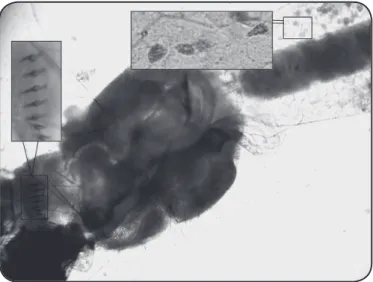
FIGURE 1 - Larvae of Aedes albopictus infected with Ascogregarina taiwanensis. The comb-scales on the eighth abdominal segment and the trophozoites inside the mid gut are evident. The larvae were dissected under a stereomicroscope.
immature larvae of Ae. albopictus and Ae. aegypti. The
larvae were identiied based on the comb-scales of the eighth
abdominal segment and were brought to the laboratory to assess the ascogregarine infection.
The aim of this study was to verify whether the Aedes species could be infected with the parasite. For this purpose, 20% of third and/or fourth instars oflarvae (100 larvae per species) from each municipality were dissected in the laboratory under
a stereoscopic microscope (OLYMPUS CX31-P). To conirm
the presence of Ascogregarina sp., the entire digestive tracts of the larvae were removed. The most effective method involved the application of pressure between the cephalic and thoracic parts of the body and of a counterforce on the respiratory siphon. Once the intact intestine of larva was removed, an accurate
search was made to conirm the presence of trophozoites in the lumen, epithelium, and peritrophic membrane. When the presence of these parasites was conirmed in the larvae, they were photographed and/or ilmed using an optical microscope
(ZEISS STEMI 200 C) with VMS3.5 program.
After conirming Ascogregarina infection in Ae. aegypti
and Ae. albopictus larvae, genomic DNA was extracted from
these protozoans using a slightly modiied salting-out protocol of Morales10. Briely, a pool of 10 dissected intestines from
each Ae. aegypti and/or Ae. albopictus larvae was placed separately in 1.5ml tubes. The digestive tracts were then macerated in 60µl buffer (0.16M NaCl, 0.06M sucrose, 0.5% Ethylenediaminetetraacetic acid (EDTA), 0.1M Sodium dodecyl sulfate (SDS), Tris-HCl, pH 8.6) and incubated at 65°C, for 30 min. Subsequently, 40µl of 8 M potassium acetate was added, and the homogenate was incubated at 4°C for 30 min. The solution was then centrifuged at 12,000rpm for 10 min. The supernatant was transferred to a new 1.5ml tube containing 100µl of absolute ethanol and centrifuged for 10 min at 12,000rpm. The DNA was washed with 70% ethanol and resuspended in 50µl of sterile distilled water.
The PCR ampliication was performed as described by
Morales10, who developed primers, namely AT and AC,
to specifically distinguish A. taiwanensis from A. culicis, respectively. A universal primer (AU) was also used. The
primer AT (5′-GAG AAG CCG TCG TCA ATA CAG C-3′) binds to the ITS2 region of rDNA and the primer AC (5′-CAC TTA GTG TTT TGT TTG ATG TC-3′) binds to the ITS1 region of rDNA. The primer AU (5′-ACC GCC CGT CCG TTC AAT CG-3′) binds to the 18S DNA of both the species
of Ascogregarina. Each PCR was performed in a total volume of 25µl containing 2µl of genomic DNA from the dissected intestine of Ae. albopictus or Ae. aegypti, 8.5µl of nuclease-free water (Promega), 125µl of GoTaq® Green Master mix (Promega), and 1µl each of 2mM primers AC or AT and AU.
The PCR included an initial denaturation at 94°C for 1 min, followed by 30 ampliication cycles (94°C for 1 min, 50°C for 1 min, 72°C for 2 min) and a inal extension step at 72°C for
10 min. To avoid any false positive or negative results, the PCR was performed thrice on different days.
The amplified fragments were electrophoresed on 2% agarose gel, stained with ethidium bromide, and visualized
under UV light and photographed. After the conirmation of
Ascogregarina sp., the DNA products were puriied using
QIAquick® extraction kit (QIAGEN GmbH, Hilden, Germany), according to the recommendations of the manufacturer. The
genomic fragments were sent to Ludwig Biotec (Alvorada – Rio
Grande do Sul) for sequencing on a MegaBace 1000 automated sequencer.
In the surveys of 2011 and 2012, only the Ae. albopictus
larvae were found in the four cities that were included in the study. The immature larvae of Ae. aegypti were found only in Tubarão in 2013, coexisting with Ae. albopictus. The larvae of
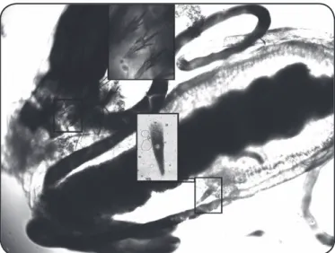
Ae. albopictus were found to be infected with Ascogregarina taiwanensis (Figure 1), in the four municipalities. The larvae of Ae. aegypti (Figure 2) collected from Tubarão were also infected with this parasite species.
All the trophozoites of A. taiwanensis detected in
Ae. albopictus and Ae. aegypti larvae were, in general,
morphologically similar to a comma and had a brown pigment when observed under a phase contrast microscope. The trophozoites differed in size, ranging from 50 to 170µm. Additionally, giant trophozoites (223, 337, and 621-µm in length) were sometimes found in the midgut of Ae. albopictus
larvae.The trophozoites, matured extracellularly, were found migrating through or in the Malpighian tubules, in some larvae.
We used molecular techniques to avoid ambiguity or error in the identiication based only on the morphology of gregarine. Using PCR, we conirmed Ae. albopictus harboring
A. taiwanensis in the four cities, as well as Ae. aegypti in
Tubarão. No ampliication was obtained from the DNA of
A. culicis (Figure 3). The 275- and 212-bp sequences of the
ampliied products from two A. taiwanensis samples obtained from Ae. aegypti and Ae. albopictus were aligned with the sequences present in NCBI using BLAST, revealing an identity
237
Rev Soc Bras Med Trop 50(2):235-238, March-April, 2017
FIGURE 2 - Larvae of Aedes aegypti infected with Ascogregarina taiwanensis. The comb-scales on the eighth abdominal segment and trophozoites inside the mid gut are evident. The larvae were dissected under a stereomicroscope.
FIGURE 3 - Visualization of the PCR ampliied products on 2% agarose gel. The primers AC and AT were used for the identiication of Ascogregarina culicis
and Ascogregarina taiwanensis, respectively. Lane M: 100-bp ladder; Lane 2:
negative control (water); Lane 3: Aedes albopictus negative (AC); Lane 4: Aedes aegypti negative (AC); Lanes 5-7: Aedes albopictus positive for Ascogregarina taiwanensis (AT); Lane 8: Aedes aegypti negative (AT); Lane 9: Aedes albopictus
negative (AT); Lanes 10-12: Aedes aegypti positive for Ascogregarina taiwanensis
(AT). PCR: Polymerase chain reaction; AC: (5′-CAC TTA GTG TTT TGT TTG
ATG TC-3′); AT: (5′-GAG AAG CCG TCG TCA ATA CAG C-3′).
M 2 3 4 5 6 7 8 9 10 11 12
600 pb
O u r o b s e r v a t i o n s r e g a r d i n g t h e m o r p h o l o g i c a l characterization of Ascogregarina were similar to those of Reyes-Villanueva et al.11 who also used these methods for identiication of A. culicis and A. taiwanensis obtained from
Ae. aegypti and Ae. albopictus, respectively, collected from Tampa, Florida, USA. However, Morales et al.10 reported that
morphological differentiation may be involved in the process
of desiccation of insects. The larvae collected in the ield could
have ingested oocysts of both the gregarine species at different times during the larval development, resulting in variations in the ages of gamonts and trophozoites in the samples.
Based on the morphological characters, Passos and Tadei8
found that Ae. aegypti were infected with A. culicis and Ae. albopictus were infected with A. taiwanensis in the same breeding place, in the Amazon region of Brazil. However, according to Blackmore4, in studies related to competition between Ae. aegypti and Ae. albopictus for breeding places, an accurate
identiication of the gregarines is important for better estimation
of the prevalence of infection among these mosquito species.
We found some bigger trophozoites than those reported by
Albicocco and Vezzani7 (148.35-217.58μm) in the midgut and
Malpighian tubules of Ae. aegypti, infected with A. culicis. Chen and Yang12 demonstrated that the size of A. taiwanensis
trophozoites depends on water temperature. In the mosquito larvae collected in this study, we expected to find bigger trophozoites than those reported by these authors.
We also observed trophozoites, matured extracellularly,
migrating through, or in the Malpighian tubules. According to Chen and Fan-Chiang13, migration toward these tubules is
normally unidirectional and usually occurs among trophozoites that are liberated in the midgut of early pupae.
Desportes3 described that the gregarines, A. culicis and A. taiwanensis, are host-specific parasites of Ae. aegypti
and Ae. albopictus, respectively. Further, Albicocco and
Vezzani7 presumed that only A. culicis is found infecting Ae.
aegypti, worldwide. However, we found a possible
cross-infection between Ascogregarina and Aedes. We conirmed Ae. albopictus, as well as Ae. aegypti, harboring A.taiwanensis, using PCR and DNA sequencing. This species of Ascogregarina
has been also found in other mosquito species, such as Aedes epactius and Culex restuans14. Vezzani and Wisnivesky14
described that gregarine infection in a non-natural mosquito host could be harmful. However, some studies have reported that
A. taiwanensis can infect Ae. aegypti and in some circumstances can cause high mortality, whereas A. culicis is not pathogenic to Ae. albopictus4,11,14. Therefore, additional studies must be
conducted on Ae. aegypti harboring this parasite, once it is considered a non-natural host.
Acknowledgments
We thank Vinicius José Maschio, M.Sc. and Dr. Rene Darela Blazius from
Universidade Federal do Rio Grande do Sul, for helping with molecular characterization.
Conlict of interests
The authors declare that there is no conlict of interest.
REFERENCES
1. Marcondes CB, Ximenes MF. Zika virus in Brazil and the danger of infestation by Aedes (Stegomyia) mosquitoes. Rev Soc Bras
Med Trop. 2016;49(1):4-10.
2. Prophiro JS, Silva OS, Luna JED, Piccoli CF, Kanis LA, Silva MAN. Aedes aegypti and Aedes albopictus (Diptera: Culicidae): coexistence and susceptibility to temephos, in municipalities with occurrence of dengue and differentiated characteristics of urbanization. Rev Soc Bras Med Trop. 2011;44(3):300-5.
3. Desportes I. Systematics of terrestrial and freshwater gregarines.
238
Taxonomy, Biology, The Gregarines. vol. 2. Netherlands: Brill Academic Publishers; 2013, p. 377-710.
4. Tseng M. Ascogregarine parasites as possible biocontrol agents of mosquitoes. J Am Mosq Control Assoc. 2007;23(suppl 2):30-4.
5. Dellapé ME, Marti GA, Tranchida MC, Garcia JJ. First record of
Aedes aegypti (L.) (Diptera: Culicidae) infected by the parasite
Ascogregarina culicis (Ross) (Apicomplexa: Lecudinidae) in Argentina. Entomol Vectores. 2005;12(1):111-5.
6. Albicócco AP, Vezzani D. Further study on Ascogregarina culicis
in temperate Argentina: prevalence and intensity in Aedes aegypti
larvae and pupae. J Invertebr Pathol. 2009;101(3):210-4.
7. Dos Passos RA, Tadei WP. Parasitism of Ascogregarina taiwanensis
and Ascogregarina culicis (Apicomplexa: Lecudinidae) in larvae of
Aedes albopictus and Aedes aegypti (Diptera: Culicidae) from Manaus,
Amazon region, Brazil. J Invertebr Pathol. 2008;97(3):230-6.
8. Instituto Brasileiro de Geograia e Estatistica (IBGE). Cidades
(internet). IBGE; 2014. (updated 2016 Jan 19) Available at: http:// www.cidades.ibge.gov.br/xtras/uf.php?lang=&coduf=42&search=s anta-catarina
9. Morales ME, Ocampo CB, Cadena H, Copeland CS, Termini M,
Wesson DM. Differential identiication of Ascogregarina species
(Apicomplexa: Lecudinidae) in Aedes aegypti and Aedes albopictus
(Diptera: Culicidae) by polymerase chain reaction. J Parasitol.
2005;91(6):1352-6.
10. Reyes-Villanueva F, Becnel JJ, Butler JF. Morphological traits for distinguishing extracellular gamonts of Ascogregarina culicis and
Ascogregarina taiwanensis in Aedes aegypti and Aedes albopictus.
J Invertebr Pathol. 2001;77(3):227-9.
11. Chen WJ, Yang CH. Developmental synchrony of Ascogregarina taiwanensis (Apicomplexa: Lecudinidae) within Aedes albopictus
(Diptera: Culicidae). J Med Entomol. 1996;33(2):212-5.
12. Chen WJ, Fan-Chiang MH. Directed migration of Ascogregarina taiwanensis (Apicomplexa: Lecudinidae) in its natural host Aedes albopictus (Diptera: Culicidae). J Eukaryot Microbiol 2001; 48(5):537-41.
13. Munstermann LE, Wesson DM. First record of Ascogregarina taiwanensis (Apicomplexa: Lecudinidae) in north American Aedes albopictus. J Am Mosq Control Assoc. 1990;6(2):235-43.
14. Vezzani D, Wisnivesky C. Prevalence and seasonality of
Ascogregarina culicis (Apicomplexa: Lecudinidae) in natural populations of Aedes aegypti (Diptera: Culicidae) from temperate
Argentina. J Invertebr Pathol. 2006;91(3):183-7.