Analysis of adhesions resulted from mesh fixation with fibrin
Analysis of adhesions resulted from mesh fixation with fibrin
Analysis of adhesions resulted from mesh fixation with fibrin
Analysis of adhesions resulted from mesh fixation with fibrin
Analysis of adhesions resulted from mesh fixation with fibrin
sealant and suture – experimental intraperitoneal model
sealant and suture – experimental intraperitoneal model
sealant and suture – experimental intraperitoneal model
sealant and suture – experimental intraperitoneal model
sealant and suture – experimental intraperitoneal model
Análise das aderências resultantes da fixação de telas cirúrgicas com selantes
Análise das aderências resultantes da fixação de telas cirúrgicas com selantes
Análise das aderências resultantes da fixação de telas cirúrgicas com selantes
Análise das aderências resultantes da fixação de telas cirúrgicas com selantes
Análise das aderências resultantes da fixação de telas cirúrgicas com selantes
de fibrina e sutura – modelo experimental intraperitoneal
de fibrina e sutura – modelo experimental intraperitoneal
de fibrina e sutura – modelo experimental intraperitoneal
de fibrina e sutura – modelo experimental intraperitoneal
de fibrina e sutura – modelo experimental intraperitoneal
JULIANO HERMES MAESO MONTES1; ANDRÉ VICENTE BIGOLIN1; RENATA BAÚ2; ROBERTO NICOLA2; JOÃO VICENTE MACHADO GROSSI3;
CLÁUDIA JULIANA LOUREIRO4; LEANDRO TOTTI CAVAZZOLA, TCBC-RS5
A B S T R A C T A B S T R A C T A B S T R A C T A B S T R A C T A B S T R A C T
Objective Objective Objective Objective
Objective: To compare surgical fixation of polypropylene mesh (PP) and coated polypropylene mesh (PCD) using polypropylene suture and fibrin glue, as for the formation of intraperitoneal adhesions. MethodsMethodsMethodsMethodsMethods: A sample of 46 female Wistar rats were randomized into six groups: two control groups, with five rats each, were subjected to one medial incision (MI) and the other to a U-shaped incision (UI), none of these groups received the mesh. Two groups of PP mesh, with ten rats, fixed with suture (PPF), the other with six rats, fixed with biological glue (PPC). And two groups of PCD mesh, at first, with ten animals, the mesh was fixed with sutures (PCDFs) and the second with ten animals with biological glue (PCDC). ResultsResultsResultsResults: After 21 days, the control groupsResults showed no significant adhesions. The PPC group showed a lower degree of adhesion than the PPF group (p = 0.01). There was no difference between the groups with PCD. ConclusionConclusionConclusionConclusionConclusion: Comparison of fixation was statistically different only with PP mesh, with lesser degrees of adherence when using the glue. Adhesions were predominantly located at the extremities of the meshes studied.
Key words Key words Key words Key words
Key words: Hernia. Surgical mesh. Fibrin tissue adhesive. Suture techniques. Tissue adhesion.
Research conducted at the Experimental Laboratory, Institute for Basic Health Sciences, Lutheran University of Brazil (ULBRA), Brazil.
1. Physician, Canoas Lutheran University of Brazil (ULBRA); 2. Medical School Graduate, Medicine, ULBRA; 3. Resident, General Surgery, Porto Alegre Emergency Room; 4. Resident, Ophthalmology Porto Alegre Eye Bank Hospital; 5. Postdoctoral, Minimally Invasive Surgery, Case Western Reserve University, Cleveland, Ohio. Associate Professor, ULBRA and Federal Universidade of Rio Grande do Sul.
INTRODUCTION
INTRODUCTION
INTRODUCTION
INTRODUCTION
INTRODUCTION
T
he weakness of the abdominal wall associated with increased pressure of the intra-abdominal cavity can cause the displacement of its contents through a defect, setting the herniation. It is a disease with high incidence; it is estimated that approximately 500,000 operations were performed in Brazil between 1993 and 1996, representing 5.3% of all surgical procedures performed by the National Health System, with an estimated cost of 100 million reais1.The most common hernia type is the inguinal, corresponding to approximately 75%, followed by the ven-tral type, with 10%. Studies have shown that the formation of hernia is associated with reduction of total collagen production, and in particular the reduction of collagen type I2. The only effective treatment is surgery. The main
encountered complications are bowel obstruction, hernia incarceration and/or strangulation, inflammation, chronic pain, fistula, adhesions and infertility3.
Risk factors for adhesion formation are trauma (surgical), foreign body reaction, infection, and ischemia4.
The prevalence of incisional hernia (IH) has many variations. Some studies suggest IH incidence in the general population between 2 and 20%5; in other studies this frequency has
shown to be greater, between 30 and 40%6. The primary
surgical incision is associated with 25-52% chance of IH formation7,8. Incisions larger than 10cm in diameter in the
longitudinal direction display frequency of IH in 20-54% when no surgical meshes are used9 -11.
Currently, the use of surgical prostheses for correction of all types of inguinal hernias is advocated by most surgical societies. Most patients benefit from the use of this technique based on the results of the concept “no tension”. In the present experiment we used meshes polypropylene (PP) and meshes with more complex coated polypropylene (PCD), the latter consisting of a net of oxidized regenerated cellulose and a PP mesh, a non-absorbable polypropylene mesh which is encapsulated by a polymer12.
The biological glue Tissucol ® (TSS) is composed of lyophilized fibrinogen, a solution of thrombin and other of aprotinin, used to produce adhesion between tissues in animals, including humans. It is mainly used in the adjuvant treatment to achieve hemostasis in diffuse bleeding, filling and adherence of tissues during surgical procedures13. Its
use requires precision and skill in order to obtain the best results. To date, there is no report in the literature that the biological glue poses any risk of infectious diseases transmission14. Different studies using fibrin glue showed
less adhesion formation15-17.
This study compared the surgical mesh fixation with sutures to fibrin glue fixation with the meshes PP and PCD.
METHODS
METHODS
METHODS
METHODS
METHODS
This project was approved by the Ethics Committee of the Lutheran University of Brazil. The experiments followed the regulations and protocols required by the institution. Protocol - 2009-003-A.
S a m p l e S a m p l e S a m p l e S a m p l e S a m p l e
The sample was composed of 46 female Wistar rats weighing 200-300 grams each. We used two types of abdominal incisions, a midline (MI) and another in a “U” (UI). For fixation of meshes with suture we used the incision MI, and in groups using glue the incision was UI. The animals were randomly divided into six different groups.
MI group: rats were submitted to five MI incisions and sutured continuously to wall closure, without using the mesh; UI group: rats sustained five incisions UI, and then sutured continuously to wall closure without mesh use; PPF Group: in ten rats PP meshes were implanted and fixed with suture; PCDF Group: in ten rats we used PCD meshes, fixed with suture; PCDC Group: ten rats were submitted to PCD meshes implantation, fixed with fibrin glue; PPC Group: in six rats PP meshes we used, fixed by fibrin glue.
Procedures Procedures Procedures Procedures Procedures
This study was a prospective longitudinal trial. The sample size was calculated based on the database of literature16,18-20. All calculations were made with a significance
level of 5% and a statistical power of 80%.
The standard procedure performed was abdomi-nal wall closure with 4-0 polypropylene surgical suture. The same type of material was used to secure the meshes. The skin closure was done with 3-0 nylon sutures.
All meshes used measured 2x2cm2. The animals
were anaesthetized using an intramuscular injection of xylazine (0.1 ml of 2% diluted in 0.2 ml of 0.9% saline) at a dose of 5mg/kg and an intramuscular injection of ketamine (0.35 ml of 50 mg/ml) at a dose of 50mg/kg. Next, an 3-4cm-length incision was made in the midline,
followed by dissection of the subcutaneous tissue, exposure and incision of the linea alba and access to the peritoneal cavity.
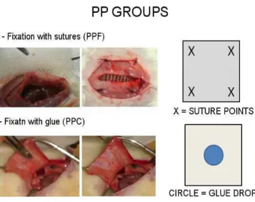
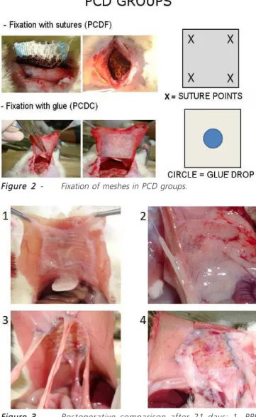
In groups with fixation with sutures, PTT and PCDFs (Figure 1), after exposure of intra-abdominal organs by medial incision, the meshes were placed on the parietal peritoneum on the inner surface of the abdominal wall, and fixed with a stitch in each quadrant.
The same procedure was reproduced in PCDC and PPC groups (Figure 2), except for the UI incision in the abdominal wall, for which a single drop of glue was applied in the center of the mesh for wall fixation.
After surgery, the animals received subcutaneous rehydration with injection of 0.5 mL of 0.9% saline solution, and placed separately in warm environment for postoperative recovery. When fully recovered from anesthesia, they were placed back in the pre-operative environment, with food and water ad libitum.
All animals were euthanized with carbon monoxide in the twenty-first day postoperatively. They were immediately evaluated for adhesion formation using a wider U-shaped abdominal incision. The variables studied were: a) adhesion: present or absent b) degree of adherence: mild, moderate and severe, c) intensity of adhesions: 0-5 Newtons; d) percentage covered: less than 50% or over 50%; and e) location of adhesions: peripheral or central.
The degree of adhesions can be classified according to a pre-standardized table described in other studies (Table 1)21.
Continuous variables were expressed as mean and standard deviation. Categorical variables were described with numbers and percentages. The Fisher exact test was used to determine associations between categorical variables. To check the differences in strength (minimum, average, maximum) between the groups we used analysis of variance (ANOVA).
Figure 1 Figure 1Figure 1
RESULTS
RESULTS
RESULTS
RESULTS
RESULTS
All animals survived the immediate postoperative period and were amenable to analysis. The results were evaluated after 21 days of the procedure (Figure 3). The procedures performed in the MI group did not result in the formation of adhesions. In the UI group, the control group of the incision, only one animal had adhesions, exactly at the site of suture Wall.
Comparing the PP meshes fixed with glue with the ones fixed with sutures, the PPS group showed the lowest degree of adherence when compared to the PTT group, p = 0.01 (Table 2). Adhesions of PP meshes occurred mainly in the peripheral region, but no statistically significant difference was observed between the types of fixation used, p = 0.250 (Table 3).
There was no significant difference in adhesion formation (p = 0.665) in the groups using PCD meshes. There were predominantly mild to moderate adhesions (Table 4).
PCD meshes, whether fixed with sutures or with fibrin glue, showed adhesions, especially in the peripheral region, although no statistical significance was found between techniques, p = 1 (Table 5).
DISCUSSION
DISCUSSION
DISCUSSION
DISCUSSION
DISCUSSION
The total collagen formation starts on the third day when using polypropylene sutures for fixation. This formation increases progressively up around the twenty-first day. Collagen type III also reaches its peak around the 21st day22.
Initially, a medial incision was chosen for use as standard. Nevertheless, the difficulty of cavity size, placement, positioning and fixation was used in an incision shape of the letter U. Using this new incision was best possible exposure of the abdominal wall. For this reason it was necessary to include a control group using a second incision technique. The incisions UI and MI did not form adhesions and were statistically similar to other studies. Regarding the abdominal incision, both the medial and the longer U-shaped, there was no significant adhesion formation.
The findings of this study are consistent with si-milar works 16,18-20,23-25 on the use of fibrin glue as the
method of fixation of surgical meshes. The fixing of PP meshes with glue formed fewer adhesions when compared to suture fixation, with significance level of 0.01. There was no statistical difference between the fixation methods of groups using PCD meshes.
Table 1 Table 1 Table 1 Table 1
Table 1 - Description of the type of adhesion 21
Type of Adhesion Type of Adhesion Type of Adhesion Type of Adhesion
Type of Adhesion D e f i n i t i o nD e f i n i t i o nD e f i n i t i o nD e f i n i t i o nD e f i n i t i o n
0 No adhesion.
1 Thin Adhesions of easy division.
2 Adhesions requiring blunt dissection to be divided.
3 Firm adhesions where the lyses can only be done with application of significant strength, resulting in partial or total injury of the viscera involved.
Figure 3 Figure 3 Figure 3 Figure 3
Figure 3 - Postoperative comparison after 21 days: 1- PPF Group; 2- PPC group; 3- PCDF group; and 4- PCDC group.
Figure 2 Figure 2 Figure 2 Figure 2
Fibrin glue used only in the center of both meshes promoted a proper attachment to the abdominal wall. There was no full attachment of the meshes on the edges, probably by the use of meshes cut to suit the size of the abdomen of the animals. The combination of this factor, cut meshes, with the location of the glue in the center of the mesh, caused increased exposure of the cut edges of the mesh to adhesion formation, especially in the coated model. These findings are supported by published data26
The group of animals with PP mesh fixed with glue showed a lower degree of adhesion when compared to surgical suture. Other variables, such as adhered structures, coated surface, breaking force and retraction of the mesh, showed no relevant significance. The results of the PCD meshes showed no differences between the types of fixation. The predominant location of adhesions formed was observed mainly in the lateral edges of the meshes for all groups.
Table 2 Table 2Table 2 Table 2
Table 2 - Degree of adhesion: PP meshes with sutures and glue.
S u t u r e S u t u r e S u t u r e S u t u r e
S u t u r e G l u eG l u eG l u eG l u eG l u e p (*)p (*)p (*)p (*)p (*) N
N N N
N %%%%% NNNNN %%%%%
Degree of adhesion 0.010
Thin 1 10.0 5 83.3
Moderate 5 50.0 0 0.0
Firm 4 40.0 1 16.7
* Fischer Exact Test
Table 3 Table 3Table 3 Table 3
Table 3 - Location of adhesions: PP meshes with suture and glue.
S u t u r e S u t u r e S u t u r e S u t u r e
S u t u r e G l u eG l u eG l u eG l u eG l u e p (*)p (*)p (*)p (*)p (*) N
N N N
N %%%%% NNNNN %%%%%
PeripheralCentral 7 70.0 6 100.0 0.250
3 30.0 0 0.0
Total 10 100.0 6 100.0
* Fischer Exact Test
Table 4 Table 4Table 4 Table 4
Table 4 - Degree of adhesion: PCD meshes with sutures and glue.
S u t u r e S u t u r e S u t u r e S u t u r e
S u t u r e G l u eG l u eG l u eG l u eG l u e p (*)p (*)p (*)p (*)p (*) N
N N N
N %%%%% NNNNN %%%%%
Degree of adhesion 0.665
Thin 7 70.0 6 60.0
Moderate 2 20.0 1 10.0
Firm 1 10.0 3 30.0
* Fischer Exact Test
Table 5 Table 5Table 5 Table 5
Table 5 - Location of adhesions: PCD meshes with suture and glue.
S u t u r e S u t u r e S u t u r e S u t u r e
S u t u r e G l u eG l u eG l u eG l u eG l u e p (*)p (*)p (*)p (*)p (*) N
N N N
N %%%%% NNNNN %%%%%
PeripheralCentral 9 90.0 8 80.0 1.000
1 10.0 2 20.0
Total 10 100.0 10 100.0
R E S U M O R E S U M O R E S U M O R E S U M O R E S U M O
Objetivo: Objetivo: Objetivo: Objetivo:
Objetivo: Comparar fixação cirúrgica de telas de polipropileno (PP) e telas de polipropileno revestido (PCD), usando fio de sutura de polipropileno e cola biológica, quanto à formação de aderências intraperitoneais. Métodos: Métodos: Métodos: Métodos: Métodos: Amostra de 46 ratas Wistar, randomizadas em seis grupos: dois grupos-controle, com cinco ratas cada, que foram submetidos um à incisão medial (IM) e o outro à uma incisão em forma de U (IU); nenhum desses grupos recebeu tela. Dois grupos com tela de PP, um com dez ratas, fixada com sutura (PPF), e o outro, com seis ratas, fixada com cola biológica (PPC). E Dois grupos com tela de PCD, no primeiro, com dez animais, a tela foi fixada com sutura (PCDF), e no segundo, com dez animais, com cola biológica (PCDC). Resultados: Resultados: Resultados: Resultados: Resultados: Após o prazo de 21 dias, os grupos-controle não apresentaram aderências significantes. O grupo PPC apresentou menor grau de aderência do que o grupo PPF (p=0,01). Não houve diferença entre as fixações nos grupos com PCD. Conclusão:Conclusão:Conclusão:Conclusão:Conclusão: A comparação da fixação apresentou diferença estatística significativa apenas à tela de PP, com menor grau de aderência utilizando a cola. As aderências se localizaram predominantemente nas extremidades das telas estuda-das.
Descritores: Descritores: Descritores: Descritores:
Descritores: Hérnia. Telas cirúrgicas. Adesivo tecidual de fibrina. Técnicas de sutura. Aderências teciduais.
REFERENCES
REFERENCES
REFERENCES
REFERENCES
REFERENCES
1. Melo RM, Cozadi AO, Matias IS, Moreira CC. Reparo pela técnica de shouldice modificada nas hérnias inguinais primárias. Rev Col Bras Cir. 1998;25(3):167-71.
2. Casanova AB, Trindade EN, Trindade MR. Collagen in the transversalis fascia of patients with indirect inguinal hernia: a case-control study. Am J Surg. 2009;198(1):1-5.
3. Matapurkar BG, Bhargave A, Dawson L, Sonal B. Regeneration of abdominal wall aponeurosis: new dimension in Marlex peritonial sandwich repair of incisional hernia. World J Surg. 1999;23(5):446-51; discussion 451.
4. Zong X, Li S, Chen E, Garlinck B, Kim KS, Fang D, et al. Prevention of postsurgery-induced abdominal adhesion by electrospun bioabsorbable nanofibrous poly (lactide-co-glycolide)-based membranes. Ann Surg. 2004;240(5):910-5.
5. van´t Riet M, de Vos van Steenwijk PJ, Bonthuis F, Marquet RL, Steyerberg EW, Jeekel J, et al. Prevention of adhesion to prosthetic mesh: comparison of different barriers using an incisional hernia model. Ann Surg. 2003;237(1):123-8.
6. Höer J, Lawong G, Klinge U, Schumpelick V. Factors influencing the development of incisional hernia. A retrospective study of 2,983 laparotomy patients over a period of 10 years. Chirurg. 2002(5);73(5):474-80.
7. Goldstein HS. Selecting the right mesh. Hernia. 1999;3(1):23-6. 8. Cobb WS, Harris JB, Lokey JS, McGill ES, Klove KL. Incisional
herniorrhaphy with intraperitoneal composite mesh: a report of 95 cases. Am Surg. 2003;69(9):784-7.
9. Luijendijk RW, Lemmen MH, Hop WC, Wereldsma JC. Incisional hernia recurrence following “vest-over-pants” or vertical Mayo repair of primary hernias of the midline. World J Surg. 1997;21(1):62-5; discussion 66.
10. van der Linden FT, van Vroonhoven TJ. Long-term results after surgical correction of incisional hernia. Neth J Surg. 1988;40(5):127-9.
11. Geçim IE, Koçak S, Ersoz S, Bumin C, Aribal D. Recurrence after incisional hernia repair: results and risk factors. Surg Today. 1996;26(8):607-9.
12. Schreinemacher MH, Emans PJ, Gijbels MJ, Greve JW, Beets GL, Bouvy ND. Degradation of mesh coatings and intraperitoneal adhesion formation in an experimental model. Br J Surg. 2009;96(3):305-13.
13. Schug-Pass C, Lippert H, Köckerling F. Mesh fixation with fibrin glue (Tissucol/Tisseel) in hernia repair dependent on the mesh structure—is there an optimum fibrin-mesh combination?--investigations on a biomechanical model. Langenbecks Arch Surg. 2010;395(5):569-74.
14. Pizzol MMD, Roggia MF, Kwitko S, Marinho DR, Rymer S. Utiliza-ção de adesivo de fibrina em cirurgias oftalmológicas. Arq Bras Oftalmol. 2009;72(3):308-12.
15. Zieren J, Castenholz E, Baumgart E, Müller JM. Effects of fibrin glue and growth factors released from platelets on abdominal hernia repair with a resorbable PGA mesh: experimental study. J Surg Res. 1999;85(2):267-72.
16. de Virgilio C, Elbassir M, Hidalgo A, Schaber B, French S, Amin S, et al. Fibrin glue reduces the severity of intra-abdominal adhesion in a rat model. Am J Surg. 1999;178(6):577-80.
17. Martín-Cartes JA, Morales-Conde S, Suárez-Grau JM, Bustos-Jiménez M, Cadet-Dussort JM, López-Bernal F, et al. Role of fibrin glue in the prevention of peritoneal adhesions in ventral herina repair. Surg Today. 2008;38(2):135-40.
18. Burger JW, Halm JA, Wijsmuller AR, ten Raa S, Jeekel J. Evaluation of new prosthetic meshes for ventral hernia repair. Surg Endosc. 2006;20(8):1320-5.
19. Avital S, Bollinger TJ, Wilkinson JD, Marchetti F, Hellinger MD, Sands LR. Preventing intra-abdominal adhesions with polylactic acid film: an animal study. Dis Colon Rectum. 2005;48(1):153-7. 20. Alimoglu O, Akcakaya A, Sahin M, Unlu Y, Ozkan OV, Sanli E, et
al. Prevention of adhesion formation following repair of abdomi-nal wall defects with prosthetic materials (an experimental study). Hepatogastroenterology. 2003;50(51):725-8.
21. Lontra MB, Bigolin AV, Costa RG, Grossi JV, Scalco P, Roll S, et al. Efetividade do uso combinado de filme de ácido lático e tela de polipropileno na formação de aderências intraperitoneal: um mo-delo experimental em ratos. Rev Col Bras Cir. 2010;37(5):364-9. 22. Vaz M, Krebs RK, Trindade EN, Trindade MRM. Fibroplasia after
polypropylene mesh implantation for abdominal wall hernia repair in rats. Acta Cir Bras. 2009;24(1): 19-25.
23. Petter-Puchner AH, Walder N, Redl H, Schwab R, Ohlinger W, Gruber-Blum S, et al. Fibrin sealant (Tissucol) enhances tissue integration of condensed polytetraflouroethylene meshes and reduces early adhesion formation in experimental intraabdominal peritoneal onlay mesh repair. J Surg Res. 2008;150(2):190-5. 24. Prieto-Díaz-Chávez E, Medina-Chávez JL, Ramírez-Barba EJ,
Trujillo-Hernández B, Millán-Guerrero RO, Vásquez C. Reduction of peritoneal adhesion to polypropylene mesh with the application of fibrin glue. Acta Chir Belg. 2008;108(4):433-7.
Received on 25/04/2012
Accepted for publication 09/07/2012 Conflict of interest: none
Source of funding: no
How to cite this article:
Montes JHM, Bigolin AV, Baú R, Nicola R, Grossi JVM, Loureiro CJ, Cavazzola LT. Analysis of adhesions resulting from mesh fixation with
fibrin sealant and suture – experimental intraperitoneal model. Rev Col Bras Cir. [periódico na Internet] 2012; 39(6). Disponível em URL: http://www.scielo.br/rcbc