w w w . j c o l . o r g . b r
Journal of
Coloproctology
Original Article
Effects of
Cupressus sempervirens
extract on the
healing of acetic acid-induced ulcerative colitis in
rat
Masood Sepehrimanesh
a, Nastaran Samimi
b,c, Omid Koohi-Hosseinabadi
d,
Maral Mokhtari
e, Saeed Amiri-Zadeh
f, Mojtaba Farjam
b,∗aGastrointestinal and Liver Diseases Research Center, Guilan University of Medical Sciences, Rasht, Iran
bNoncommunicable Diseases Research Center, Fasa University of Medical Sciences, Fasa, Iran
cStudent Research Committee, Fasa University of Medical Sciences, Fasa, Iran
dCenter of Comparative and Experimental Medicine, Shiraz University of Medical Science, Shiraz, Iran
eDepartment of Pathology, School of Medicine, Shiraz University of Medical Sciences, Shiraz, Iran
fGastroenterohepatology Research Center, Shiraz University of Medical Sciences, Shiraz, Iran
a r t i c l e
i n f o
Article history: Received 10 April 2018 Accepted 8 July 2018
Available online 7 August 2018
Keywords:
Cupressus sempervirens Ulcerative colitis Histopathology Antioxidant Healing
a b s t r a c t
Ulcerative colitis is a chronic inflammatory condition of the colon with an unknown etiology. In this study, we aimed to evaluate the therapeutic effects ofCupressus sempervirensextract on the healing of acetic acid-induced ulcerative colitis in rat. Fifty-five male rats divided into five equal treatment groups were used for this study and received the following treatments: Group 1, 250 mg/kg asacol; Group 2, 1 ml gel base (carboxymethyl cellulose); Group 3, 0.5% gel form ofC. sempervirensextract; Group 4, 1% gel form ofC. sempervirensextract, and; Group 5, considered as negative control and received 1 ml of normal saline. Body weight changes, histopathological and antioxidant changes in the colon tissue were evaluated. Significant weight gain was observed in rats that received 1% gel extract ofC. sempervirens. Significant superoxide dismutase activity was also detected in 0.5 and 1% gel extract groups compared toC. sempervirensextract, Asacol and in 1% gel extract groups compared to the gel base group. Furthermore, both gel extract groups had significant lower total antioxidant capac-ity compared to Asacol group. Several histopathological lesions including inflammation, ulceration, crypt disarray, and goblet cell depletion were detected in the different groups, however, the mean rank of pathological changes showed no significant difference among the five groups. In summary, our results showed that hydroalcoholic extracts ofC. sempervirens leaves produces healing effects in acetic acid induced ulcerative colitis.
© 2018 Sociedade Brasileira de Coloproctologia. Published by Elsevier Editora Ltda. This is an open access article under the CC BY-NC-ND license (http://creativecommons.org/ licenses/by-nc-nd/4.0/).
∗ Corresponding author.
E-mails:[email protected],[email protected](M. Farjam). https://doi.org/10.1016/j.jcol.2018.07.002
Efeitos do extrato de
Cupressus sempervirens
na cicatrizac¸ão de colite
ulcerativa induzida por ácido acético em ratos
Palavras-chave: Cupressus sempervirens Colite ulcerativa Histopatologia Antioxidante Cicatrizac¸ão
r e s u m o
A colite ulcerativa é uma doenc¸a inflamatória crônica do cólon com uma etiologia descon-hecida. O objetivo deste estudo foi avaliar os efeitos terapêuticos do extrato deCupressus sempervirens na cicatrizac¸ão de colite ulcerativa induzida por ácido acético em ratos. Cinquenta e cinco ratos machos divididos em cinco grupos de tratamento iguais foram utilizados para este estudo e receberam os seguintes tratamentos: Grupo 1: 250 mg/kg de asacol; Grupo 2: 1 mL de gel base (carboximetilcelulose); Grupo 3: extrato deC. sempervirens a 0,5% em gel; Grupo 4: extrato deC. sempervirensa 1% em gel e; Grupo 5: considerado con-trole negativo que recebeu 1 mL de soluc¸ão salina normal. Alterac¸ões no peso corporal, alterac¸ões histopatológicas e antioxidantes no tecido do cólon foram avaliadas. Ganho de peso significativo foi observado em ratos que receberam extrato em gel deC. sempervirens a 1%. Atividade significativa de superóxido dismutase também foi detectada em grupos de extrato em gel de 0,5 e 1% em comparac¸ão com o extrato deC. sempervirens, Asacol e em grupos de extrato em gel a 1% em comparac¸ão com o grupo base de gel. Além disso, ambos os grupos de extrato em gel apresentaram capacidade antioxidante total significati-vamente menor em comparac¸ão ao grupo Asacol. Várias lesões histopatológicas, incluindo inflamac¸ão, ulcerac¸ão, desarranjo da cripta e deplec¸ão de células caliciformes foram detec-tadas nos diferentes grupos; no entanto, a classificac¸ão média de alterac¸ões patológicas não apresentou diferenc¸a significativa entre os cinco grupos. Em resumo, nossos resulta-dos mostraram que extratos hidroalcoólicos de folhas deC. sempervirensproduzem efeitos cicatrizantes em colite ulcerativa induzida por ácido acético.
© 2018 Sociedade Brasileira de Coloproctologia. Publicado por Elsevier Editora Ltda. Este ´e um artigo Open Access sob uma licenc¸a CC BY-NC-ND (http://creativecommons.org/ licenses/by-nc-nd/4.0/).
Introduction
Ulcerative colitis (UC) is a type of inflammatory bowel disease (IBD) characterized by continuous mucosal inflammation and ulceration.1The etiology of UC is not completely known but there are several possible causes such as immune dysfunction, genetic susceptibility, invasion of the intestinal microbiota and alteration of the autophagy pathway.2–4 Despite the underlying causes, UC treatment and healing of its related ulcers are significant medical issues. These include using 5-amino salicylic acid drugs, immunosuppressive agents, iron supplements, bacterial recolonization, surgical approaches, and alternative treatments.5Nowadays, the use of medicinal plants and their derivatives as alternative treatment agents for many diseases such as UC have been studied by physicians and scientists. However, this requires previous confirmed results in animal models.
Cupressus sempervirens, also called Mediterranean cypress, is a herb belonging to the Cupressaceae family. It is found in the northern half of the planet, in places such as the northwest and central regions of the United States, northwest Africa, the eastern Mediterranean, Iran, Turkey, southern China, and north of Wintam.6The leaves and fruits of this plant are rich in flavonoids and tannins but lacking in alkaloids and low lev-els of saponin. In traditional medicine, the plant is used as a diuretic, gastrointestinal stimulants, and disinfectants for the treatment of common cold and wound healing.7In 2013, Asgary and his colleagues reported high antioxidant activity
for the essential oil produced from the branches and fruits of C. sempervirens.8
Due to the non-availability of previous studies on the antioxidant and healing properties of this plant in UC, the study aimed to evaluate the histopathological and antioxidant changes in the colon tissue of rat with experimental-induced UC afterC. sempervirensextract consumption.
Materials and methods
Plant material
The leaves ofC. sempervirenswere collected from Shiraz, Fars province, Iran (GPS coordinates: Latitude: 29.591768 and Lon-gitude: 52.583698) during the spring season (May 2017). To prepare the hydroalcoholic extract, the leaves were dried away from direct sunlight at an ambient temperature of 25–30◦C. Then, 1 kg of powdered plant was transferred into ratio 80:20 ethanol:water solution and extracted using perco-lation method for 72 h. The gel form of the extract (37.7%, w/w) were obtained after filtration and evaporation under reduced pressure in a rotary evaporator.9
Animals
The study was approved by the Ethics Committee of Fasa University of Medical Sciences under registration number: IR.FUMS.REC.1395.88. All animals were fed ad libitum with the standard laboratory chow and water and housed in a restricted-access room, maintained at 23◦C and a 55% relative humidity with a 12:12 h light:dark cycle. All of the procedures in this study were carried out in accordance with the ethical standards of the Helsinki Declaration of 2008 and approved by the Ethics committee of Fasa University of Medical Sciences.
Induction of UC and interventions
The bowels of the animals were cleaned as they were fasted 24 h prior to UC induction. A 2 mm diameter polypropylene cannula was inserted through the rectum into the colon and placed 8 cm proximal to the anus verge for injection of 2 ml of 3% acetic acid under ether anesthesia. For 30 s, the rats were maintained in a supine Trendelenburg position to pre-vent early leakage of the intracolonic instillate and for proper colitis induction.10 Rats were randomly divided into 5 equal groups as follows and treatments were started immediately:
• Group 1: received 250 mg/kg asacol
• Group 2: received 1 ml gel base carboxymethyl cellulose • Group 3: received 0.5% gel extract ofC. sempervirens • Group 4: received 1% gel extract ofC. sempervirens
• Group 5: considered as negative control and received 1 ml of normal saline
Weighing and sampling
Body weight changes were recorded prior to the study on day 0 and on the 3rd, 5th and 7th days, with the use of a digital scale with 0.1 g precision. All animals were euthanized under deep anesthesia after seven days of therapy. Laparotomy was performed and the 8 cm of the distal colon was removed for histopathological examination and biochemical investigation.
Histopathological evaluations
For histopathological evaluation, 2 cm of the severed colonic tissue was fixed in 10% buffered formalin. They were embed-ded in paraffin and cut into 5m thick sections and stained with hematoxylin and eosin (H&E) for proper study under a light microscope. All the slides were reviewed by a single blinded pathologist and the degree of inflammation sever-ity and extent, crypt damage, percentage of involvement and regeneration were evaluated on a scale of 0–4.
Oxidative stress evaluation
Colon samples were stored in liquid nitrogen immediately until analysis. About 0.5 g of each tissue sample was homog-enized in 5 ml of 0.05 M phosphate buffer saline pH 7.4. Then, the samples were centrifuged at 3500 rpm for 15 min. The supernatants were then collected and stored at−20◦C. These samples were used to evaluate antioxidant indices as follows:
• Total antioxidant capacity (TAC) was evaluated by ELISA (ZB-TAC-96A, ZellBio GmbH, Germany) and considered as the amount of antioxidant in the sample compared with ascor-bic acid action as a standard. This method can determine TAC with 0.1 mM sensitivity (100mol L−1) colorimetrically
at 490 nm.
• Superoxide dismutase (SOD) was evaluated by ELISA (ZB-SOD-96A, ZellBio GmbH, Germany). In this assay, SOD activity unit was considered as the amount of the sample that will catalyze the decomposition of 1mol of O2- and
H2O2and O2in 1 min colorimetrically at 420 nm. The SOD
activity was expressed as unit per g of tissue (U/g). • Glutathione peroxidase (GPx) activities were measured by
ELISA (BXC0551, Biorexfars, Iran). GPx exist in the cytoplasm and mitochondria of cells and catalyzes the oxidation of glu-tathione (GSH) by cumene hydroperoxide. One unit of GPx activity was defined as the amount of enzyme that converts 1mol of NADPH to NADP+ per minute. The GPx activity was expressed as unit per g of tissue (U/g).
Statistical analysis
Data were expressed as mean and standard deviation (SD) or mean rank. SPSS version 21 for statistical analysis and GraphPad Prism 7.0 for drawing the figure were used. Between group differences in weight and antioxidant statuses were analyzed by One-way ANOVA and Tukey test as Posthoc test. Differences in histopathological scores were analyzed using non-parametric Kruskal–WallisHtest.Pvalue lesser than 0.05 was considered as significant difference.
Results
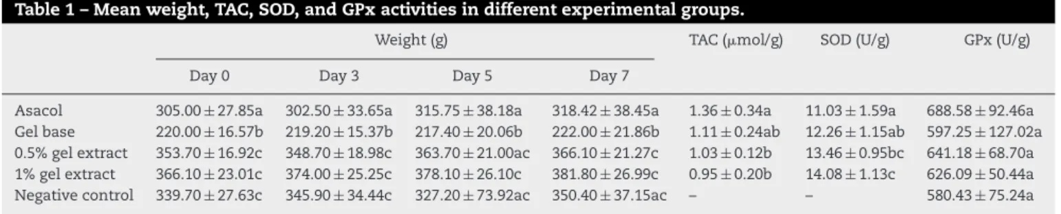
The mean weight of rats (in each group at days 0 and 7), TAC, SOD, and GPx activities are presented inTable 1. As shown, rats treated with 1%gel extract ofC. sempervirensextract showed significant weight gain. As regards the antioxidant assess-ment, no significant difference was observed in the colon GPx
Table 1 – Mean weight, TAC, SOD, and GPx activities in different experimental groups.
Weight (g) TAC (mol/g) SOD (U/g) GPx (U/g)
Day 0 Day 3 Day 5 Day 7
Fig. 1 – Histopathological lesion in the colon tissue of different groups. A (Asacol), ulceration with crypt disarray and goblet cell depletion; B (gel base) and C (negative control), surface ulceration, goblet depletion, irregularity in crypt architecture and inflammation; D (0.5% extract), regeneration of colonic mucosa with irregularity in crypt architecture, decreased goblet cells and increased inflammation; E (1% extract), restoration of normal colon structure in 1% gel extract (H&E×400).
Table 2 – Comparison of mean rank of different score of pathological changes in different experimental groups.
Lesions Asacol Gel base Gel extract Negative control p-Value
0.5% 1%
Inflammation severity 29.45 29.45 19.90 29.70 26.09 0.442
Inflammation extent 28.73 30.18 19.30 29.50 26.82 0.395
Crypt damage 28.59 28.59 20.00 28.75 28.59 0.580
Percent of involvement 27.77 30.68 20.00 28.85 27.23 0.527
Regeneration 28.27 26.50 20.55 30.00 29.36 0.592
Total pathology score 28.50 29.68 19.15 29.50 27.68 0.470
activity among all the groups (p> 0.05). However, a significantly higher SOD activity was detected as follows: 0.5 and 1% gel extract groups compared to the Asacol group and in 1% gel extract groups compared to the gel base group. In addition, both the gel extract groups showed significantly lower TAC compared to the Asacol group.
Fig. 1 shows the histopathological features of colon
tis-sues and related lesions. In addition, scoring of the degree of inflammation severity, inflammation extent, crypt damage, percentage of involvement and regeneration are presented in Table 2. Although, several histopathological lesions including inflammation, ulceration, crypt disarray, and goblet cell deple-tion were identified in the different groups, the mean rank of pathological changes showed no significant difference among the 5 groups.
Discussion
In the present study, the antioxidant and histopathological features of colon tissue in experimental-induced UC and their
changes in response to rectal use of C. sempervirensextract were evaluated and compared. Beneficial effects ofC. semper-virensextract in healing ulcerated mucosa especially in terms of increased SOD activity was reported. Moreover, our treat-ment had significant positive effects on weight gain, especially at higher doses.
C. sempervirens plant is a good source of antioxidants.20,21 Moreover, it has been reported that this plant possesses antimicrobial properties.7Since it is known that one of the causative agents of UC is microbial invasion, it is possible that C. sempervirensand its derivatives modulate microbial activity and combat ROSs in the intestine and thus, help to heal lesions in the UC. Such antioxidant and antimicrobial properties have also been reported in previousin vivoandin vitrostudies22–24 of which this study is in agreement.
Despite the results obtained, this study has two limitations. First, the lack of molecular evaluation of the ulceration and healing changes in the intestinal mucosa such as apoptosis and autophagy pathways. Second, no assessment of inflam-matory indices such as interleukins or tumor necrosing factors in the tissue. However, the results of this study were confirmed by histological and antioxidant evaluations which showed that the hydroalcoholic extracts ofC. sempervirensleaves have several beneficial effects in acetic acid-induced UC. These therapeutic effects were due to the presence of flavonoid com-pounds, especially cupressuflavone. Conclusively, the findings of this study will provide new opportunities for the develop-ment of novel therapeutic alternative agents for UC.
Conflicts of interest
The authors declare no conflicts of interest.
Acknowledgements
The authors acknowledge Fasa University of Medical Sciences for supporting this work (grant number: 95062).
r e f e r e n c e s
[1]. Hosseini SV, Taghavi SA, Jafari P, Rezaianzadeh A, Moini M, Mehrabi M, et al. Incidence of ulcerative colitis relapse: a prospective cohort study in southern Iran. Ann Colorectal Res. 2016;4.
[2]. Lankarani KB, Sepehrimanesh M, Seghatoleslam SF, Hoseini SE, Ghavami S. Autophagy-related protein 7 level in patients with ulcerative colitis. Scand J Gastroenterol. 2017;52:468. [3]. Taghavi SA, Majd SK, Sianati M, Sepehrimanesh M.
Prevalence of IgG-4-associated cholangiopathy based on serum IgG-4 levels in patients with primary sclerosing cholangitis and its relationship with inflammatory bowel disease. Turk J Gastroenterol. 2016;27:547–52.
[4]. Tanideh N, Nematollahi SL, Hosseini SV, Hosseinzadeh M, Mehrabani D, Safarpour A, et al. The healing effect of hydroalcoholic extract ofHypericum perforatumon acetic acid-induced ulcerative colitis in male rats. J Fasa Univ Med Sci. 2017;6:530–7.
[5]. Sepehrimanesh M, Poorbaghi SL. Ulcerative colitis: a phytomedical technical note. Compar Clin Pathol.
2017;26:1237–9,http://dx.doi.org/10.1007/s00580-017-2552-x. [6]. Tumen I, Süntar I, Keles¸ H, Küpeli Akkol E. A therapeutic
approach for wound healing by using essential oils of CupressusandJuniperusspecies growing in Turkey. Evid Based Complement Altern Med. 2012.
[7]. Selim SA, Adam ME, Hassan SM, Albalawi AR. Chemical composition, antimicrobial and antibiofilm activity of the essential oil and methanol extract of the Mediterranean
cypress (Cupressus sempervirensL.). BMC Complement Altern Med. 2014;14:179.
[8]. Asgary S, Naderi GA, Shams Ardekani MR, Sahebkar A, Airin A, Aslani S, et al. Chemical analysis and biological activities ofCupressus sempervirensvar. horizontalis essential oils. Pharm Biol. 2013;51:137–44.
[9]. Koohi-Hosseinabadi O, Moini M, Safarpoor A, Derakhshanfar A, Sepehrimanesh M. Effects of dietaryThymus vulgaris extract alone or with atorvastatin on the liver, kidney, heart, and brain histopathological features in diabetic and hyperlipidemic male rats. Comp Clin Pathol. 2015;24:1311–5. [10]. Millar A, Rampton DS, Chandler CL, Claxson AW, Blades S,
Coumbe A, et al. Evaluating the antioxidant potential of new treatments for inflammatory bowel disease using a rat model of colitis. Gut. 1996;39:407–15.
[11]. Koriem KMM, Gad IB, Nasiry ZK. Protective effect ofCupressus sempervirensextract against indomethacin-induced gastric ulcer in rats. Interdiscip Toxicol. 2015;8:25–34,
http://dx.doi.org/10.1515/intox-2015-0006.
[12]. Waisel Y, Epstein V. How to reduce air pollution byCupressus pollen? Allerg Immunol (Paris). 2000;32:141–2.
[13]. Ibrahim NA, El-Seedi HR, Mohammed MM. Phytochemical investigation and hepatoprotective activity ofCupressus sempervirensL. leaves growing in Egypt. Nat Prod Res. 2007;21:857–66,http://dx.doi.org/10.1080/14786410601132477. [14]. Chiej R. Encyclopaedia of medicinal plants. London:
MacDonald; 1984. p. 54–62.
[15]. Safarpour AR, Kaviani F, Sepehrimanesh M, Ahmadi N, Hosseinabadi OK, Tanideh N, et al. Antioxidant and anti-inflammatory effects of gel and aqueous extract of Melilotus officinalisL. in induced ulcerative colitis: aRattus norvegicusmodel. Ann Colorectal Res. 2015;3.
[16]. Saito R, Tamura M, Matsui H, Nagano Y, Suzuki H, Kaneko T, et al. Qing Dai attenuates nonsteroidal anti-inflammatory drug-induced mitochondrial reactive oxygen species in gastrointestinal epithelial cells. J Clin Biochem Nutr. 2015;56:8–14,http://dx.doi.org/10.3164/jcbn.14-59.
[17]. Tanideh N, Jamshidzadeh A, Sepehrimanesh M, Hosseinzadeh M, Hosseinabadi OK, Najibi A, et al. Healing acceleration of acetic acid-induced colitis by marigold (Calendula officinalis) in male rats. Saudi J Gastroenterol. 2016;22:50–6.
[18]. Tanideh N, Nematollahi SL, Hosseini SV, Hosseinzadeh M, Mehrabani D, Safarpour A, et al. The healing effect of Hypericum perforatumextract on acetic acid-induced ulcerative colitis in rat. Ann Colorectal Res. 2014;2.
[19]. Wang Z, Li S, Cao Y, Tian X, Zeng R, Liao DF, et al. Oxidative stress and carbonyl lesions in ulcerative colitis and associated colorectal cancer. Oxid Med Cell Longev. 2016:9875298,http://dx.doi.org/10.1155/2016/9875298. [20]. Asgary S, Naderi GA, Shams Ardekani MR, Sahebkar A, Airin
A, Aslani S, et al. Chemical analysis and biological activities ofCupressus sempervirensvar. horizontalis essential oils. Pharm Biol. 2013;51:137–44,
http://dx.doi.org/10.3109/13880209.2012.715168.
[21]. Senol FS, Orhan IE, Ustun O. In vitro cholinesterase inhibitory and antioxidant effect of selected coniferous tree species. Asian Pac J Trop Med. 2015;8:269–75,
http://dx.doi.org/10.1016/s1995-7645(14)60329-1.
[22]. Afsharzadeh M, Naderinasab M, Tayarani Najaran Z, Barzin M, Emami SA. In-vitro antimicrobial activities of some Iranian conifers. Iran J Pharm Res. 2013;12:63–74.
[23]. Emami SA, Asili J, Mohagheghi Z, Hassanzadeh MK. Antioxidant activity of leaves and fruits of Iranian conifers. Evid Based Complement Alternat Med. 2007;4:313–9, http://dx.doi.org/10.1093/ecam/nem011.