BASIC RESEARCH
1Department of Pathology, São Paulo University Medical School- São Paulo/
SP, Brazil
2Department of Medicine, Rheumatology, São Paulo University Medical
School - São Paulo/SP, Brazil EMAIL: [email protected] Received for publication on March 29, 2007 Accepted for publication on April 26, 2007
NASAL TOLERANCE WITH COLLAGEN V PROTEIN
REVERTS BRONCHOVASCULAR AXIS REMODELING
IN EXPERIMENTAL BRONCHIOLITIS OBLITERANS
Ana Garippo1, Edwin Parra1, Walcy Teodoro2, Dolores Rivero1, Francisca Souza2,
Natalino Yoshinari2, Vera Capelozzi1
Garippo A; Parra E; Teodoro W; Rivero D; Souza F; Yoshinari N; Capelozzi V. Nasal tolerance with collagen v protein reverts bronchovascular axis remodeling in experimental bronchiolitis obliterans. CLINICS. 2077;62(4):499-506.
INTRODUCTION: The precise role of the remodeling process and possible therapies for bronchiolitis obliterans remain to be established.
OBJETIVE: In the present study, we sought to validate the importance of nasal collagen V tolerance to verify whether bronchovascular axis remodeling could be reverted by this therapeutic approach when compared to steroid treatment.
METHODS: Mice were randomly divided into 4 groups: control, bronchiolitis obliterans, collagen V tolerance, and prednisone groups. Morphometry was employed to evaluate bronchovascular axis dimensions, collagen density, and immune cell response. Collagen V nasal tolerance and steroid-treated mice showed significantly lower values of terminal bronchiole wall thickness and reduction in peribronchovascular cells; bronchioalveolar lymphoid tissue; and CD3+, CD4+, CD8+, and CD20+ lymphocytes. A
significant decrease in CD68+ macrophage density was found in prednisone-treated mice. In addition, a strong quantitative relationship
was found between collagen V tolerance, and reduction in density of immune cells and collagen.
RESULTS: Our results indicate that bronchovascular axis remodeling in bronchiolitis obliterans can be reverted by collagen V nasal tolerance, possibly as the result of T-cell suppression.
CONCLUSION: We concluded that the tolerance effects in this model were strongly related to the improvement in bronchovascular remodeling, and these may be an appropriate targets for further prospective studies on nasal collagen V tolerance.
KEYWORDS:Bronchiolitis obliterans.Chemical injury. Nasal immunization. Collagen V. Morphometry.
INTRODUCTION
Only a few patients with brochovascular axis remodeling process following bronchiolitis obliterans respond to treat-ment.1 The recognition of possible pathological mechanisms
involved in the manifestation of bronchiolitis obliterans might help establish the adequate treatment or definitively block
lung remodeling. Thus, the understanding of the mechanisms in bronchiolitis obliterans remodeling and the treatment ef-fects aiming at preventing irreversible damage and decreased survival are of great interest. Although the exact mechanism of the steroid treatment effect remains unknown, steroid treat-ment seems to diminish the affluence of immune inflamma-tory cells and their mediators in animal models of bronchi-olitis obliterans, thus affecting collagen synthesis and deg-radation, interfering with the remodeling of the extracellu-lar matrix. Because active remodeling of the extracelluextracellu-lar matrix has been thought to be important in prompting bronchovascular axis restoration, a group of collagens have also been targeted as potentially useful indicators.2
interstitium, and capillary basement membranes and is surrounded by vascular smooth cells.3,4 It is a very
pre-served protein found in many different animal species,
and it maintains the NH2 terminal end, making this
mol-ecule more immunogenic.5 Normally, this protein is not
found exposed in the extracellular matrix, as it is hid-den among collagen types I and III, composing
hetero-typic fibers.4,5 Collagen V tolerance has shown
promis-ing results in experimental lung allograft rejection, pre-venting bronchovascular axis remodeling.6,7 Other
clini-cal studies have also shown that oral tolerance induced by collagen I, II, and IV has meaningful clinical efficacy in rheumatoid arthritis,8–10 systemic sclerosis,11 and
glomerulonephritis.12
Recently, we have shown an important inflammatory re-sponse in bronchiolitis obliterans produced by chemical in-jury, followed by remodeling of the bronchovascular axis.13
The aim of promoting mucosal tolerance is to suppress specific immune reactivity without affecting the entire im-munological system by deleting or inactivating specific an-tigen-driven T-cell clones. We have recently reported im-mune cell infiltration and broncovascular axis remodeling after nasal instillation of nitric acid in a mouse model of bronchiolitis obliterans.13 In the present study, we proposed
to establish nasal tolerance with collagen V protein in an attempt to ascertain whether the main aspects of bronchi-olitis obliterans, including bronchovascular axis remodeling, could be reverted by this therapeutic approach. To evaluate the significance of the effect of nasal tolerance with collagen V protein in bronchiolitis obliterans, we also assessed the effect of steroid treatment on bronchovascular axis remodeling.
MATERIALS AND METHODS
Experimental groups
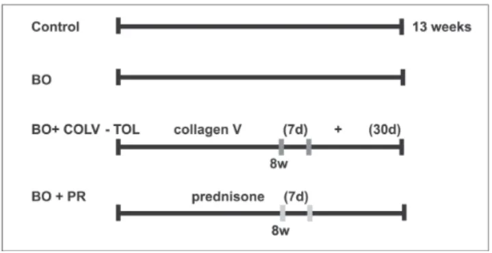
Forty normal female BALB/c mice (4-6 weeks old and 17-26 g) were randomly divided into 4 groups: (i) in the control group, animals did not undergo any procedures (CTRL, n = 10); (ii) in the bronchiolitis obliterans group (BO, n = 10), mice received a single nasal instillation of a
solution of 4.8 mmol/mL of HNO3 2%, pH 0.1, as
previ-ously described;13 (iii) nasal tolerance was induced in the
animals with 8 weeks of bronchiolitis obliterans by daily nasal administrations of 20 ml (0.5 mg/mL/animal/day) col-lagen V for 7 days and, subsequently, after 30 days on
al-ternate days7 (BO + COLV-TOL group; n = 10); (iv)
ani-mals with 8 weeks of bronchiolitis obliterans were treated with 0.002 mg/animal/day of prednisone for 7 days (BO+PR group; n = 10). Figure 1 shows the experimental design.
Animal preparation
At the end of the experiments, animals were anaesthe-tized with an intraperitoneal injection of ketamine 1 mL, xylazine 0.25 µl suspended in 3.75 mL saline solution at a concentration of 0.015 mL/g body weight. The animals were exsanguinated, and the thoracic and abdominal cavi-ties were opened and the lungs removed in bloc.
All mice received humane care in compliance with the “Guide for care and use of laboratory animals” (NIH pub-lication 85-23, revised 1985). The study was approved by the institutional review board of our institution, the Coun-cil for Research of the School of Medicine, University of São Paulo (CAPPesq).
Lung histology
Sections representing peripheral areas of the lung were cut and subsequently fixed with 10% formaldehyde for 24 h. After fixation, tissues were paraffin-embedded. Histologi-cal sections (3 mm in thickness) were cut and stained with hematoxylin and eosin (H&E) and were evaluated by re-searchers blinded to the protocol design. As previously de-scribed,10 bronchiolitis obliterans was histologically
char-acterized by bronchovascular axis remodeling that resulted in reduction or total obliteration of the terminal bronchi-ole lumen, increased wall thickness, deposition of collagen fibers at the peribronchovascular extracellular matrix, and immune cell infiltration.
To evaluate the deposition of collagen fibers at the peribronchovascular axis, collagen was stained in a 0.2% solution of Sirius red (Direct Red 80, C.I. 35780, Aldrich, Milwaukee, WI) dissolved in a saturated aqueous solution
Figure 1 - Experimental Design: (i) group (CTRL): no procedures; (ii) bronchiolitis obliterans group (BO): single nasal HNO3solution instillation;
of picric acid, and observed under polarized light microscopy.
Immunohistochemistry analysis was used to evaluate immune cell airway infiltration of the bronchovascular axis as previously described.13 For the immune cell infiltration
markers, we used the following antibodies: CD20, B-Cell (Clone L26, Dako Corporation Carpinteria, USA, 1:600);
CD3 (Leu-4, T3, 1:600), T-Cell CD4 (CD45RO, clone
OPD4, 1:400); CD8 T-Cell (Clone C8/144B, 1:100); Mac-rophage, CD68 (Clone KP1, 1:3200), Neutrophil Elastase (Clone NP57, 1:800) from Dako A/S Denmark. A
second-ary procedure using the VECTASTAIN® ABC kit (Vector
Technologies, Burlingame, CA), which produces a streptavidin-biotin complex, was then performed.
The area of the terminal bronchiole and artery lumen was expressed in µm2and comprised the area of wall
thick-ness, including the internal border lumen, epithelial cell height, and basement membrane extending to the outer smooth muscle border. The wall thickness of the terminal bronchiole and artery was determined by the difference
be-tween the total area and the lumen area in µm2. Immune
cell infiltration was evaluated by determining the presence
of bronchiole-associated lymphoid tissue (BALT); CD3+,
CD4+, CD8+, and CD20+ lymphocytes; CD68+
macrophages; neutrophils; and total cells. Collagen density and immune cell infiltration were expressed as percentages. Morphometric analysis was performed to evaluate the ex-tent and distribution of histological changes that resulted in bronchiolitis obliterans after bronchovascular axis remodeling. The bronchovascular axis includes the terminal bronchiole, terminal artery, and correspondent extracellular matrix. These were measured regarding diameter, wall thickness, collagen, and immune cell densities using an image analysis system (LEICA Qwin Imaging Systems Ltd., Cambridge, England).
The terminal bronchiole and artery diameter present in each sample at (x100) and (x400) magnification, respec-tively, were measured in 10 random noncoincident micro-scopic fields using the lengths of the total and transversal areas when the minimum and maximum diameter ratio was greater than 0.6. The wall thickness of the terminal bron-chiole and artery was determined by the difference between the total area and the lumen area in mm2. Immune cell
in-filtration was evaluated for the presence of bronchiole-as-sociated lymphoid tissue (BALT); CD3+, CD4+, CD8+, and
CD20+ lymphocytes; CD68+ macrophages; neutrophils; and
total cells. Collagen density and immune cell infiltration were expressed as percentages.
Statistical analysis
Measurements were expressed as mean ± standard
de-viation. All statistical procedures were performed using SPSS software v.13.0 (SPSS, Inc., Chicago, IL 2004). Data
were assessed by t test and ANOVA with Tukey-HSD or
Dunnett-T3 post-hoc tests for multiple comparisons. A P
value less than .05 was considered statistically significant.
RESULTS
Lung histology
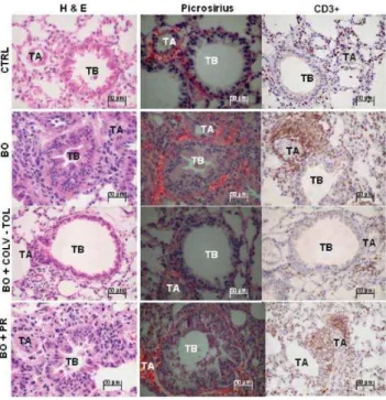
Figure 2 shows the bronchovascular axis (terminal chiole and matched artery) in the control (CTRL), bron-chiolitis obliterans (BO), nasal tolerance with collagen V (BO + COLV-TOL), and prednisone-treated (BO+PR) ani-mals. Tissue is shown treated with H&E stain, picrosirius red under polarized light microscopy, and an immunohis-tochemical reagent (CD3). In control mice, the bronchovascular axis architecture is preserved, and there is a weak red-orange birefringence of bronchovascular axis tissue sections, coincident with the maintenance of the peribronchovascular extracellular matrix density. In
con-Figure 2 - Lung histology from the CTRL, BO, BO+COL-TOL, BO+PR groups respectively, stained with H&E and picrosirius, observed under polarized light microscopy, with immunohistochemistry for CD3+. CTRL
trast, bronchiolitis obliterans animals show a distortion of the bronchovascular axis, with reduction or total oblitera-tion of the lumen, diffuse increase of birefringence, and in-tense immune cell infiltration in the peribronchovascular extracellular matrix, shown in H&E preparations. Pred-nisone and collagen V treatments induce a remarkable res-toration of the bronchovascular axis architecture and de-crease collagen deposition and immune cell infiltration around the bronchovascular axis, which is more intense in animals induced to nasal tolerance with collagen V.
The extension and distribution of the lesions along the bronchovascular axis differed in quantitative terms accord-ing to the animal group.
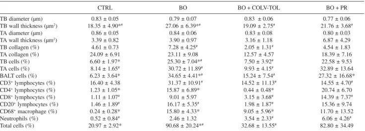
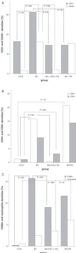
Measurements of the terminal bronchiole, terminal ar-tery, peribronchovascular collagen, and immune cells are shown in Table 1 and illustrated in Figures 3A-C and 4A-C. Both collagen V and prednisone treatments affect the wall thickness of the bronchovascular axis (terminal bron-chiole and artery) (Figure 3A). Reductions were found in the following parameters in the COLV-TOL group com-pared with the BO group: terminal bronchiole wall thick-ness (P = .002); collagen density in the terminal bronchi-ole (P = .01, Figure 3B); density of peribronchovascular cells (P = .002); density of BALT in both the terminal bron-chiole and the matched artery when compared to bronchi-olitis obliterans animals (P = 0.002 and P = .01, respec-tively, Figure 3C); density of CD3+ cells (P = .03, Figure
4A); density of CD20+ cells (P = .005, Figure 4A); and
den-sity of CD4+ cells (P = .001, Figure 4B). Notable was the
increase in density of CD8+ cells after collagen V and
pred-nisone treatments when compared with control animals (P
= .004 and P = .01, respectively, Figure 4B). Densities of CD68+ cells and neutrophils, which were increased in the
BO group, did not differ between the collagen V nasal tol-erance and prednisone groups (Figure 4C). The total cell density decreased along the bronchovascular axis after col-lagen V nasal treatment (P= .003).
Inter- and intra-observer comparisons were performed in 20% of the slides by 2 observers (ALG and ERP) or by doubling the time of the same observer. The coefficient of variation for the inter- and intra-observer was less than 5%.error
DISCUSSION
Systemic steroids have been widely employed for infants with acute viral bronchiolitis and young children with acute viral bronchiolitis or adults with bronchiolitis obliterans af-ter transplantation, but the actual benefit of this inaf-terven- interven-tion requires clarificainterven-tion.14,15 In 13 studies involving 1,198
affected children, no benefits were found for duration of hospital stay, clinical score, or other outcomes.
Patel et al16 demonstrated that no benefits were found
in either bronchiolitis or clinical score in infants and young children treated with systemic steroids as compared with placebo. In addition, marked study heterogeneity and oc-casionally, conflicting direction of benefit between trials suggest that these results should be interpreted with
cau-Table 1 - Measurements of terminal bronchioles, terminal arteries, peribronchovascular collagen, and immune cells in the 4 experimental groups of animals (mean ± SD).
CTRL BO BO + COLV-TOL BO + PR
TB diameter (µm) 0.83 ± 0.05 0.79 ± 0.07 0.83 ± 0.06 0.77 ± 0.06
TB wall thickness (µm2) 18.35 ± 4.90*# 27.06 ± 6.39*# 19.09 ± 2.75# 21.76 ± 3.68#
TA diameter (µm) 0.86 ± 0.05 0.84 ± 0.06 0.83 ± 0.08 0.80 ± 0.03
TA wall thickness (µm2) 3.39 ± 0.82 3.90 ± 0.97 3.16 ± 1.18 6.87 ± 4.29
TB collagen (%) 4.61 ± 0.73 7.28 ± 4.25# 2.05 ± 1.31# 4.54 ± 1.83
TA collagen (%) 24.09 ± 6.91 23.11 ± 9.08 12.57 ± 4.57 18.39 ± 7.16
TB cells (%) 6.60 ± 1.97* 25.30 ± 7.04*# 7.50 ± 3.92# 22.58 ± 9.53
TA cells (%) 8.14 ± 1.65# 30.72 ± 11.89# 9.93 ± 4.15# 32.89 ± 13.64
BALT cells (%) 6.23 ± 3.64* 34.65 ± 4.41*# 15.24 ± 7.54# 27.32 ± 16.68*
CD3+ lymphocytes (%) 16.40 ± 4.38 31.37 ± 10.91# 14.52 ± 11.13# 14.55 ± 4.70#
CD4+ lymphocytes (%) 1.23 ± 1.05* 15.87 ± 6.89* 0.44 ± 0.48* 20.74 ± 6.70
CD8+ lymphocytes (%) 1.11 ± 1.07# 9.01 ± 5.97 3.15 ± 3.68# 14.39 ± 7.37#
CD20+ lymphocytes (%) 1.46 ± 1.89# 16.17 ± 5.35# 1.98 ± 1.87# 15.36 ± 9.74
CD68+ macrophage (%) 0.24 ± 0.28* 15.80 ± 4.33* 9.05 ± 5.96* 11.70 ± 13.52
Neutrophils (%) 0.52 ± 0.84# 2.46 ± 1.32 3.54 ± 2.33# 6.06 ± 4.26#
Total cells (%) 20.97 ± 2.92* 90.68 ± 20.24*# 32.68 ± 13.55# 82.80 ± 34.49
Figure 3- Quantitative data of TB wall thickness (A), expressed in µm2.
Collagen density (B) and bronchovascular axis immune cells (C), expressed in (%). Decreased TB area and wall thickness seen in BO+COLV-TOL animals compared to the BO group. Collagen density (B) in TB decreased in the BO+COLV-TOL group compared to BO. Peribronchovascular cell density (D) in TB and matched artery reduced in BO+COLV-TOL animals compared to BO. TB- terminal bronchiole; TA- terminal artery.
Figure 4 - Quantitative data of densities (%) of CD3+ and CD20+ (A), CD4+
and CD8+ (B), and CD68+ and macrophage (C) immune cells in CTRL, BO,
BO+COLV-TOL and BO+PR lungs. Compare CD3+ cells for TB (A) from
BO+COLV-TOL and BO+PR to BO animals. CD20+(A) and CD4+ (B) cells
reduced after BO+COLV-TOL, when compared to BO. Note increase in CD8+ cells (B) in animals receiving the BO+COLV-TOL and BO+PR
treatments, vs. CTRL. Density of CD68+ cells and neutrophils, increased in
tion. The variable response to anti-inflammatory treatment could be attributed to the heterogeneous biochemical and molecular mechanisms activated in response to different initial insults.17,18 Moreover, the inflammatory mechanisms
are continuous and cyclic, sometimes causing deterioration or improvement in lung function. Other major continuing problems include determining when to administer pred-nisone, monitoring its use, and determining what other therapy it should be compared with.16 Hence, the aim of the
present study was to compare collagen V nasal tolerance with a traditional therapeutic approach, such as prednisone, in an experimental bronchiolitis obliterans model.
Clearly, the likely reason for failure of treatment to cure some patients with bronchiolitis obliterans is due to the ir-reversible scarring of the bronchovascular axis, which is not detected by either routine imaging or routine pathologi-cal analysis.
The question of interest is whether additional, more technological information obtained from histopathological assessment can help us identify patients with bronchiolitis obliterans who are likely to present scarring of the bronchovascular axis and, consequently, poor response to treatment, thus helping improve long-term function and prevent the cumulative morbidity.19
The inflammation/repair processes that occur in bron-chiolitis obliterans undoubtedly comprise a series of com-plex, sequential steps; however, among these, the immune system is thought to be especially important, due to its in-teraction with the extracellular matrix remodeling.15
Type V collagen fulfills all the criteria of a fibrillar col-lagen: both N- and C-termini are processed, and it consists of an approximately 1000-amino acid chain, thus control-ling fibrillogenesis and making it more immunogenic.20
Despite being a quantitatively minor collagen, collagen V is fundamental not only for regulation of fibril diameter of other collagen types, but also for the integrity of con-nective tissues.21 Whereas prior studies4–7 were able to show
a significant relationship between lung transplantation and collagen V tolerance, our results suggested that nasal col-lagen V tolerance could be used to interrupt the bronchi-olitis obliterans process, reducing collagen deposition and T- and B-cell reactivity.
Although the precise mechanisms for daily sampled an-tigens in the bronchial epithelia induction are still unclear, collagen V treatment of lung diseases that develop with fi-brosis may be promising. Presumably, when collagen V im-munization is carried out by the nasal route, it may reach the lymphoid tissue associated with the nasal mucosa, where it is processed and then locally presented to T cells so they can induce pro-inflammatory as well as suppressive immune responses4–7in the pre-acinar axis.
In a variety of situations, oral and nasal induction to collagen may stabilize experimental,8,22 and human
arthri-tis,10 systemic sclerosis,23and osteoarthrosis.24
Our data are both interesting and important and con-tinue the studies of Dr. David Wilkes’ group, which have previously documented that collagen V-induced oral toler-ance prevents bronchiolitis obliterans after rat lung allografts.4–7
Considering these facts, we were not be surprised to learn that collagen V nasal tolerance could revert the bronchovascular axis remodeling in bronchiolitis obliterans, and our results have now confirmed the importance of this therapeutic approach.
A beneficial effect of low doses of orally or nasally ad-ministered collagen types I and II has also been demon-strated as an immunotherapeutic intervention for arthritic joints.8–10
We have also observed that collagen V and prednisone treatments reduced peribronchovascular cells and BALT in both the terminal bronchiole and matched artery when com-pared to control animals.
Regarding collagen fibers in the terminal bronchiole, we observed that collagen V treatment resulted in a decrease in this response compared with control animals. Most in-teresting was the strong, quantitative relationship observed between tolerance and reduction in density of immune cells and collagen. Although a variety of associations between prednisone and lung remodeling have been described re-cently,15,25 we observed that its relationship with immune
cells and collagen density was not as strong as that observed for collagen V tolerance.
In fact, we found that collagen V treatment reduced den-sities of CD3+, CD4+, CD20+, and CD68+ cells. Not only
has tolerance been thought to dramatically reduce immune cell response and thus protect the host from deleterious cytokine effects, but it has also been thought to regulate ex-tracellular matrix synthesis and degradation.8
Of interest was our finding of an increase in density of CD8+ lymphocytes in animals with untreated bronchiolitis
obliterans when compared with control animals and a de-crease after collagen V treatment, suggesting that collagen V nasal tolerance induces significant, prompt restoration of the bronchovascular axis, possibly as the result of T-cell suppression. It has been postulated that CD8+ T
suppres-sor cells can be induced by exposure to an antigen. When stimulated, CD8+ T suppressor cells also suppress
immune system responses. The B-cell population is “toler-ated” through clonal deletion, anergy and/or sequestering of antigens within an organ.26 Where T-cell help is required
pre-vent possible autoantibody production.27,28 It is well known
that a decrease in B- and T-cell reactivity induced by col-lagen V tolerance can prevent bronchiolitis obliterans in lung transplantation.4,29
At present, we have demonstrated that there is an asso-ciation between tolerance and reduction in immune cell in-filtration and restoration of the bronchiole dimension in bronchiolitis obliterans caused by chemical injury. Thus, decreased immune cell infiltration may be more of a pri-mary event, and bronchovascular axis remodeling, more of a secondary event. Regardless of the mechanism, immune cell infiltration and collagen density provide important mor-phological information in experimental bronchiolitis oblit-erans.
Finally, randomized and prospective trials will be re-quired, since we believe it is important to validate our thera-peutic assessment of collagen V nasal tolerance as well as to extend it to other lung diseases by studying additional
patients. We conclude that the effects of collagen V nasal tolerance in bronchiolitis obliterans induced by chemical injury were strongly related to the improvement in bronchovascular axis remodeling. Lungs with increased immune cell and collagen density around the peribronchovascular axis comprise a subset with a high risk of irreversible scarring and may be an appropriate target for prospective studies of collagen V tolerance. The colla-gen V therapeutic approach may be especially important in patients who are resistant to steroids.
ACKNOWLEDGMENTS
The authors would like to thank the Laboratory Animal Facility, Lim-51, and Maria Cristina Medeiros for expert assistance in the histological preparation and the Depart-ment of Pathology of the University of São Paulo School of Medicine for excellent technical assistance.
RESUMO
Garippo A; Parra E; Teodoro W; Rivero D; Souza F; Yoshinari N; Capelozzi V. Tolerância nasal com a proteína colágeno V reverte o remodelamento no eixo bronco-vascular na bronquiolite obliterante experimental. CLINICS. 2007;62(4):499-506.
INTRODUÇÃO: A participação precisa do processo de remodelamento e possíveis implicações no tratamento da bronquiolite obliterante ainda não está estabelecida.
OBJETIVOS: Estabelecer a importância da tolerância na-sal induzida pelo colágeno do tipo V e verificar se o pro-cesso de remodelamento do eixo broncovascular pode ser revertido com esta estratégia terapêutica comparada ao efei-to do tratamenefei-to com esteróides.
MATERIAL E MÉTODO: Camundongos foram divididos em quatro grupos: controle, bronquiolite obliterante, tolerân-cia nasal com colágeno do tipo V e prednisona. Morfometria foi realizada para avaliar as dimensões do eixo broncovas-cular, densidade de colágeno e resposta imunocelular. Ca-mundongos submetidos à tolerância nasal com colágeno do tipo V e tratados com prednisona exibiram significativas
re-duções da espessura da parede de bronquíolos terminais, da densidade de células inflamatórias ao redor do eixo peribroncovascular e da resposta imunocelular às custas de linfócitos CD3, CD4, CD8 e CD20. Houve também signifi-cativa redução da densidade de macrófagos CD68 nos ca-mundongos tratados com prednisona. Adicionalmente, hou-ve uma forte associação entre tolerância nasal induzida pelo colágeno do tipo V, resposta imunocelular e redução do con-teúdo de colágeno peribroncovascular.
RESULTADOS: O remodelamento do eixo broncovascular na bronquiolite obliterante pode ser revertido pela indução de tolerância nasal com o colágeno do tipo V, possivelmen-te como resultado de supressão de linfócitos T.
CONCLUSÃO: Os efeitos da tolerância nasal no presen-te modelo estiveram forpresen-temenpresen-te relacionados à melhora no remodelamento do eixo broncovascular, despontando como um alvo promissor para estudos prospectivos.
REFERENCES
1. King VJ, Viswanathan M, Bordley WC, Jackman AM, Sutton SF, Carey TS. Pharmacologic treatment of bronchiolitis in infants and children: a systematic review. Arch Pediatr Adolesc Méd. 2004;158:127-37. 2. Niyibizi C, Fietzek, Rest M. Human placenta type V collagens. Evidence
for existence of a a1(V), a2(V), a3(V). J Biol Chem. 1984;259:14170-4. 3. Madri J A and Furthmayr H. Collagen polymorphism in the lung. An immunochemical study of pulmonary fibrosis. Hum Pathol. 1980;11:353-66.
4. Yoshida S, Haque A, Mizobuchi T, Iwata T, Chiyo M, Webb TJ, et al. Anti-type V collagen lymphocytes that express IL-17 and IL-23 induce rejection pathology in fresh and well-healed lung transplants. Am J Transplant. 2006; 6:724-35.
5. Mares DC, Heidler KM, Smith Jr GN, Cummings OW, Harris ER, Foresman BH, et al. Type V collagen modulates alloantigen-induced pathology and immunology in the lung. Am J Respir Cell Mol Biol. 2000;23:62-70.
6. Haque MA, Mizobuchi T, Yasufuku K, Fujisawa T, Brutkiewicz R, Zheng Y, et al. Evidence for immune responses to a self-antigen in lung transplantation: role of type V collagen-specific T cells in the pathogenesis of lung allograft rejection. J Immunol. 2002;169:1542-9.
7. Yasufuku K, Heidler KM, Woods KA, Smith Jr GN, Cummings OW, Fujisawa T, et al. Prevention of bronchiolitis obliterans in rat lung allograft by typo V collagen-induced oral tolerance. Transplantation. 2002;73:500-5. 8. Garcia G, Komagata K, Slavin AJ, Maron Ruth, Weiner, HL. Suppression
of collagen-induced arthritis by oral or nasal administration of type II collagen. J Autoimmun. 1999;13:315-24.
9. Meyer O. Oral immunomodulation therapy in rheumatoid arthritis. Joint Bone Spine 2000;67:384-92.
10. Myers LK, Higgins GC, Findel TH, Reed AM, Thompson JW, Walton RC, et al. Juvenile arthritis and autoimmunity to type II collagen. Arthritis Rheum. 2001;44:1775-81.
11. Mckown KM, Carbone LD, Bustillo J, Seyer JM, Kang AH & Postlethwaite AE. Induction of immune tolerance to human type I collagen in patients with systemic sclerosis by oral administration of bovine type I collagen. Arthritis Rheum 2000;43:1054-61.
12. Reynolds J, Prodromidi EI, Juggapah JK, Abbott DS, Holthaus KA, Kalluri R, et al. Nasal administration of recombinant rat alpha3(IV) NC1 prevents the development of experimental autoimmune glomerulonephritis in the WKY rat. J Am Soc Nephrol. 2005 May;16:1350-9.
13. Garippo AL, Parra ER, Teodoro WR, Veloza AP, Yoshinari NH, Capelozzi VL. Immune cell infiltration and broncovascular remodeling after nitric acid nasal instillation in a mouse bronchiolitis obliterans model. Lung. 2006;184:229-38.
14. Daniels CE, Myers JL, Utz JP, Markovic SN, Ryu JH. Organizing pneumonia in patients with hematologic malignancies: A steroid-responsive lesion. Respir Med. Respir Med. [published online ahead of print May 15, 2006] 2007;101:162-8.
15. Fernandes ABS, Zin WA, Rocco PMR. Corticosteroids in acute respiratory distress syndrome. Braz J Med Biol Res. 2005;38:147-159 . Review.
16. Patel H, Platt R, Lozano JM, Wang EE. Glucocorticoids for acute viral bronchiolitis in infants and young children. Cochrane Database of Systematic Reviews 2004, Issue 3. Art. No.: CD004878. DOI: 10.1002/ 14651858.CD004878.
17. Halna M, Leblond P, Aissi E, Dumonceaux A, Delepoulle F, El Kohen R, Hue V, Martinot A. Impact of the consensus conference on the ambulatory treatment of bronchiolitis in infants. Presse Med. 2005;34:277-81. 18. Scarfone RJ. Controversies in the treatment of bronchiolitis. Curr Opin
Pediatr. 2005;17:62-6.
19. Smyth RL and Openshaw PJ. Bronchiolitis. Lancet. 2006;368:312-22. 20. Wenstrup RJ, Florer JB, Brunskill EW, Bell SM, Chervonevai I, Birk
DE. Type V collagen control the initiation collagen fibril assembly. J Biol Chem. 2004;279:53331-7.
21. Kelley J. Collagen. Lung Cell Biology. New York: Marcel Dekker Inc; 1991. Cap 17, p. 821-58.
22. Higuchi K, Kweon MN, Fujihashi K, Mcghee JR, Kiyono H. Comparison of nasal and oral tolerance for the prevention of collagen induced murine arthritis. J Rheumatol. 2000;27:1039-44.
23. Wan-Uk K, Woo-Kyoung L, Jae-Woong R, Seung-Hoon K, Ho-Youn K. Suppression of collagen-induced arthritis by single administration of poly(lactin-co-glycolic acid) nanoparticles entrapping type II collagen. Arthritis Rheum. 2002;46:119-20.
24. Stancikova M, Stancik R, Gubzova Z, Rovensky J. Collagen in the treatment of rheumatic diseases-oral tolerance. Bratisl Lek Listy. 1999;100:567-71.
25. Kurland G and Michelson P. Bronchiolitis in children. Pediatr Pulmonol. 2005;39:193-208.
26. Faria AMC and Weiner HL. Oral tolerance: mechanisms and therapeutic applications. In: Dixon, FG, editor. Advances in Immunology, 73. Elsevier; Amsterdam: 1999. p. 153-264.
27. Hoyne GF. Immunological tolerance to inhaled antigen. Am J Respir Crit Care Med. 2000;162(4 Pt 2):S169-74.
28. Novak N, Allam JP, Betten H, Haberstok J, Bieber T. The role of antigen presenting cells at distinct anatomic sites: they accelerate and they slow down allergies. Allergy. 2004;59:5-14.