Ocular risk management in patients undergoing general
anesthesia: an analysis of 39,431 surgeries
Newton Kara-Junior,I,IIRodrigo Franc¸a de Espindola,II ,*Joao Valverde Filho,IChristiane Pellegrino Rosa,I Andre Ottoboni,IEnis Donizete SilvaI
ISı´rio-Libaneˆs Hospital, Sa˜o Paulo/SP, Brazil.IIFaculdade de Medicina da Universidade de Sa˜o Paulo, Ophthalmology Department, Sa˜o Paulo/SP, Brazil.
OBJECTIVE: This study sought to describe and analyze ocular findings associated with nonocular surgery in patients who underwent general anesthesia.
METHODS:The authors retrospectively collected a series of 39,431 surgeries using standardized data forms. RESULTS:Ocular findings were reported in 9 cases (2.3:10,000), which involved patients with a mean age of 58.9±
19.5 years. These cases involved patients classified as ASA I (33%), ASA II (55%) or ASA III (11%). General anesthesia with propofol and remifentanil was used in 4 cases, balanced general anesthesia was used in 4 cases, and regional block was used in combination with balanced general anesthesia in one case. Five patients (55%) underwent surgery in the supine position, one patient (11%) underwent surgery in the lithotomy position, two patients (22%) underwent surgery in the prone position, and one patient (11%) underwent surgery in the lateral position. Ocular hyperemia was detected in most (77%) of the 9 cases with ocular findings; pain/burning of the eyes, visual impairment, eye discharge and photophobia were observed in 55%, 11%, 11% and 11%, respectively, of these 9 cases. No cases involved permanent ocular injury or vision loss.
CONCLUSION:Ophthalmological findings after surgeries were uncommon, and most of the included patients were relatively healthy. Minor complications, such as dehydration or superficial ocular trauma, should be prevented by following systematic protocols that provide appropriate ocular occlusion with a lubricating ointment and protect the eye with an acrylic occluder. These procedures will refine the quality of anesthesia services and avoid discomfort among patients, surgeons and anesthesia staff.
KEYWORDS: Blindness; Anesthesia; Eye Injuries.
Kara-Junior N, Espindola RF, Valverde Filho J, Rosa CP, Andre Ottoboni, Silva ED. Ocular risk management in patients undergoing general anesthesia: an analysis of 39,431 surgeries. Clinics. 2015;70(8):541-543
Received for publication onJanuary 22, 2015;First review completed onApril 7, 2015;Accepted for publication onMay 12, 2015 E-mail: [email protected]
*Corresponding author
’ INTRODUCTION
Postoperative visual loss (POVL) following general sur-gery is a relatively uncommon but devastating complication that is most frequently associated with cardiac, spine, head and neck operations. Estimates have indicated that POVL occurs in up to 0.2% (1) and 4.5% (2) of spine and cardiac surgeries, respectively.
Although studies of 65,000 and 400,000 patients who underwent anesthesia for all types of surgery at two large academic institutions suggested a low prevalence of perio-perative vision loss in surgeries other than cardiac and spinal fusion procedures, the actual prevalences of perioperative vision loss for the most common types of operations remain unknown (3,4).
Because the frequency of ocular complications is very low, few peer-reviewed studies have analyzed ocular symptoms and vision loss after surgeries under general anesthesia. The aim of this retrospective study was to contribute to the prevention of ocular complications during anesthesia by determining and analyzing the ocular findings from a large series of cases involving general anesthesia.
’ MATERIALS AND METHODS
This retrospective study included 39,431 nonocular sur-geries. We began the study by reviewing the documented cases of ocular findings after surgical procedures performed at our institution between January 2007 and December 2010. The preoperative variables included age, sex, American Society of Anesthesiologists (ASA) physical status classifica-tion, urgency of surgery (emergency or elective), duration of the procedure, ocular findings (signs and symptoms) and surgical position during surgery. Other variables included the use of ocular lubricant during anesthesia, the required treatment and the final diagnosis.
DOI:10.6061/clinics/2015(08)02
Copyright&2015CLINICS–This is an Open Access article distributed under the terms of the Creative Commons License (http://creativecommons.org/licenses/by/ 4.0/) which permits unrestricted use, distribution, and reproduction in any medium or format, provided the original work is properly cited.
541
All demographic variables were analyzed using descrip-tive statistics; in particular, means and SDs were determined for continuous variables, and frequencies (in percentages) were calculated for categorical variables.
’ RESULTS
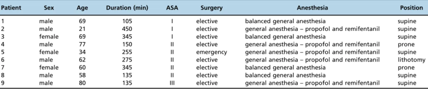
This retrospective study included 39,431 nonocular surgeries. Ocular findings were reported in 9 cases (2.3:10,000), which involved patients with a mean age of 58.9±19.5 years. Table 1 presents the characteristics of all 9 cases. Examinations of individual variables revealed that male patients (66%), ASA II status (55%), elective surgery (88%) and the supine position (66%) were each involved in the majority of these cases.
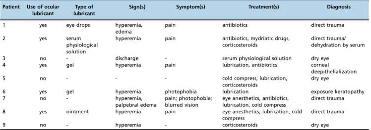
For patients with ocular findings who were subjected to general anesthesia (9 cases), pain (55%) and photophobia (22%) were the main symptoms, and hyperemia (77%) was the main sign (Table 2). Only one patient presented with blurred vision (11%).
The main diagnoses in these cases were direct trauma (44%) and dry eye (33%) (Table 2). All 9 patients experienced ocular occlusion during surgery, and 5 patients (55%) also received lubricant. No patient exhibited permanent ocular injury or significant visual loss.
’ DISCUSSION
Perioperative ischemic optic neuropathy (PION) has been reported after spine (5-7), orthopedic (8), neck (9-13), heart, and abdominal surgeries (14,15). Intraoperative variables that reportedly play roles in the pathogenesis of PION include hypotension, anemia, and elevated intraocular pressure associated with the prone position during spinal surgery (16). Vascular risk factors, such as diabetes, coronary artery disease, and hypertension, are present in many patients who experience PION (17,18), although vision loss has been reported in children and healthy adults who exhibit none of these factors (6).
Given that the mechanisms and risk factors for PION are poorly understood, the risks of vision loss should be considered in preoperative discussions with patients who expect to undergo spine surgery or surgery requiring cardiopulmonary bypass because such procedures are associated with the highest incidences of this rare complication (19).
In the present study, the incidence of ocular findings was 2.3:10.000. No patient experienced permanent ocular injury or significant visual loss. However, certain of the observed symptoms/signs could significantly impact eye health.
All 9 of the patients with ocular findings experienced ocular occlusion during their procedures, and 55% of these
patients received ocular lubricant (in the form of eye drops, serum or gel). These findings indicated the precautions implemented by the staff to prevent ocular injury. However, these actions cannot avoid ocular complications in all cases. The most common diagnoses found in our study were direct trauma and dry eye. Preventive strategies are the only option to reduce the effect of ocular complications during general anesthesia. Ocular occlusion and the use of eye-lubricating ointment are important strategies to prevent dehydration of the ocular surface during long surgeries. In these situations, the mechanisms of aggravation can include not only corneal exposure if the eyelids remain open but also decreased tear secretion induced by the anesthesia. After the surgical procedure, the maintenance of the patient’s ocular occlusion is recommended during the postanesthetic period, when the blink reflex is poor and the patient remains sleepy. The main symptoms of dry eye are pain, redness and tearing. With respect to the treatment of corneal deepithelialization due to exposure keratopathy, eye lubrication with artificial tears and occlusion with ocular lubricating ointment are frequently recommended in severe cases. The ocular admin-istration of saline should be avoided due to the risk of further dehydration of the cornea. Frequent review of dry eye cases by an ophthalmologist is necessary due to the risk of progression to ulceration of the cornea, which can lead to permanent vision loss.
Direct trauma to the eye is generally caused by pressure exerted by the surgeon’s arm or hand on the patient’s eye during surgery or by direct corneal injury with instruments or components of the surgical field. During intubation, the anesthetist himself may cause trauma to the patient’s eye. Ocular trauma can be prevented by the systematic use of acrylic eye protection similar to the postoperative protection used after eye surgery to prevent patients from exerting pressure on their eyes; the use of such protection may be particularly important for patients whose surgical sites are near the head (20).
In the present study, there were no cases of permanent vision loss. Many strategies can be used to prevent blindness, particularly PION-related blindness. Maneuvers to keep the head at or above heart level to reduce venous congestion in the head have been recommended in the ASA practice advisory for perioperative visual loss associated with spine surgery (21). Minimizing the duration of time in the prone position and maximizing hemostasis may also be beneficial. In summary, an understanding of the risk factors and characteristics that promote the occurrence of perioperative ocular lesions is extremely important for the development of prevention strategies. Despite the low incidence of these complications, the potential for serious and permanent visual
Table 1-Patient characteristics.
Patient Sex Age Duration (min) ASA Surgery Anesthesia Position
1 male 69 105 I elective balanced general anesthesia supine
2 male 21 450 I elective general anesthesia – propofol and remifentanil supine
3 female 69 345 I elective balanced general anesthesia supine
4 male 77 150 II elective general anesthesia – propofol and remifentanil prone
5 female 34 255 II emergency general anesthesia – propofol and remifentanil supine
6 male 62 275 II elective general anesthesia – propofol and remifentanil lithotomy
7 female 60 345 II elective balanced general anesthesia prone
8 male 58 135 II elective balanced general anesthesia supine
9 male 80 135 III elective general anesthesia – propofol and remifentanil supine
542 Ocular risk in general anesthesia
injuries such as retinal ischemia and PION justify appro-priate care and the active pursuit of high-quality anesthesia services. Since 2010, a protocol involving ocular occlusion with the instillation of lubricant eye drops during relatively complex procedures has been systematically adopted by the anesthesia services of Sírio Libanês Hospital. Beginning in 2014, guided by the results and insights of this study, which was conducted and analyzed in collaboration with ophthal-mologists, lubricating ointment and ocular occlusion with an acrylic occluder for eye protection have been used for all surgeries involving general anesthesia. It is recommended that these procedures, which have been implemented to achieve the objective of further improving patient safety during surgery, should be followed from the induction of anesthesia to the complete awakening of the patient in the postanesthesia recovery room.
Minor complications, such as dehydration or superficial ocular trauma, which can generally be rapidly resolved during the postoperative period, should be prevented by following systematic protocols that include appropriate ocular occlusion with lubricating ointment and protection of the eye with an acrylic occluder. These protocols will refine the quality of anesthesia services and avoid discomfort among patients, surgeons and anesthesia staff.
’ AUTHOR CONTRIBUTIONS
Kara-Junior N: study conception and design; drafting of the manuscript; and critical revision. Espindola RF: drafting of the manuscript; critical revision; and analysis and interpretation of study data. Valverde Filho J, Rosa CP, Ottoboni A, and Silva ED: study conception and design; data acquisition; and analysis and interpretation of study data.
’ REFERENCES
1. Stevens WR, Glazer PA, Kelley SD, Lietman TM, Bradford DS. Ophthal-mic complications after spinal surgery. Spine. 1997;22(12):1319–24. 2. Shaw PJ, Bates D, Cartlidge NE, Heaviside D, French JM, Julian DG, et al.
Neuro-ophthalmological complications of coronary artery bypass graft surgery. Acta Neurol Scand. 1987;76(1):1–7.
3. Roth S, Thisted RA, Erickson JP, Black S, Schreider BD. Eye injuries after non ocular surgery: a study of 60,965 anesthetics from 1988 to 1992. Anesthesiology 1996;85(5):1020–7.
4. Warner ME, Warner MA, Garrity JA, MacKenzie RA, Warner DO. The frequency of perioperative vision loss. Anesth Analg. 2001;93(6):1417–21. 5. Katz DM, Trobe JD, Cornblath WT, Kline LB: Ischemic optic neuropathy
after lumbar spine surgery. Arch Ophthalmol. 1994;112(7):925–31. 6. Alexandrakis G, Lam BL. Bilateral posterior ischemic optic neuropathy
after spinal surgery. Am J Ophthalmol. 1999;127(3):354–5.
7. Cheng MA, Sigurdson W, Tempelhoff R, Lauryssen C. Visual loss after spine surgery: A survey Neurosurgery. 2000;46(3):625–31.
8. Roth S, Nunez R, Schreider BD. Unexplained visual loss after lumbar spinal fusion. J Neurosurg Anesthesiol. 1997;9(4):346–8.
9. Bhatti MT, Enneking FK. Visual loss and ophthalmoplegia after shoulder surgery. Anesth Analg. 2003;96(3):899–902.
10. Marks SC, Jaques DA, Hirata RM, Saunders JR Jr. Blindness following bilateral radical neck dissection. Head Neck. 1990;12(4):342–5. 11. Nawa Y, Jaques JD, Miller NR, Palermo RA, Green WR. Bilateral posterior
optic neuropathy after bilateral radical neck dissection and hypotension. Graefes Arch Clin Exp Ophthalmol. 1992;230(4):301–8.
12. Schobel GA, Schmidbauer M, Millesi W, Undt G. Posterior ischemic optic neu- ropathy following bilateral radical neck dissection. Int J Oral Maxillofac Surg. 1995;24(4):283–7.
13. Worrell L, Rowe M, Petti G. Amaurosis: A complication of bilateral radical neck dissection. Am J Otolaryngol. 2002;23(1):56–9.
14. Pazos GA, Leonard DW, Blice J, Thompson DH. Blindness after bilateral neck dissection: Case report and review. Am J Otolaryngol. 1999; 20(5):340–5.
15. Asensio JA, Forno W, Castillo GA, Gambaro E, Petrone P. Posterior ischemic optic neuropathy related to profound shock after penetrating thoracoabdominal trauma. South Med J. 2002;95(9):1053–7.
16. Johnson MW, Kincaid MC, Trobe JD. Bilateral retrobulbar optic nerve infarctions after blood loss and hypotension. A clinicopathologic case study. Ophthalmology. 1987; 94(12):1577–84.
17. Cheng MA, Todorov A, Tempelhoff R, McHugh T, Crowder CM, Lauryssen C. The effect of prone positioning on intraocular pressure in anesthetized patients. Anesthesiology. 2001;95(6):1351–5.
18. Kim JW, Hills WL, Rizzo JF, Egan RA, Lessell S. Ischemic optic neuro-pathy following spine surgery in a 16-year-old patient and a ten-year-old patient. J Neuroophthalmol. 2006;26(1):30–3.
19. Holy SE, Tsai JH, McAllister RK, Smith KH. Perioperative Ischemic Optic Neuropathy. A Case Control Analysis of 126,666 Surgical Procedures at a Single Institution. Anesthesiology. 2009; 110(2):246–53.
20. Carvalho RS, Kara-Jose N, Temporini ER, Kara-Junior N, Noma RK. Self-medication: the first attempt in patients seen in an ophthalmologic emergency room. Clinics. 2009;64(8):735–41.
21. American Society of Anesthesiologists Task Force on Perioperative Blindness: Practice advisory for perioperative visual loss associated with spine surgery: A report by the American Society of Anesthesiologists Task Force on Perioperative Blindness. Anesthesiology. 2006;104(6):1319–28.
Table 2-Description of ocular findings (signs and symptoms), treatments and final diagnoses for patients subjected to general anesthesia.
Patient Use of ocular lubricant
Type of lubricant
Sign(s) Symptom(s) Treatment(s) Diagnosis
1 yes eye drops hyperemia,
edema
pain antibiotics direct trauma
2 yes serum
physiological solution
hyperemia pain antibiotics, mydriatic drugs, corticosteroids
direct trauma/ dehydration by serum
3 no - discharge - serum physiological solution dry eye
4 yes gel hyperemia pain lubrication, antibiotics corneal
deepithelialization
5 no - - - cold compress, lubrication,
corticosteroids
dry eye
6 yes gel hyperemia photophobia lubrication exposure keratopathy
7 no - hyperemia,
palpebral edema
pain; photophobia; blurred vision
eye anesthetics, antibiotics, lubrication, cold compress
direct trauma
8 yes ointment hyperemia pain eye anesthetics, lubrication, cold
compress
direct trauma
9 no - hyperemia - corticosteroids dry eye
543
CLINICS 2015;70(8):541-543 Ocular risk in general anesthesia