The effect of elemene on lung adenocarcinoma A549
cell radiosensitivity and elucidation of its mechanism
Kun Zou, Caigang Liu, Zhuo Zhang*, Lijuan Zou*
2nd Affiliated Hospital of Dalian Medical University, Radiotherapy Department, Dalian/Liaoning, China.
OBJECTIVE:To investigate the effect of elemene on the radiosensitivity of A549 cells and its possible molecular mechanism.
METHODS:Apoptosis of A549 cells was detected by flow cytometry and fluorescence microscopy. The effect of double-strand break (DSB) damage repair in A549 cells was evaluated using the neutral comet assay. Protein expression levels were detected using western blotting, and the correlation between protein levels was analyzed.
RESULTS:Elemene exhibited a radiosensitizing effect on A549 cells. The level of apoptosis induced by elemene combined with radiation was significantly greater (po0.01) than that elicited by either radiation or elemene
alone. Following radiation and subsequent repair for 24 h, the tail intensity of A549 cells treated with a combination of elemene and radiation was greater than that of cells treated with either elemene or radiation alone (po0.01). This result indicates that elemene inhibits cellular DSB repair. Both elemene combined with
radiation and radiation alone decreased the protein expression of DNA-PKcs and Bcl-2 compared to elemene alone (po0.01), while p53 protein expression was increased (po0.01). A negative correlation was observed
between DNA-PKcs and p53 expression (r=-0.569,p=0.040), while a positive correlation was found between
DNA-PKcs and Bcl-2 expression (r=0.755,p=0.012).
CONCLUSIONS: Elemene exhibits a radiosensitizing effect on A549 cells, and its underlying molecular mechanism of action may be related to the downregulation of DNA-PKcs gene expression.
KEYWORDS: Elemene; Radiosensitivity; A549 cells; DNA-PKcs; Bcl-2; p53.
Zou K, Liu C, Zhang Z, Zou L. The effect of elemene on lung adenocarcinoma A549 cell radiosensitivity and elucidation of its mechanism. Clinics. 2015;70(8):556-562
Received for publication onJanuary 6, 2015;First review completed onFebruary 27, 2015;Accepted for publicationJune 1, 2015
E-mail: [email protected], [email protected]
*Corresponding authors
’ INTRODUCTION
Lung cancer is one of the most common malignant tumors, and radiotherapy is the prevailing method of choice for treatment (1), especially for middle- to late-stage lung cancer. Radiation works by damaging the DNA of cancerous cells and altering apoptosis-related genes or proteins, leading to cell death. Improving the radiosensitivity of tumor cells is a significant factor that would improve the efficacy of radiotherapy.
DNA-dependent protein kinase (DNA-PK) is an important enzyme that participates in DNA damage repair and has become the main target of radiation sensitivity interventions (2-4). The catalytic subunit (cs) of DNA-PK affects cellular radiosensitivity by regulating the phosphorylation of DNA
damage repair-related proteins (5). Thus, inhibition of DNA-PKcs gene expression can block DNA double-strand break (DSB) repair and improve cellular radiosensitivity.
Cellular apoptosis is the core characteristic of radio-therapy, and regulation of this process thus plays an important role in cellular radiosensitivity (6,7). Previous studies have shown that apoptosis-related genes, such as phosphoprotein (p53), p16, B-cell lymphoma-2 (Bcl-2), and erythroblastic leukemia viral oncogene homolog 2 (erbB-2), are associated with tumor radiosensitivity (8,9), especially p53 and Bcl-2. It has also been reported that elemene interacts with the frontier orbitals of DNA bases to form complexes between DNA molecules. Specifically, Jiang et al. (10) showed that elemene increases the radiosensitivity of A549 cells, the mechanism for which may be related to the upregulation of p53, downregulation of Bcl-2, and induction of cellular apoptosis.
Elemene, which is extracted from Zingiberaceae plants (Curcuma aromatica Salisb.), is a non-cytotoxic antitumor compound that can improve the radiosensitivity of tumor cells (11). Results of anin vitrostudy showed that elemene increased the radiosensitivity of renal carcinoma cells, tongue squamous cancer cells, and non-small cell lung DOI:10.6061/clinics/2015(08)05
Copyright&2015CLINICS–This is an Open Access article distributed under the terms of the Creative Commons License (http://creativecommons.org/licenses/by/ 4.0/) which permits unrestricted use, distribution, and reproduction in any medium or format, provided the original work is properly cited.
cancer cells (10,12,13). Animal experiments further showed that elemene exhibited radiotherapy-sensitizing effects in many types of tumor cells, such as transplanted murine U14 tumors, kidney cancer GRC-1 cells, and tongue squamous carcinoma Tca-8113 cells (13-15). In addition, beta elemene enhances A549 cell radiosensitivity through enhancement of DNA damage and suppression of DNA repair (16).
In the current study, A549 cells were irradiated following elemene treatment, and the changes in expression of the apoptosis-related genes Bcl-2 and p53 as well as the double-stranded DNA damage repair-related gene DNA-PKcs were evaluated. These experiments were conducted to further understand elemene’s molecular mechanism of action in enhancing radiation sensitivity of A549 cells.
’ MATERIALS AND METHODS Cell culture
The human lung adenocarcinoma A549 cell line was purchased from the Chinese Academy of Medical Sciences (CAMS) Cell Center and passaged at the Second Affiliated Hospital of Dalian Medical University Center Laboratory. The cells were cultured in RPMI 1640 medium containing 10% inactivated fetal bovine serum (FBS) at 37˚C under an atmosphere of 5% CO2 and saturated humidity. The cells were subcultured when they reached the exponential phase.
Reagents and instruments
Elemene (0.1 g/20 mL), which was obtained from DaLian JinGang Pharmaceutical Co. Ltd. (China), was dissolved in RPMI 1640 medium to final working concentrations of 10 and 20mg/mL before use. RPMI 1640 medium was obtained from Gibco (USA); FBS was obtained from TianJin TBD Biotechnology Company (China); p53 and Bcl-2 antibodies were obtained from Santa Cruz (USA, 1:1,000); and DNA-PKcs antibody (1:2,000), anti-human b-actin mouse mono-clonal antibody and histone H1 internuclear internal reference antibodies (1:200) were obtained from Neomarker (USA). The Jim-X half-dry transfer electrophoresis apparatus was obtained from DaLian JingMai Biotechnology Co. Ltd. (China). The flow cytometer was purchased from the Gene Company (USA). The CK2 type inverted microscope was obtained from Olympus (Japan). The BX51 type fluorescent microscope was also obtained from Olympus (Japan).
Irradiation conditions
Cell irradiation was performed using the Varian 2300C/D medical linear accelerator (Varian Companies, USA) with a coverage field of 20 cm 20 cm. The culture dish was placed in the radiation field above 1.5 cm of organic glass. Cells with irradiated with 6 MV X-ray irradiation at a dosage rate of 300 cGy/min, a rack angle of 180˚, and source-to-surface distance (SSD) of 100 cm.
Clonogenic assay
Logarithmic-growth phase cells were inoculated in a 60-mm culture dish. After adherence, the cells in the drug and combined irradiation groups were cultured in the presence of 10 or 20mg/mL elemene and seeded in culture plates at 100 cells/well for 24 h. The cells were exposed to 0, 2, 4, 6, 8, and 10 Gy of irradiation and cultured for another 14 days. The number of cell clones viewed under a low magnification microscope was 50. The plating efficiency (PE) was calculated relative to the control group (0 Gy), and
the survival fraction (SF) of each group was calculated as follows:
SER=control group (D0, Dq)/experimental group (D0, Dq).
Morphological assessment of apoptosis
The cells were treated as follows: the control group received RPMI 1640 medium; the radiation group received a radiation dose of 4 Gy; the drug group was treated with 10 or 20mg/mL elemene; and the drug plus radiation group was treated with 10 or 20mg/mL elemene followed by a radiation dose of 4 Gy. Following incubation with elemene, the exponentially growing cells were irradiated as described above. Samples of 3 105 cells were then collected from each group, treated with pancreatic enzyme digesting cells, rinsed twice with PBS, and centrifuged at 1,000 rpm for 5 min. The SF was determined using the following equation:
SF=colony number/(plating cell number PE).
The dose survival curve was fitted using the linear-quadratic (LQ) function modelS=e-(ad+bd2)
(17) for calculat-ing radiobiological parameters, includcalculat-ing the sensitivity enhancement ratio (SER), SER of the mean lethal dose (D0) SERDq, and SER of the quasi-threshold dose (Dq) SERD0. Nuclear morphology was examined using fluorescence microscopy following Hoechst 33342 staining (final concen-tration 8 mg/mL) for 15 min at 37˚C. Imaging was performed using an Olympus BX-51 fluorescent microscope with appropriate filter cubes. The excitation and emission wavelengths were 350 nm 460 nm, respectively.
Apoptosis standard
Normal cells demonstrated uniform dispersion of low-density fluorescence, while apoptotic cells showed high-density fluorescence, characterized by a bright blue hue.
Assessment of apoptosis
The cells used were grouped and treated as specified above. Then, samples of 3 105cells were collected from each group, treated with pancreatic enzyme digesting cells, rinsed twice with PBS, and centrifuged at 1,000 rpm for 5 min. The cells were treated with 100mL 2% Triton X-100 for 20 min, rinsed twice with PBS, and centrifuged at 1,000 rpm for 5 min. Next, 200mL of DNA-Prep LPR reagent (Beckman-Coulter Ltd) was added for 20 min, and the cells were rinsed twice with PBS, followed by centrifugation at 1,000 rpm for 5 min. The cells were resuspended in PBS and 50mg/mL propidium iodide (PI) reagent containing 480mL of PBS, 5mL of PI (5 mg /mL), and 5mL of RNase (10 mg /mL), and 10mL of Triton X-100 (10%) was added 30 s later. Single-cell suspensions were analyzed by flow cytometry to determine the cellular apoptosis rate.
Neutral comet assay
acid (EDTA) and 0.5% sodium dodecyl sulfate (SDS, pH 8.3) for 1.5 h at 37˚C. Following lysis, the slides were rinsed three times in Tris-borate-EDTA (TBE) buffer consisting of 90 mM Tris, 90 mM boric acid, and 2 mM EDTA, pH 8.5, and stored overnight in TBE buffer at 4˚C. Slides were transferred to an electrophoresis unit with TBE buffer and electrophoresed at 1 V/cm for 20 min. Following electrophoresis, the slides were neutralized with 0.4 M Tris buffer (pH 7.5) and stained with ethidium bromide (20mg/mL). Finally, the slides were viewed using an Olympus BX-51 fluorescent microscope (excitation filter 549 nm, barrier filter 590 nm). Images of 50 randomly selected cells from each slide were analyzed with Comet Assay Software Project casp-1.2.2 (University of Wroclaw, Poland). The tail moment was used as a parameter to assess DNA damage. The assay was completed three separate times, and 50 cells were evaluated per experiment.
Western blot assay
Western blotting was performed to detect the expression levels of DNA-PKcs, p53, and Bcl-2. The cells were grouped and treated as specified above. After 24 h, western blot analysis was performed using cytosolic fractions as pre-viously described (19). Equal amounts of cytosolic protein were separated on 8–12% SDS polyacrylamide denaturing
gels and transferred to nitrocellulose membranes. The membranes were blocked in TBS-Tween (TBS-T, 10 mM Tris-HCl, pH 7.4; 150 mM NaCl; and 0.1% Tween-20) with 5% non-fat milk for 2 h and incubated with specific primary antibodies overnight at 4˚C. Finally, the membranes were incubated with horseradish peroxidase (HRP)-conjugated secondary antibodies at 37˚C for 2 h and assayed using an enhanced chemiluminescence plus detection system.
Statistical analysis
Data were analyzed using the statistical package for the social sciences (SPSS) v13.0 software. Data are expressed as the mean ± standard deviation (SD). The statistical significance of differences between groups was determined by one-way analysis of variance (ANOVA), followed by post hoc analysis using the least significant difference (LSD) for multiple comparisons. The Spearman test was used for the correlation analysis of the relationships between the expres-sion levels of genes. The level of significance was set at po0.05 andpo0.01 for all statistical analysis.
’ RESULTS
Effects of elemene on cell radiosensitivity
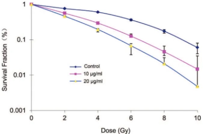
The survival fraction of A549 cells decreased following treatment with different doses of radiation and the same concentration of elemene. Conversely, the A549 cell survival fraction decreased in the groups treated with the same dose of radiation in combination with increasing concentrations of elemene (Table 1). Following treatment with 10 or 20mg/mL elemene, the A549 cell survival curve shifted to the left, the shoulder area was diminished, and the steepness of the curve increased (Figure 1). Based on the cell survival curve, the radiobiological parameters and radiosensitization ratio were obtained and are listed in Table 1. These data show that, compared with the control group, the SERD0and SERDqvalues for the 10 and 20mg/mL elemene treatment groups were greater than 1. Furthermore, the ratio gradually increased with an increasing drug concentration (Table 2).
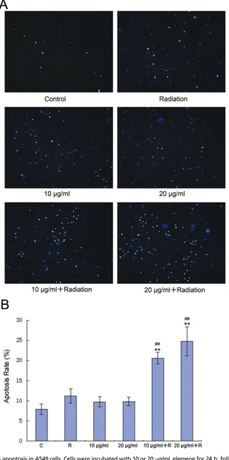
Effect of elemene on A549 cell apoptosis
Fluorescence microscopy showed that compared with the control group, the groups treated with radiation alone and elemene alone contained more apoptotic cells (Figure 2). Furthermore, significantly higher apoptotic levels were observed in the groups treated with radiation and elemene at 10 or 20mg/mL (Figure 2A). Flow cytometry revealed that
Table 1-Survival fraction (%).
Group 0 Gy 2 Gy 4 Gy 6 Gy 8 Gy 10 Gy
Control 100 84.1±13.2 70.2±10.5 66.3±1.9 47.3±2.5 26.0±1.3
10mg/ml 97.7±20.2 76.2±10.4 49.5±6.4 22.5±2.3 7.5±1.8 3.4±0.2 20mg/ml 95.4±18.8 63.6±7.5 30.4±3.0 5.7±1.1 2.7±0.6 0.7±0.1
Cells were incubated with 10 or 20mg/mL concentrations of elemene for 24 h and were then irradiated with 0, 2, 4, 6, 8, and 10 Gy and cultured for another 14 days. The number of cells forming more than 50 clones was counted under the inverted microscope to calculate the cloning efficiency (CE): CE (%)=(Clone formation average of treatment group/inoculated cell number)100%. Survival fraction (SF)=(irritated group CE/non-irradiated group
PE) 100%. The experiment was repeated 3 times to calculate the average.
Figure 1 -Cell survival curves of A549 cells following treatment
with 0, 10 or 20mg/mL elemene and exposure to x-ray doses from 0 to 10 Gy.
Table 2-Radiation parameters for a single-hit multi-target model.
Group D0(Gy) Dq(Gy) SF2(%) SERD0 SERDq
Control 2.54±0.24 2.68±0.25 84.6±20.9 - -10mg/mL 1.64±0.15 1.87±0.22 56.3±14.9 1.54±0.20 1.43±0.15 20mg/mL 1.55±0.13 1.53±0.11 43.2±10.7 1.63±0.32 1.75±0.19
SPSS17.0 was used to calculate survival parameters including SF2 (surviving fraction of 2 Gy), D0 (mean lethal dose or final slope), and
Dq (quasi-thres-hold dose). SER (sensitization enhancement ratio). SER D0
(Dq)=control group D0(Dq)/drug group D0(Dq) . Three parallel samples
compared with the control group, the apoptosis rate of the group treated with radiation alone appeared to increase, but this was not found to be statistically significant (p40.05). The apoptosis rate of the group treated with elemene alone did not change (p40.05), while the apoptosis rate of the elemene plus radiation group increased significantly (po0.01). The rate of apoptosis increased with increasing concentrations of elemene (Figure 2B).
Effect of elemene on DSB repair in A549 cells Figure 3 (0 h) shows that both elemene and irradiation alone increased the tail intensity in A549 cells, and the degree of DSB was augmented as the elemene concentration increased. In particular, the results showed a higher number of DSB in the combination group than the irradiation alone and elemene alone groups (po0.01). As shown in Figure 3 (24 h), following incubation for 24 h, the tail intensity in the irradiation alone group returned to background levels, while there was a significant increase in the amount of remaining tail intensity in the combination group compared with the irradiation alone and elemene alone groups (po0.01).
Effects of elemene on DNA-PKcs, Bcl-2, and p53 protein expression in A549 cells
The results shown in Figure 4A revealed that in the 10 and 20mg/mL combined treatment groups, a significant decrease in the protein expression of DNA-PKcs (po0.01, Figure 4B) and Bcl-2 (po0.01, Figure 4C) was observed, while the p53 protein expression level was significantly increased (po0.01, Figure 4C).
Protein expression correlation analysis
Spearman correlation analysis showed that DNA-PKcs and p53 protein expression was negatively correlated (r=-0.569, po0.05), while DNA-PKcs expression was posi-tively correlated with Bcl-2 protein expression (r=0.755, po0.05).
’ DISCUSSION
Basic research in radiation biology has shown that radiation therapy works mainly by damaging tumor cell DNA and altering the expression of apoptosis-related genes and proteins. The radiosensitivity of tumor cells relates to their capacity to repair DSB via the related genes DNA-PKcs, Ku70/80, and ataxia telangiectasia mutated (ATM). Other genes known to be involved in radiosensitivity and responsible for apoptosis regulation include p53, Bcl-2, c-myc proto-oncogene (c-myc), and survivin (9). Beta elemene, which is the active component of elemene, has recently been demonstrated to enhance the radiosensitivity of human cancer cell linesin vitro and in an animal tumor modelin vivo(16,20). In particular, beta elemene was found to enhance radiosensitivity by influencing the cell cycle distribution of gastric cancer MKN28 cells, and the mechanisms responsible for this effect include the induction of G2/M phase arrest, inhibition of sublethal damage repair, and induction of cell apoptosis, which enhances the killing effects of radioactive rays (21). The results of the current study show that the SERD0and SERDqvalues of A549 cells exposed to a low concentration of cytotoxic elemene were greater than 1. In addition, elemene enhanced the sensitivity
Figure 3 -Influence of elemene on radiation-induced DSB. A549 cells were irradiated with 4 Gy X-rays in the absence or presence of
elemene (10 or 20mg/mL) and assayed immediately after radiation or returned to the incubator for 24 h to permit repair. Comet assays were performed immediately after incubation with elemene treatment, irradiation, or combination treatment. Columns represent the means of the tail moments from three independent experiments, and the bars represent the SD,**
po0.01 vs. control, ##po0.01
of A549 cells to radiotherapy. Cellular apoptosis is funda-mental to radiotherapy, and its regulatory mechanism plays an important role in cellular radiosensitivity. Apoptosis-related genes such as p53 and Bcl-2 have important regulatory functions in the progression of rapid apoptosis induced by radiation therapy. For instance, a previous study showed that the levels of the antiapoptotic genes Bcl-2 and Bcl-xl in A549 cells decreased, while p53 expression and the production of exosomes increased, following elemene treatment (22). This result demonstrates that both p53 and Bcl-2 have important regulatory actions in cervical cancer cell apoptosis induced by radiation. A number of experi-mental studies have further shown that elemene is involved in regulating the expression of Bax, c-myc, p53, poly (ADP-ribose) polymerase (PARP), survivin, and livin as well as inducing tumor cell apoptosis (23-26). Our results showed that, compared with the exposure alone group, the group that received elemene combined with irradiation exhibited increased p53 gene expression and significantly decreased Bcl-2 gene expression, and the expression of both genes was significantly correlated. Furthermore, elemene was shown to regulate expression of the apoptosis-related genes Bcl-2 and
p53 and induce A549 cell apoptosis, thereby increasing cell radiosensitivity.
Interestingly, when Bcl-2 and p53 gene expression was significantly altered, DNA-PKcs protein expression was significantly decreased in the combined treatment group. This result indicates that elemene is also involved in regulating DNA damage repair pathways. Protein kinase activation leads to the phosphorylation of downstream DNA repair proteins, which initiate DNA chain fracture repair (27), and the relationship between DNA-PKcs and radiotherapy sensitivity has been a topic of significant research in recent years. It is well established that inhibiting tumor cell expression of DNA-PKcs increases radiation sensitivity. Panet al. (28) studied the relationship between DNA-PKcs expression and radiation sensitivity in non-small cell lung cancer cell lines, and in adenocarcino-mas and large cell carcinoadenocarcino-mas, DNA-PKcs was shown to be an important component regulating cellular radio-sensitivity. This result indicates that DNA-PKcs may be predictive of non-small cell lung cancer cell radiosensitiv-ity. Zou et al. (29) silenced the DNA-PKcs gene of human mammary epithelial cells (MCF10F) using small interfer-ing RNA (siRNA) technology. Simultaneously, the expres-sion of DNA repair-related proteins, such as DNA-PKcs, Ku80, ATM, and p53, was decreased in these cells, while their sensitivity increased with low doses of radiation. Small molecule inhibitors of DNA-PKcs were also shown to enhance radiation sensitivity of cervical cancer cells (30). Our experimental results showed that elemene inhibited DNA-PKcs expression in A549 cells, reduced DNA damage repair, and increased cellular radiosensitivity.
DNA-PKcs is a protein with a wide range of functions and is involved in DNA damage repair, apoptosis, and V(D)J recombination (31). Yu et al. (32) found that in non-small cell lung cancer, high expression of DNA-PKcs increased the activity of the DNA damage repair system. In addition, apoptosis inhibition caused by mutant p53 and Bcl-2 expression exhibited a combined effect, which may explain the development of resistance to radiotherapy in small-cell lung cancer. Daido et al. (33) indicated that following exposure to low doses of radiation, human malignant glioma M059J cells that lack DNA-PKcs underwent massive autop-hagic cell death that was significantly increased after exposure to DNA-PKcs inhibitors. Furthermore, DNA-PKcs inhibitors exert radiotherapy-sensitizing effects on glioma cells by enhancing type II programmed cell death. Lee et al. (34) found that p53-inducible gene 3 (PIG3) is involved in apoptosis caused by p53 activation and that this molecule can regulate DNA-PKcs expression. Moreover, knockdown of PIG3 was shown to decrease the level of DNA-PKcs in cells. Our study further addressed the correlation between DNA-PKcs, Bcl-2, and p53 expression, and the results showed that DNA-PKcs expression was significantly posi-tively correlated with that of Bcl-2 (r=0.755, po0.05) and significantly negatively correlated with p53 (r=0.569, po0.05). We further showed that DNA-PKcs was closely related to apoptosis and that elemene increased apoptosis of A549 cells and strengthened cellular radiosensitivity by inhibiting DNA-PKcs expression.
In summary, elemene exhibits radiotherapy-sensitizing effects on lung adenocarcinoma A549 cells, and its mechan-ism of action involves the upregulation of p53 and
down-Figure 4 -Western blot analysis of the protein levels of Bcl-2, p53
and DNA-PKcs. A549 cells were irradiated with 4 Gy X-rays following treatment with 10 or 20mg/mL elemene for 24 h. Proteins were extracted and separated by SDS-PAGE.(A)Levels of Bcl-2, p53 and DNA-PKcs were quantitated by densitometry, and the ratios of the three proteins are displayed. Values represent the mean ± SD, n=3, **po0.01 compared to the control,
##
po0.01 compared to radiation alone. Elemene inhibited the
protein expression of(B)DNA-PKcs and(C)Bcl-2 and promoted the protein expression of p53. Key: 1, Control; 2, irradiation; 3, 10mg/mL; 4, 20mg/mL; 5, 10mg/mL+irradiation; 6, 20mg/mL+
regulation of Bcl-2 to promote cell apoptosis, as well as the downregulation of DNA-PKcs to inhibit DSB repair. How-ever, the specific mechanism of action of elemene requires further elucidation.
’ ACKNOWLEDGEMENT
This study was supported by grants from the National Natural Science Foundation of China (No. 81473452).
’ AUTHOR CONTRIBUTIONS
Zou K, Zhang Z and Zou L designed the research study and wrote the paper. Zou K and Liu C performed the research. Zou K and Zhang Z analyzed the data.
’ REFERENCES
1. Maciejczyk A, Skrzypczynska I, Janiszewska M. Lung cancer. Radio-therapy in lung cancer: Actual methods and future trends. Rep Pract Oncol Radiother. 2014;19(6):353-60, http://dx.doi.org/10.1016/j.rpor.2014.04.012. 2. Collis SJ, Swartz MJ, Nelson WG, DeWeese TL. Enhanced radiation and chemotherapy-mediated cell killing of human cancer cells by small inhi-bitory RNA silencing of DNA repair factors. Cancer Res. 2003; 63(7):1550-4. 3. Marples B, Cann NE, Mitchell CR, Johnston PJ, Joiner MC. Evidence for the involvement of DNA-dependent protein kinase in the phenomena of low dose hyper-radiosensitivity and increased radioresistance. Int J Radiat Biol. 2002;78(12):1139-47, http://dx.doi.org/10.1080/09553000210166606. 4. Sak A, Stuschke M, Wurm R, Schroeder G, Sinn B, Wolf G, et al. Selective inactivation of DNA-dependent protein kinase with antisense oligo-deoxynucleotides: consequences for the rejoining of radiation-induced DNA double-strand breaks and radiosensitivity of human cancer cell lines. Cancer Res. 2002;62(22):6621-4.
5. Jackson SP. Sensing and repairing DNA double-strand breaks. Carcino-genesis. 2002;23(5):687-96, http://dx.doi.org/10.1093/carcin/23.5.687. 6. Kurdoglu B, Cheong N, Guan J, Corn BW, Curran WJ, Jr, Iliakis G.
Apoptosis as a predictor of paclitaxel-induced radiosensitization in human tumor cell lines. Clin Cancer Res. 1999;5(9):2580-7.
7. Wang LP, Liang K. Apotosis and Radiosensitivity of Cells. Foreign Med-ical Science. 2000;24(2):75-8.
8. Rosen EM, Fan S, Rockwell S, Goldberg ID. The molecular and cellular basis of radiosensitivity: implications for understanding how normal tis-sues and tumors respond to therapeutic radiation. Cancer Invest. 1999; 17(1):56-72, http://dx.doi.org/10.3109/07357909909011718.
9. Rosen EM, Fan S, Goldberg ID, Rockwell S. Biological basis of radiation sensitivity. Part 2: Cellular and molecular determinants of radiosensitivity. Oncology (Williston Park). 2000;14(5):741-57; discussion 757-8, 761-6. 10. Jiang H, Ma S, Feng J. In vitro study of radiosensitization ofb-elemene in
A549 cell line from denocarcinoma of lung. Chinese-German J of Clin Oncol. 2009;8(1):12-5, http://dx.doi.org/10.1007/s10330-008-0139-3. 11. Zou K, Tong E, Xu Y, Deng X, Zou L. Down regulation of mammalian
target of rapamycin decreases HIF-1aand survivin expression in anoxic lung adenocarcinoma A549 cell to elemene and/or irradiation. Tumor Biol. 2014;35(10):9735-41, http://dx.doi.org/10.1007/s13277-014-2226-0. 12. Cheng W, Li JP, Wang ZM, Song W, Huang C. The variation of gene
expression profile in radiosensitivity to kidney cancer cells induced by elemenen. Chin J Urol. 2007;28(2):87-90.
13. Wu DP, Li XM, Zhao JF, Wang HB, Zhao DQ. A Study of Radiosensitivity ofb-Elemene to Squamous Cell Carcinoma of Tongue Tca-8113 Cell Line in Vitro. Journal of Basic and Clinical Oncology. 2006;19(2):116-7. 14. Zou LJ, Sun XH, Xu XY. The study of theb-elemene effect on the augment
of radiation treatment in mouse inoculated U14 strain. Chin J Radiological Medicine and Protection. 2004;24(3):254-5.
15. She JJ, Wang ZM, Che XM, Pan CE. Radiosensitization of beta-elemene on VX2 carcinoma transplanted on kidney in rabbits in vivo. Zhong Xi Yi Jie He Xue Bao. 2006;4(4):392-6, http://dx.doi.org/10.3736/jcim20060415. 16. Li LJ, Zhong LF, Jiang LP, Geng CY, Zou LJ.b-Elemene radiosensitizes lung
cancer A549 cells by enhancing DNA damage and inhibiting DNA repair. Phytother Res. 2011; 25(7):1095-7, http://dx.doi.org/10.1002/ptr.v25.7. 17. Lindblom E, Dasu A, Lax I, Toma-Dasu I. Survival and tumour control
probability in tumours withheterogeneous oxygenation: A comparison between the linearquadratic and the universal survival curve models for high doses. Acta Oncol. 2014;53(8):1035-40, http://dx.doi.org/10.3109/ 0284186X.2014.925582.
18. Forchhammer L, Johansson C, Loft S, Moller L, Godschalk RW, Langie SA, et al. Variation in the measurement of DNA damage by comet assay measured by the ECVAG inter-laboratory validation trial. Mutagenesis. 2010;25(2):113-23, http://dx.doi.org/10.1093/mutage/gep048.
19. Li Z, Berk M, McIntyre TM, Gores GJ, Feldstein AE. The lysosomal-mitochondrial axis in free fatty acid-induced hepatic lipotoxicity. Hepa-tology. 2008;47(5):1495-503, http://dx.doi.org/10.1002/(ISSN)1527-3350. 20. Li G, Xie B, Li X, Chen Y, Wang Q, Xu Y, et al. Down-regulation of
survivin and hypoxia-inducible factor-1 alpha by beta-elemene enhances the radiosensitivity of lung adenocarcinoma xenograft. Cancer Biother Radiopharm. 2012;27(1):56-64, http://dx.doi.org/10.1089/cbr.2011.1003. 21. He S, Liu J, Zhang Z, Che X, Fan L, Chang S, et al. Enhancement of gastric
cancer MKN28 cell line radiosensitivity induced by beta-elemene. Zhon-ghua Wai Ke Za Zhi. 2014;52(6):442-5.
22. Li J, JunYu, Liu A, Wang Y.b-Elemene against human lung cancer via up-regulation of P53 protein expression to promote the release of exosome. Lung Cancer. 2014;86(2):144-50, http://dx.doi.org/10.1016/j.lungcan.2014.08.015. 23. Zhang Y, Zhao MF, Liu YP. The effects ofb-elemene on the activation of
Akt and the expressions of apoptosis-related proteins in gastric cancer. Shanxi Oncology Medicine. 2012;3:451-4.
24. Zhou XQ, Qiu XJ, Zhao HY. Influence on apoptosis relevant protein expression of human lung adenocarcinoma A549 cell line managed by Elemene. Journal of Modern Oncology. 2012; 20(10):2027-30.
25. Yang WZ, Chen CM, Shi SS. The Study on inhibition of the expression of c-myc gene of human glioma U251cell line and the effect mechanism of effect on cellular apoptosis by elemene. Chin J Clin Oncol. 2005; 32(13):763-6.
26. Zhao QT, Yang Y, Sun CB. Effect ofb-elemene combined with radio-therapy on the expression of Livin mRNA and apoptosis of lung adeno-carcinoma cell line A549. Journal of Modern Oncology. 2013; 21(2):257-60. 27. Calsou P, Delteil C, Frit P, Drouet J, Salles B. Coordinated assembly of Ku and p460 subunits of the DNA-dependent protein kinase on DNA ends is necessary for XRCC4-ligase IV recruitment. J Mol Biol. 2003;326(1):93-103, http://dx.doi.org/10.1016/S0022-2836(02)01328-1.
28. Pan Y, Li WX, Li JM, Zhu JQ, Liang YQ, Guo AL. Correlation of DNA-dependent protein kinase catalytic subunit expression to radiosensitivity of non-small cell lung cancer cell lines. Ai Zheng. 2009;28(7):714-7. 29. Zou W, Che J, Wang CJ. DNA-PKcs silencing inhibit the DNA repair
induced by low dose radiation on human breast epithelial cells. Chin J Biotech. 2009;25(5):727-32.
30. An J, Sui JL, Xu QZ. Inhibition of DNA-PKcs by siRNA and its effect on the growth of HeLa cells. Carcinogenesis, Teratogenesis & Mutagenesis. 2005;17(6):327-31.
31. Yan YQ, Zhou PK. Mechanism of non-homologous end joing and its bilolgical implications. Journal of Medical Molecular Biology. 2006; 3(1):69-72.
32. Yu S, Xiong Y, Tian S. The expression of DNA-PKcs in non-small cell lung cancer and its relationship with apoptosis associated proteins. Zhongguo Fei Ai Za Zhi. 2003;6(5):356-9.
33. Daido S, Yamamoto A, Fujiwara K, Sawaya R, Kondo S, Kondo Y. Inhi-bition of the DNA-dependent protein kinase catalytic subunit radio-sensitizes malignant glioma cells by inducing autophagy. Cancer Res. 2005;65(10):4368-75, http://dx.doi.org/10.1158/0008-5472.CAN-04-4202. 34. Lee JH, Kang Y, Khare V, Jin ZY, Kang MY, Yoon Y, et al. The