The negative prognostic impact of bone metastasis
with a tumor mass
Birsen Yu¨cel,I,* Mustafa Gu¨rol Celasun,IBilge O¨ ztoprak,II Zekiye Hasbek,IIISeher Bahar,ITurgut Kac¸an,
IV Aykut Bahc¸eci,
IVMehmet Metin S
¸eker
IV
IDepartment of Radiation Oncology IIDepartment of Radiology IIIDepartment of Nuclear Medicine IVDepartment of Medical Oncology, Sivas,
Turkey.
OBJECTIVE:Typically, bone metastasis causes osteolytic and osteoblastic lesions resulting from the interactions of tumor cells with osteoclasts and osteoblasts. In addition to these interactions, tumor tissues may grow inside bones and cause mass lesions. In the present study, we aimed to demonstrate the negative impact of a tumor mass in a large cohort of patients with bone metastatic cancer.
METHODS:Data from 335 patients with bone metastases were retrospectively reviewed. For the analysis, all patients were divided into three subgroups with respect to the type of bone metastasis: osteolytic, osteoblastic, or mixed. The patients were subsequently categorized as having bone metastasis with or without a tumor mass, and statistically significant differences in median survival and 2-year overall survival were observed between these patients (the median survival and 2-year overall survival were respectively 3 months and 16% in patients with a tumor mass and 11 months and 26% in patients without a tumor mass;po0.001).
RESULTS:According to multivariate analysis, the presence of bone metastasis with a tumor mass was found to be an independent prognostic factor (p=0.011, hazard ratio: 1.62, 95% confidence interval: 1.11–1.76). Bone
metastasis with a tumor mass was more strongly associated with osteolytic lesions, other primary diseases (except for primary breast and prostate cancers), and spinal cord compression.
CONCLUSION:Bone metastasis with a tumor mass is a strong and independent negative prognostic factor for survival in cancer patients.
KEYWORDS: Bone metastasis; Bone metastasis with a tumor mass; Prognostic factor; Survival.
Yu¨cel B, Celasun MG, O¨ ztoprak B, Hasbek Z, Bahar S, Kac¸an T, et al. The negative prognostic impact of bone metastasis with a tumor mass. Clinics. 2015;70(8):535-540
Received for publication onFebruary 6, 2015;First review completed onMarch 23, 2015;Accepted for publication onMarch 31, 2015 E-mail: [email protected]
*Corresponding author
’ INTRODUCTION
Bone metastasis is the most frequent complication of cancer, occurring in up to 70% of patients with breast or prostate cancer and in approximately 15–30% of patients with carcinoma of the
lung, colon, stomach, bladder, rectum, thyroid, or kidney (1). Although the exact incidence of bone metastasis remains unknown, this type of metastasis is an attractive area of study given its high prevalence in cancer patients.
Bone metastases develop as a result of interactions between tumor cells and bone cells. Cancer cells can induce various metastatic bone lesions through different mechanisms that depend on the primary disease, and two types of metastatic
bone lesions have been described (2,3). The first is an osteolytic lesion that progresses with bone resorption as a result of osteoclast activation; the second is an osteoblastic lesion that triggers bone formation and osteoblastic cell activation. These two types of lesions may be present concomitantly in certain patients (mixed type) following stimulation of the two different types of bone cells. Alternatively, the tumor itself may grow inside the bone tissue and destroy the bone directly (4). These mass lesions may cause an increase in complications (e.g., spinal cord compression, pathologic fracture) due to metastasis-related bone destruction and suggest the presence of a significant tumor burden. Examples of computerized tomography images of osteolytic lesions, osteoblastic lesions, and bone metastasis with a tumor mass are shown in Figure 1.
Although the duration of survival varies according to the primary tumor, bone metastases are usually incurable (5). General treatment procedures for patients with bone metastasis include bisphosphonate administration, che-motherapy, and palliative radiation therapy. However, responses to these treatment modalities are relatively poor, and the patient’s quality of life is generally impaired.
DOI:10.6061/clinics/2015(08)01
Copyright&2015CLINICS–This is an Open Access article distributed under the terms of the Creative Commons License (http://creativecommons.org/licenses/by/ 4.0/) which permits unrestricted use, distribution, and reproduction in any medium or format, provided the original work is properly cited.
Prognosis may vary among patients depending on factors such as the primary disease type, age, the patient’s performance status, the metastatic interval, and the number of metastatic sites (6,7). Nevertheless, these factors are not particularly helpful with respect to decision making in routine clinical practice. Moreover, data on both the prognostic impact of the mechanism type on bone metastasis and the additional role of tumor masses in these patients are lacking.
Therefore, we designed a retrospective analysis to evaluate the impact of bone metastasis-related tumor mass on patient survival. We also evaluated differences in the response to radiation therapy, in complications, and in the pain response in our cohort according to the type of metastasis.
’ MATERIALS AND METHODS
This study was conducted at the Department of Radiation Oncology at Cumhuriyet University Hospital in Sivas, Turkey, in accordance with the principles of the Declaration of Helsinki. A total of 335 cancer patients with bone metastasis who were admitted to the department between 2007 and 2013 were evaluated retrospectively.
All patients were treated with palliative radiotherapy and bisphosphonate. During the treatment period, all patients were examined by a radiation oncologist immediately before and 1 month after radiotherapy. The physical examination findings as well as body weight; Eastern Cooperative Oncology Group (ECOG) performance scores; and histo-pathological, radiological, and laboratory data (alkaline phosphatase [ALP] and calcium levels) were recorded. The patients’survival data were obtained from hospital records, and patients lost to follow-up were contacted to obtain information about their condition. Survival was defined as the time between the date of the first detection of bone metastasis and the date of last contact or death.
The cancer type was classified based on the primary site: head and neck, lung, breast, prostate, gastrointestinal system, genitourinary system, or other. Prior to palliative radiotherapy, each patient’s performance status was scored according to the ECOG scoring system (8). Weight loss was defined as loss of410% of body weight in 1 month.
Bone metastasis was revealed by computerized tomogra-phy or magnetic resonance imaging and was confirmed by bone scintigraphy and positron emission tomography.
All patients were divided into three subgroups with respect to the type of bone metastasis: osteolytic, osteo-blastic, or mixed type. All patients were subsequently
recategorized into two groups: bone metastasis with or without a tumor mass.
Pain intensity was evaluated using visual analog scales in 139 (41%) of the cases (9). Patients were routinely asked to rate their pain intensity by placing a mark on a 10-mm visual analog scale at the start of radiotherapy and at 1 month after radiotherapy. This scaling system was used to evaluate the intensity of pain only in the radiotherapy-affected region. The response to radiotherapy was determined by calculating the difference between the pain intensity on the visual analog scale before and 1 month after the initiation of radiotherapy. Statistical Package for Social Sciences (SPSS) for Windows 14.0 (SPSS, Inc., Chicago, IL, USA) was used for the statistical analysis. For descriptive statistics, the mean, standard deviation, frequency, and median were used. Categorical data were compared statistically using the chi-square test or Fisher’s exact test. Survival rates were calculated according to the Kaplan-Meier method. A multivariate analysis (Cox regression analysis) was used to evaluate independent risk factors affecting survival. P-valuesp0.05 were accepted as
statistically significant.
’ RESULTS
The study group comprised 234 (70%) men and 101 (30%) women. The median age at the time of cancer diagnosis was 59 years (range, 21–82 years). The primary disease
distribu-tion was as follows: lung cancer in 107 (32%) patients, breast cancer in 64 (19%), prostate cancer in 62 (19%), gastro-intestinal system tumors in 40 (12%), genitourinary system tumors in 20 (6%), head and neck tumors in 11 (3%), and tumors in other organs in 31 (9%).
Osteolytic bone metastasis was observed in 99 (30%) patients, whereas 155 (46%) had osteoblastic bone metastasis, and 71 (21%) had mixed-type bone metastasis. Ten (3%) patients had bone metastasis and only a tumor mass, without any other lesions; these 10 patients were excluded when categorizing the patients with respect to the type of bone lesion (i.e., osteolytic, osteoblastic, or mixed). Bone metastasis with a tumor mass was present in 73 (22%) cases. Eleven (3%) patients had a single bone metastatic lesion, and 324 (97%) had two or more lesions. The 11 patients with single bone lesions had no metastases in other organs. The locations and frequencies of bone metastases were as follows: vertebral column metastasis in 283 (84%) patients, pelvic bone metastasis in 246 (73%), long bone metastasis in 189 (56%), costal metastasis in 189 (56%), and skull metastasis in 63 (19%).
Spinal cord compression was observed in 20 patients, or 7% of all patients with vertebral column metastases (N: 283), whereas 49 (15%) patients had pathologic fractures, 26 (8%) had neurological deficits, and 16 (5%) had hypercalcemia. Surgical interventions were performed for pathologic frac-tures in 19 (39%) patients with pathologic fracfrac-tures (N: 49).
The types of bone metastasis with respect to primary disease were as follows. Among patients with lung cancer, 42 (39%) had osteolytic lesions, 44 (41%) had osteoblastic lesions, 19 (18%) had mixed lesions, and 2 (2%) had bone metastases with only tumor masses. For patients with breast cancer, 22 (34%), 19 (30%), and 22 (34%) had osteolytic, osteoblastic, and mixed lesions, respectively; 1 (2%) had a bone metastasis with only a tumor mass. Osteolytic, osteoblastic, and mixed lesions developed in 2 (3%), 53 (86%), and 7 (11%), respectively, patients with prostate cancer. Regarding patients with gastrointestinal system tumors, 9 (22%), 18 (45%), and 11 (28%) had osteolytic, osteoblastic, and mixed lesions, respectively, and 2 (5%) showed bone metastases with only tumor masses. Among patients with genitourinary system tumors, 8 (40%), 5 (25%), and 5 (25%) had osteolytic, osteoblastic, and mixed lesions, respectively, with 2 (10%) exhibiting bone metastases with only tumor masses. The incidence of osteolytic, osteoblastic, and mixed lesions was 2 (18%), 5 (46%), and 2 (18%), respectively, for the patients with head and neck tumors; 2 (18%) had bone metastases with only tumor masses.
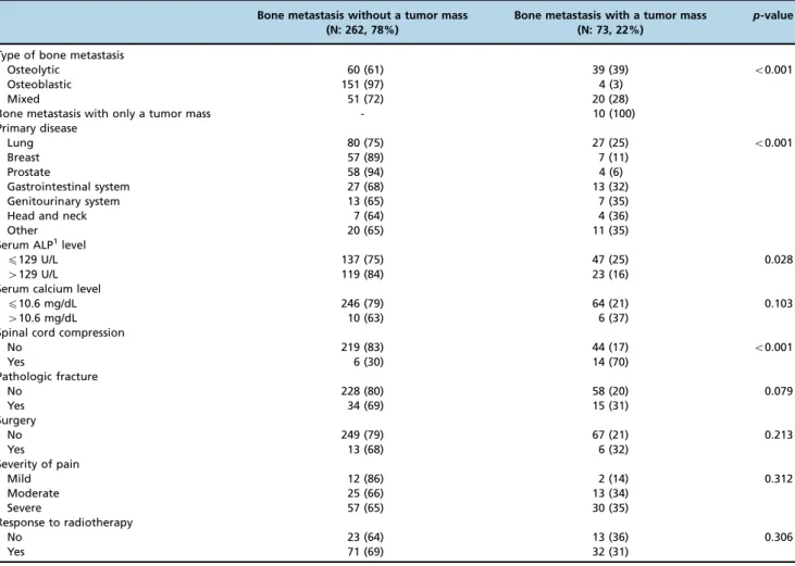
Bone metastasis with a tumor mass was observed more frequently in patients with osteolytic lesions than in those with other bone lesions. Spinal cord compression was observed more frequently in cases of bone metastasis with a tumor mass compared to cases without a tumor mass; when occurring in the latter, the compression was mostly due to compression fracture, as observed for osteolytic metastases, or to new bone formation, as observed in osteoblastic lesions. However, serum ALP levels were higher in patients without tumor masses. In addition, bone metastases with tumor masses were observed less frequently in patients with primary breast or prostate cancer compared with patients with other primary diseases, such as lung or gastrointestinal system tumors. With respect to pathologic fractures, pain severity, and responses to radiotherapy, no differences were observed between cases of bone metastases with tumor masses and cases of other bone metastases (Table 1).
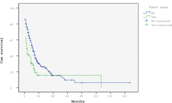
The median survival duration was 10 months (range, 1–
147 months), and the 1- and 2-year survival rates were 46% and 24%, respectively. The median survival duration was 3 months and the 1- and 2-year survival rates were 28% and 16%, respectively, among patients who had bone metastases with tumor masses and 11 months and 50% and 26%, respectively, in patients who had bone metastasis without tumor masses. The survival curves of the patients with or without a tumor mass are shown in Figure 2. Univariate
Table 1-Comparison of features associated with bone metastases with or without tumor masses.
Bone metastasis without a tumor mass (N: 262, 78%)
Bone metastasis with a tumor mass (N: 73, 22%)
p-value
Type of bone metastasis
Osteolytic 60 (61) 39 (39) o0.001
Osteoblastic 151 (97) 4 (3)
Mixed 51 (72) 20 (28)
Bone metastasis with only a tumor mass - 10 (100)
Primary disease
Lung 80 (75) 27 (25) o0.001
Breast 57 (89) 7 (11)
Prostate 58 (94) 4 (6)
Gastrointestinal system 27 (68) 13 (32)
Genitourinary system 13 (65) 7 (35)
Head and neck 7 (64) 4 (36)
Other 20 (65) 11 (35)
Serum ALP1level
p129 U/L 137 (75) 47 (25) 0.028
4129 U/L 119 (84) 23 (16)
Serum calcium level
p10.6 mg/dL 246 (79) 64 (21) 0.103
410.6 mg/dL 10 (63) 6 (37)
Spinal cord compression
No 219 (83) 44 (17) o0.001
Yes 6 (30) 14 (70)
Pathologic fracture
No 228 (80) 58 (20) 0.079
Yes 34 (69) 15 (31)
Surgery
No 249 (79) 67 (21) 0.213
Yes 13 (68) 6 (32)
Severity of pain
Mild 12 (86) 2 (14) 0.312
Moderate 25 (66) 13 (34)
Severe 57 (65) 30 (35)
Response to radiotherapy
No 23 (64) 13 (36) 0.306
Yes 71 (69) 32 (31)
analyses showed that the survival duration after metastasis was affected by the presence of bone metastasis with a tumor mass as well as by gender, weight loss, performance status, serum ALP and calcium levels, primary disease, bone metastasis type, number of bone lesions, the presence of extraosseous metastasis, and the disease-free interval. The prognostic factors that affected survival time after the development of bone metastasis are shown in Table 2.
Multivariate analyses revealed that the presence of bone metastasis with a tumor mass as well as gender, weight loss, primary disease, type of bone metastasis, and serum ALP and calcium levels were independent prognostic factors that affected survival. The independent prognostic factors that affected the duration of survival after the development of bone metastasis are shown in Table 3.
’ DISCUSSION
The prevalence of bone metastasis is higher in advanced-stage cancers. Patients diagnosed with bone metastasis usually have incurable disease, though the survival duration does vary based on the primary disease. Accordingly, it is very important to determine prognostic factors once a diagnosis of bone metastasis has been made. The present study investigated the prognostic and clinical importance of bone metastasis with a tumor mass and found that this feature was an apparently strong negative prognostic factor for survival. The higher incidence of these metastases in association with osteolytic lesions might have contributed to this result, as the presence of osteolytic lesions was found to be a poor prognostic factor in a multivariate analysis. In addition, growth of the tumor itself inside the bone might indicate a larger tumor burden, which might also contribute to a shorter survival duration. Given the soft tissue component of bone metastasis with a tumor mass, spinal cord compression was observed more frequently in these patients; nonetheless, the presence of these lesions did not
increase the pain intensity or affect the response to radiotherapy.
Certain researchers have studied prognostic factors in patients with bone metastases. In a study of 350 patients with skeletal metastases, Katagiri et al. (6) reported that the patient’s performance status, the primary lesion site, the presence of multiple skeletal metastases, the presence of visceral or cerebral metastases, and a history of previous chemotherapy were important prognostic factors. Van der Linder et al. (7) reported a median survival time of 7 months for 342 patients with vertebral metastases, and Karnofsky stated that the performance score, the primary tumor type, and absence of visceral metastasis were significant predictors of survival. In the present study, female gender, the presence of osteoblastic and/or mixed lesions, and primary breast or prostate cancer were considered to be good prognostic predictors. In contrast, the presence of bone metastasis with a tumor mass as well as male gender, weight loss, primary lung cancer, the presence of osteolytic lesions, and elevated ALP and calcium levels were found to be poor prognostic predictors. Poor performance in a single-variable analysis, a disease-free interval ofo2 years, the presence of
extraoss-eous metastasis, and multiple bone lesions were also poor prognostic factors.
Circulating metastatic cells in blood become entrapped by the bone marrow spongiosum. Cancerous bone undergoes secondary lytic or blastic changes (10), and the type of bone metastasis is determined by these changes. In the literature, osteolytic lesions have been reported to be more frequent in breast cancer cases, whereas osteoblastic lesions are observed in cases of prostate cancer. In the present study, osteoblastic lesions (46%) were more frequently observed in the overall patient population; similar to the findings of other studies, osteolytic lesions were more frequent in patients with breast cancer, with osteoblastic lesions being more common in patients with prostate cancer. In terms of the conventional classification of bone metastases, the presence of a tumor
Months
140 120 100 80 60 40 20 0 100
80
60
40
20
0
Yes-censored Yes-censore No-censored No-Yes Ye No No
Tumor mass Survival Functions
Cum survival
mass was significantly more frequent among osteolytic lesions (62%). The frequencies of bone metastasis with a tumor mass were low among patients with breast or prostate cancer and similar among those with other types of cancer. Specifically, 25–36% of patients with other types of cancer
(non-breast or prostatic) had bone lesions with tumor masses.
Bone metastases are associated with a particular set of complications, and the frequency of these complications varies depending on the features of the metastatic lesions. For example, pathologic fractures and spinal cord compres-sion are encountered more frequently with osteolytic lecompres-sions, as these lesions cause bone destruction (2,11). It is rational to expect that bone metastases with tumor masses would present more complications; indeed, spinal cord compression was more frequent among cases of bone metastasis with a tumor mass in the current study. However, an elevated serum ALP level was more frequently observed in cases of bone metastasis without a tumor mass. In terms of pathologic fractures, serum calcium levels, surgical interven-tion, pain severity, and responses to radiotherapy, no differences were observed between patients with bone metastasis with a tumor mass and those with other types of bone metastases.
The survival duration in patients with bone metastases varied quite significantly depending on the primary disease, and it is reported that the duration is generally longer for patients with breast or prostate cancer than for those with other types of cancer (1,7,6,11). Ahn et al. (12) reported a median survival time of 55.2 months among 110 breast cancer patients with only bone metastases. In contrast, survival durations as short as 5–7 months were reported
among patients with lung cancer and bone metastases (11,13,14). In our study, the longest survival durations were observed in patients with breast cancer, followed by those with prostate cancer (median survival durations of 18 months and 15 months, respectively); conversely, the survival times of patients with other cancers were relatively short.
Many studies have reported that patients with single bone lesions in the absence of metastases in other organs have a longer survival duration relative to those with multiple bone metastases (15-17). In a study of 42 patients with solitary bone metastases, Hoshi et al. (15) reported a median survival duration of 30 months and a 1-year survival rate of 76.5%. In the present study, the 11 patients with single bone lesions had a median survival duration of 32 months and a 1-year survival rate of 68%. The survival durations were shorter
Table 2-Prognostic factors affecting patient survival after the development of bone metastasis, as determined by univariate survival analysis.
No. of patients 1-year survival (%) 2-year survival (%) Median survival (months) p-value Bone metastasis with tumor mass
No 262 50 26 11 *o0.001
Yes 73 28 16 3
Gender
Male 234 39 17 8 o0.001
Female 101 61 42 17
Weight loss
No 248 53 27 12 o0.001
Yes 87 24 12 5
ECOG PS1
ECOG0-1 168 55 30 13 o0.001
ECOG2 and higher 167 36 17 7
Serum ALP2level
p129 U/L 184 50 29 12 0.004
4129 U/L 142 39 16 9
Serum calcium level
p10.6 mg/dL 310 46 24 10 0.027
410.6 mg/dL 16 - - 3
Primary disease
Lung 107 27 10 5 o0.001
Breast 64 72 47 18
Prostate 62 69 31 15
Gastrointestinal system 40 24 6 5
Genitourinary system 20 20 10 5
Head and neck 11 9 - 3
Type of bone metastasis
Osteolytic 99 29 14 4 0.004
Osteoblastic 155 53 26 12
Mixed 71 49 26 12
Number of bone lesions
1 lesion 11 68 68 32 0.040
X2 lesions 324 44 22 10
Extraosseous metastasis
No 176 51 27 12 0.032
Yes 159 40 18 8
Disease-free interval
o24 months 259 41 20 9 0.026
X24 months 76 61 35 18
among the patients with osteolytic lesions compared with patients with osteoblastic or mixed lesions. Moreover, patients with bone metastases with tumor masses had significantly shorter survival durations compared with those with bone metastases without tumor masses (median survival durations of 3 months and 11 months, respectively; 1-year survival rates of 28% and 50%, respectively).
Two major limitations of the present study were its retrospective design and its heterogeneous study population. We believe that studies of more specific groups would yield more significant results.
The presence of bone metastasis with a tumor mass appeared to be a strong negative prognostic factor and was associated with a higher incidence of spinal cord compression.
’ AUTHOR CONTRIBUTIONS
Yücel B designed the research and analyzed the data. Yücel B, Celasun MG, Öztoprak B, Hasbek Z, Bahar S, Kac¸an T, Bahc¸eci A, and S¸eker MM
performed the research. Kac¸an T and S¸eker MM contributed analytical
tools. Yücel B and Öztoprak B wrote the paper. The authors have no financial disclosures to declare, no conflicts of interest to report, and have no commercial or proprietary interest.
’ REFERENCES
1. Coleman RE, Rubens RD. The clinical course of bone metastases from breast cancer. Br J Cancer. 1987;55(1):61-6, http://dx.doi.org/10.1038/ bjc.1987.13
2. Roodman GD. Mechanisms of bone metastasis. N Engl J Med. 2004;350 (16):1655-64, http://dx.doi.org/10.1056/NEJMra030831
3. Mundy GR. Metastasis to bone: causes, consequences and therapeutic opportunities. Nat Rev Cancer. 2002;2(8):584-93, http://dx.doi.org/ 10.1038/nrc867
4. Käkönen SM, Mundy GR. Mechanisms of osteolytic bone metastases in breast carcinoma. Cancer. 2003;97(3Suppl):834-9, http://dx.doi.org/ 10.1002/(ISSN)1097-0142
5. Coleman RE. Metastatic bone disease: clinical features, pathophysiology and treatment strategies. Cancer Treat Rev.2001;27(3):165-76, http://dx. doi.org/10.1053/ctrv.2000.0210
6. Katagiri H, Takahashi M, Wakai K, Sugiura H, Kataoka T, Nakanishi K. Prognostic factors and a scoring system for patients with skeletal metas-tasis. J Bone Joint Surg Br. 2005;87(5):698-703, http://dx.doi.org/10.1302/ 0301-620X.87B5.15185
7. Van der Linder YM, Dijkstra SPDS, Vonk EJA, Marijnen CAM, Leer JWH. Prediction of survival in patients with metastases in the spinal column. Cancer.2005;103(2):320-8, http://dx.doi.org/10.1002/(ISSN)1097-0142 8. Oken MM, Creech RH, Tormey DC, Horton J, Davis T, Mc Fadden ET,
et al. Toxicity and response criteria of the Eastern Cooperative Oncology Group. Am J ClinOncol. 1982:5(6):649-55, http://dx.doi.org/10.1097/ 00000421-198212000-00014
9. Price DD, McGrath PA, Rafii A, Buckingham B. The validation of visual analogue scales as ratio scale measures for chronic and experimental pain. Pain. 1981;17(1):45-56, http://dx.doi.org/10.1016/0304-3959(83)90126-4 10. Rubin P, Brasacchio R, Katz A. Solitary metastases: illusion versus reality.
Semin Radiat Oncol. 2006;16(2):120-30, http://dx.doi.org/10.1016/j. semradonc.2005.12.007
11. Coleman RE. Clinical features of metastatic bone disease and risk of skeletal morbidity. Clin Cancer Res. 2006;66(12):6243-9, http://dx.doi. org/10.1158/1078-0432.CCR-06-0931
12. Ahn SG, Lee HM, Cho SH, Lee SA, Hwang SH, Jeong J, et al. Prognostic factors for patients with bone-only metastasis in breast cancer. Yonsei Med J. 2013;54(5):1168-77, http://dx.doi.org/10.3349/ymj.2013.54.5.1168 13. Stanley KE. Prognostic factors for survival in patients with inoperable
lung cancer. J Natl Cancer Inst. 1980;65(1):25-32.
14. Sugiura H, Yamada K, Sugiura T, Hida T, Mitsudomi T. Predictors of sur-vival in patients with bone metastasis of lung cancer. Clin Orthop Relat Res. 2008;466(3):729-36, http://dx.doi.org/10.1007/s11999-007-0051-0 15. Hoshi M, Takada J, Leguchi M, Takahashi S, Nakamura H. Prognostic
factors for patients with solitary bone metastasis. Int J Clin Oncol. 2013;18 (1):164-9, http://dx.doi.org/10.1007/s10147-011-0359-3
16. Koizumi M, Yoshimoto M, Kasumi M, Ogata E. Comparison between soli-tary and multiple skeletal metastatic lesions of breast cancer patients. Ann Oncol. 2003;14(8):1234-40, http://dx.doi.org/10.1093/annonc/mdg348 17. Hirano Y, Oda M, Tsunezuka Y, Ishikawa N, Watanabe G. Long-term
survival cases of lung cancer presented as solitary bone metastasis. Ann Thorac Cardiovasc Surg. 2005;11(6):401-4.
Table 3-Independent prognostic factors affecting the duration of survival after the development of bone metastasis, as determined by multivariate analysis.
Overall survival HR1 95% CI2
p-value Bone metastasis with a tumor mass
No 1
Yes 1.62 1.11–1.76 *0.011
Gender
Male 1
Female 0.45 0.31–0.64 o0.001
Weight loss
No 1
Yes 1.39 1.02–1.90 0.034
Primary disease
Lung 1
Breast 0.32 0.20–0.57 o0.001
Lung 1
Prostate 0.45 0.30–0.67 0.001
Type of bone metastasis
Osteolytic 1
Osteoblastic 0.56 0.39–0.81 0.002
Osteolytic 1
Mixed 0.56 0.38–0.83 0.004
Serum ALP3level
p129 U/L 1
4129 U/L 1.34 1.03–2.00 0.030
Serum calcium level
p10.6 mg/dL 1
410.6 mg/dL 2.22 1.03–4.81 0.042