Surgical amniotic membrane transplantation after conjunctival
and limbal tumor excision
Transplante de membrane amniótica após excisão de tumor limbar e conjuntival
Seniz engur goktaS1, YaSemin katircioglu2, tuba celik3, FirdevS ornek2
Submitted for pulication: July 21, 2016 Accepted for pulication: February 2, 2017
1 Department of Ophthalmology, Sanliurfa Mehmet Akif Inan Training and Research Hospital, Sanliurfa,
Turkey.
2 Department of Ophthalmology, Ankara Training and Research Hospital, Ankara, Turkey. 3 Department of Ophthalmology, Bulent Ecevit University, Zonguldak, Turkey.
Funding: No specific financial support was available for this study.
Disclosure of potential conflicts of interest: None of the authors have any potential conflict of interest to disclose.
Corresponding author: Tuba Celik. Department of Ophthalmology. Bulent Ecevit University - Zon-guldak, Turkey - E-mail: [email protected]
Approved by the following research ethics committee: Ankara Training and Research Hospital.
ABSTRACT
Purpose: To evaluate the clinical results of patients treated by amniotic membra-ne transplantation (AMT) following excision of conjunctival and limbal tumors. Methods: A total of 14 eyes of 14 patients who underwent AMT after total lesion-free tumor excision and perilesional cryotherapy were evaluated. Results: The excised tumors comprised 7 conjunctival intraepithelial neoplasia, 5 conjunctival nevus, 1 primary acquired melanosis, and 1 squamous cell carci-noma. Limbus was involved in 10 cases, whereas cornea was involved in 6 cases. The average measurement of the tumor base was 14.8 mm (range 6-20 mm, SD 16 mm). The mean follow-up time period was 17.5 months (range 6 -60 months, SD 20 months). Complete healing occurred in eight eyes, but limbal cell deficiency developed in two eyes. Four cases had recurrence and were treated with the same surgical and medical procedures; during follow-up after recurrence, superficial peripheral vascularization and corneal scar were noted to have developed. Conclusions: AMT was effective for reconstruction of tissue defect after excision and cryotherapy of limbal and conjunctival tumors. In most of these cases, complete healing was achieved with a smooth, stable, and translucent surface.
Keywords: Conjunctival neoplasm surgery; Squamous cell carcinoma; Amnion transplantation; Cryotherapy
RESUMO
Objetivo: Avaliar os resultados clínicos de pacientes tratados com transplante de membrana amniótica (TMA) após a excisão de tumores conjuntival e limbar.
Métodos: Foram avaliados 14 olhos de 14 pacientes submetidos a transplante de membrana amniótica após excisão total de tumor e crioterapia perilesional.
Resultados: Os tumores que foram excisados podem ser classificados como neoplasia intraepitelial conjuntival em 7, nevus conjuntival em 5, melanose adquirida primária e carcinoma espinocelular em 1 caso cada. O limbo foi envolvido em 10 casos e a córnea foi envolvida em 6 casos. A medida média da base do tumor foi de 14,8 mm (6-20, DP 16). O tempo médio de seguimento foi de 17,5 meses (6-60, DP 20). A cicatrização completa ocorreu em 8 olhos e deficiência de células limbares ocorreu em 2 olhos. A recorrência foi diagnosticada em 4 casos e tratada com o mesmo procedimento cirúrgico e médico e, após essa recorrência, a vascularização periférica superficial e a cicatriz corneana desenvolveram-se em 4 casos durante o seguimento.
Conclusões: O transplante de membrana amniótica é um método eficaz para a reconstrução do defeito do tecido após a excisão de tumores limbares e conjuntivais com a combinação de crioterapia. Na maioria destes casos, a cicatrização completa pode ser conseguida com uma superfície lisa, estável e transparente.
Descritores: Neoplasias da túnica conjuntiva/cirurgia; Carcinoma de células esca-mosas;Âmnio/transplante; Crioterapia
INTRODUCTION
Conjunctival and limbal tumors comprise a wide variety of condi-tions from benign to malignant lesions that may threaten the visual function and life of the patient. The most common types of ocular surface tumors are squamous cell carcinoma (SCC), primary acquired melanosis (PAM), lymphoma, and malignant melanoma(1). Older age,
male gender, prolonged ultraviolet B exposure, and human papillo-ma virus are the risk factors for developing ocular surface squa-mous neoplasia (OSSN). These neoplasms are classified as benign, pre-invasive, or invasive. Benign OSSN includes papillomas, pseudo-epitheliomatous hyperplasia, and benign hereditary intraepithelial dyskeratosis. Pre-invasive lesions, such as conjunctival intraepithelial neoplasms (CIN) grade I-III, have malignant potential and are classi-fied as mild, moderate, or severe depending on the degree of invol-vement of the dysplastic epithelium. Mild dysplasia or CIN grade I is defined as dysplasia of the lower third of the conjunctival epithelium; dysplasia that extends into the middle third is defined as CIN grade II; and dysplasia that extends into the upper third is defined as CIN
grade III or carcinoma-in situ(2). Invasive OSSN consists of SCC and,
rarely, mucoepidermoid carcinoma. These lesions were traditionally treated by complete surgical excision with or without adjunctive therapy to reduce the rate of recurrence(1). Complete resection and
histopa thological control of the borders are very important(3).
The amniotic membrane (AM), which is the innermost layer of the placenta, contains a thick basement membrane and an avas-cular stroma, which may promote epithelial cell proliferation(4). The
activity of the AM has been attributed to various mechanisms, most of which are related to the structural and biochemical composition of the membrane; however, these have not been proven. The ba-sement membrane characteristics of the AM are similar to that of the cornea and conjunctiva(5). AM transplantation (AMT) promotes
epithelial wound healing and exerts potent anti-inflammatory and anti-scarring effects on the ocular surface. These valuable properties make the AM an ideal tissue for reconstruction of ocular surface tumors(6-9). Several studies have been performed for ocular surface
was an effective technique without complications after removal of conjunctival and limbal tumors in nine patients(9). Palamar et al.
stu-died the long-term efficacy of AMT for ocular surface reconstruction in 21 OSSN patients. They found that AMT was an effective procedure even for tumors larger than 10 mm(10).
Cryotherapy has been used as an adjunctive therapy to reduce the recurrence rates. Surgical excision with wide margins had not been sufficient in all cases. Occasionally, a surgeon can miss the tu-mor cells at the margins or base of the sclera. Cryotherapy destroys these small groups by superficial and deep freezing techniques and reduces recurrence rates(11). The reported recurrence rate among
patients who underwent cryotherapy after surgical excision ranged from 7.1% to 12.3%(11-13). In this paper, we reported our long-term
experience on AMT and cryotherapy following excision of large con-junctival and limbal tumors.
METHODS
This study was approved by the institutional ethics committee and was conducted in accordance with the Declaration of Helsinki. The details of the study were explained to the patients and written informed consent was obtained. Fourteen patients who underwent AMT procedure and cryotherapy after excision of limbal and con-junctival tumors were analyzed. All surgeries were performed by one surgeon (YK) at a single center. Histopathologic analyzes were done at the pathology department of the same hospital.
During all visits, slit lamp bio-microscopy and fluorescein staining were performed. Any complications, such as scarring, vascularization, corneal opacities, and recurrences, were noted. Complete healing without any complication was defined as a successful; non-complete healing without recurrence was defined as partial success; and recur-rence was defined as failure.
E
XCISIONOFTHETUMORSAll surgeries were performed under sedation or local anesthesia with 1:1 mixture of 2% lidocaine and 0.5% bupivacaine. The tumors were successfully excised completely with lesion-free margins mea-suring 2-3 mm. The surgeon avoided any tumor contact with the surgical instruments to prevent tumor seeding into the other sites. Fresh instruments were used after removal of the tumor. The mana-gement of lesions in the limbal area involved alcohol epitheliectomy for the corneal component, with wide margins for the conjunctival component followed by freeze-thaw cryotherapy to the remaining adjacent bulbar conjunctiva.
A
MNIOTICMEMBRANETRANSPLANTATIONAfter obtaining from donors who underwent elective caesarean sections, AMs were processed and stored in a solution of glycerine and Dulbecco’s Modified Eagle Medium (DMEM) in a 1:1 ratio con-taining antibiotics at -80 °C. Donor serums were checked for micro-biologic safety at the time of donation and after six months.
A single layer of AM was placed over the defect area as stromal face down and was sutured with 8/0 vicryl one by one. No bandage contact lens was used to cover the AM. Postoperatively, all patients received fluorometholone, ofloxacin, and dextran 70 hydroxypropyl methylcellulose four times daily for one month. In three patients, sutures were removed on the fifteenth day postoperatively.
RESULTS
Of the 14 cases included in this study, 6 were men and 8 were women; mean age was 56 years (range, 15-82 years). All tumors were primary cases and did not receive any prior treatment. These excised tumors were classified as CIN (n=7), conjunctival nevus (n=5), PAM (n=1), and SCC (n=1). There was no local invasion to the orbit or local lymph node metastasis at the time of surgery.Thebulbar conjunctiva
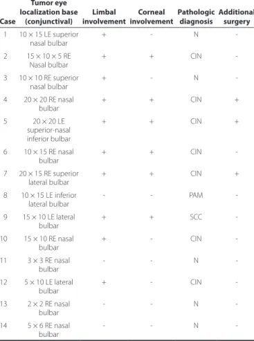
was involved in all cases, the limbus was involved in 10 cases, and the cornea was involved in 6 cases. Table 1 presents the size, localization, pathologic diagnoses, and additional surgical therapy of the study population. The average measurement of the tumor base was 14.8 mm (range 6-20 mm, SD 16 mm). The mean follow-up time period was 17.5 months (range, 6-60 months, SD 20 months).
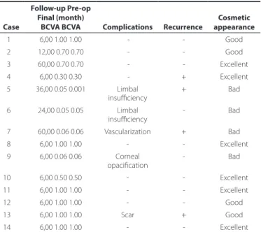
Table 2 shows the complications, recurrence, and cosmetic outcomes of the study. Complete healing was achieved in eight eyes. Limbal cell deficiency developed in two eyes. Figures 1-4 de-monstrate the preoperative and postoperative images of cases with conjunctival tumor, SCC, and CIN. Four cases developed recurrence and were treated with the same surgical and medical procedures; these cases were noted as failure because of the development of superficial peripheral vascularization and corneal scar on follow-up after recurrence. Eight cases were reported as successful and two cases were reported as partial success.
DISCUSSION
Management of conjunctival and limbal tumors is a significant therapeutic challenge. The key point in treating ocular surface tu-mors depends mainly on complete surgical removal(1) because the
presence of residual tumor cells in adjacent tissues might cause recurrence. Impression cytology (IC) has an important role in thera-peutic monitoring of residual cells after tumor removal. Although this test cannot substitute histopathological evaluation, it is safe, simple, and non-invasive, particularly in cases that require multiple repeated
Table 1. Surgical characteristics of the excised tumors
Case
Tumor eye localization base
(conjunctival)
Limbal involvement
Corneal involvement
Pathologic diagnosis
Additional surgery 01 10 × 15 LE superior
nasal bulbar
+ - N
-02 15 × 10 × 5 RE
Nasal bulbar + + CIN
-03 10 × 10 RE superior nasal bulbar
+ - N
-04 20 × 20 RE nasal bulbar
+ + CIN +
05 20 × 20 LE superior-nasal inferior bulbar
+ + CIN +
06 10 × 15 RE nasal bulbar
+ + CIN
-07 20 × 15 RE superior lateral bulbar
+ + CIN +
08 10 × 15 LE inferior
lateral bulbar - - PAM
-09 15 × 10 LE lateral bulbar
+ + SCC
-10 15 × 10 RE nasal bulbar
+ - CIN
-11 3 × 3 RE nasal bulbar
- - N
-12 5 × 10 LE lateral bulbar
+ - CIN
-13 2 × 2 RE nasal
bulbar - - N
-14 5 × 6 RE nasal bulbar
- - N
biopsies. With this technique, a cellulose acetate paper is applied to the conjunctiva, cornea, and limbal region to obtain superficial cells. IC has been widely used for diagnosis of diseases; evaluation of sequential changes in the conjunctival, limbal, and corneal surfaces; and follow-up after treatment(14).
Success of the treatment specifically rests on eradication of tumor cells from the ocular surface(1,15). Therefore, complete removal
should be accompanied by excision of a tumor-free margin, which measures 3-4 mm. Several adjunctive therapeutic measures, such as cryotherapy, mitomycin C (MMC), 5-fluorouracil, and interferon, have been used to reduce recurrence rate(2,16). MMC has significant
advantages over excision by treating tumor cells in the rapid cycle, but not the slow-cycle stem cells(17). More recently, Hanada et al.
studied eight cases to evaluate the efficacy of AMT combined with 0.04% MMC for reconstruction of conjunctival defects created during the OSSN removal; they found that complete healing can be achieved without any clinically significant complications(18). However, previous
studies reported serious complications, such as scleral melting,
Table 2. Characteristics of the cases on follow-up
Case
Follow-up Pre-op Final (month)
BCVA BCVA Complications Recurrence
Cosmetic appearance
01 6,00 1.00 1.00 - - Good
02 12,00 0.70 0.70 - - Good
03 60,00 0.70 0.70 - - Excellent
04 6,00 0.30 0.30 - + Excellent
05 36,00 0.05 0.001 Limbal insufficiency
+ Bad
06 24,00 0.05 0.05 Limbal
insufficiency - Bad
07 60,00 0.06 0.06 Vascularization + Bad
08 6,00 1.00 1.00 - - Excellent
09 6,00 0.06 0.06 Corneal opacification
- Bad
10 6,00 0.50 0.50 - - Excellent
11 6,00 1.00 1.00 - - Excellent
12 6,00 1.00 1.00 - - Good
13 6,00 1.00 1.00 Scar + Good
14 6,00 1.00 1.00 - - Excellent
BCVA= best corrected visual acuity.
Figure 1. A superior-medial conjunctival nevus case. A) Preoperative and B) postope-rative appearances.
A B
A B
D C
SCC= squamous cell carcinoma; AMT= amniotic membrane transplantation.
Figure 2. A and B) An SCC case involving the lateral bulbar conjunctiva and cornea. C. Excision of tumor with “no touch technique,” AMT is applied to the defect area and freze-thaw cryotherapy is applied to the free conjunctival margins. D) Histopathologic slide of SCC stained with hematoxilin-eosin.
B
CIN= conjunctival intraepithelial neoplasia; AMT= amniotic membrane transplantation.
Figure 3. Excision of a CIN tumor with “no touch technique.” AMT is applied to the defect area and freze-thaw cryotherapy is applied to the free conjunctival margins. B) Histopathologic slide of CIN stained with hematoxilin & eosin.
A
CIN= conjunctival intraepithelial neoplasia; AMT= amniotic membrane transplantation.
Figure 4. A) A CIN case involving the superior and medial bulbar conjunctivae and cornea. B) Excision of the tumor with “no touch technique.” AMT is applied to the defect area and freze-thaw cryotherapy is applied to the free conjunctival margins. C) Six months after the irst surgery, there was recurrence and limbal deiciency. D) Histopathologic slide of CIN.
A
C
B
limbal stem cell deficiency, cataract, and infection(19). In our study,
we used cryotherapy to avoid these serious complications of MMC. Although corneal edema, intraocular inflammation, and fibrosis have been reported with cryotherapy, we did not encounter such compli-cations in our cases.
Aside from the complications of adjunctive therapies, complica-tions caused by surgical removal of tumors are frequently seen; these include symblepharon, corneal scarring, granulation tissue forma-tion, and partial or total limbal stem cell deficiency(20). Symblepharon
is the most common complication that requires a large conjunctival flap or buccal mucosal graft to reconstruct the ocular surface(21).
The main concern in these challenging situations is to find a suita-ble technique for reconstructing the resected areas. Conjunctival, nasal-buccal mucosal grafts, and AM can be used for reconstruction of a large conjunctival surface after removal(1,2). AM has several
supe-rior features over mucosal grafts. The use of thicker mucosal grafts has cosmetic disadvantages compared with the use of AM, particu-larly when visible areas of conjunctiva are involved in the surgery(22).
Furthermore, these thicker mucosal grafts might hide tumor growth for a long time. On the other hand, the use of AM allows surgeons to observe for recurrence of tumors underneath its transparent struc-ture. AM also has the advantage of avoiding donor site morbidity, as may often be seen in patients in whom their own mucosal autografts were used(22). Neuhaus et al. encountered donor site complications,
such as submucosal scarring with contracture, after full-thickness mu-cous membrane grafting. Conjunctival autografting can also result in postoperative complications(23). Vrabec et al. reported two cases of
subconjunctival fibrosis at the graft harvest site following pterygium excision; one of them formed scar tissue that impaired extraocular mo vement and resulted to diplopia(24). In addition to donor site
scar-ring, poor cosmetics and probable infection are the disadvan taged of conjunctival autograftand mucous membrane grafts for ocular surface reconstruction(23,24). Unlike these two techni ques, the
me-thod of AMT that we used in study did not develop complications of symblepharon and granulation tissue, although scar formation was noted in one case. This result supported our hypothesis that AMT has better cosmetic outcomes. Corneoscleral dellen, cicatri-cial reaction, epithelial inclusion cysts and graft edema may occur as a complication of conjunctival auto grafting, whereas AMT was associated with transient mild edema and hyperemia, which resol-ved with topical steroids and antibiotics(25). In addition, AMT allows
preservation of the conjunctiva for patients who might necessitate a glaucoma filtering procedure in the future(26).
Currently, AM is used fresh or preserved; both were found to be equally effective, but fresh AM has the risk for transmission of infec-tious diseases. On the other hand, the preserved preparation compri-ses frozen and dehydrated options. Frozen AM is stored in a preserved medium, such as glycerol/DMEM; glycerol/Hanks’ balanced salts; and dimethyl sulfoxide, then cryopreserved at -80°C. The AM is warmed to room temperature immediately before use. The dehydrated form of AM can be stored at room temperature for up to five years by applying low-energy electron beam radiation then preserving with low heat and air(27). Cooke et al. compared the preserved forms with
the fresh form of AM and concluded that cryopreservation retained the extracellular matrix of the AM and retained the activity of biolo-gic factors, high molecular weight hyaluronic acid, heavy chain-HA complex, and pentraxin 3. However, dehydrated tissues were missing these biological components and were structurally compromised(28).
The superior benefit of the dehydrated form over frozen tissue was that it can be kept at room temperature and did not require cold chain transportation(27). The anti-inflammatory and anti-angiogenic
features of the AM make this biological membrane unique in fighting against inflammation, neovascularization, and pain(29). The stroma of
the AM suppresses the signal activity of transforming growth factor β and myofibroblast differentiation in the corneal, limbal, and con-junctival fibroblasts(30). AMT facilitates epithelial healing and restores
the entire ocular surface without formation of scar(6). Because of
this property, AM has been successfully used to reconstruct various ocular surface disorders, such as conjunctival neoplasms or scarring, chemical or thermal burns, advanced ocular cicatricial pemphigoid and Stevens-Johnson syndrome, partial or total limbal stem cell deficiency, recurrent pterygium, symblepharon, persistent epithelial defects, and corneal ulcers(31-34). Studies that have been reported on
these various disorders demonstrated that AM enabled epithelial healing and suppressed inflammation after surgical excision. These valuable features make the AM an ideal tissue for reconstruction in cases of extensive resection(6-8). Espana et al. evaluated AMT after
excision of large ocular surface neoplasms followed by adjunctive cryotherapy. They studied 16 eyes that had CIN, PAM, and malignant melanoma; after a follow-up period of 23 months, they found that ocular surface healing was rapid and complete in all cases. One complication of pyogenic granuloma was noted. Tumor recurrence occurred in 1 of 10 CIN cases, but none was observed in the patients with melanotic lesions(35).
We also evaluated the long-term results of AMT for conjunctival surface reconstruction after removal of large conjunctival and limbal tumors and demonstrated satisfactory outcomes when combined with cryotherapy. In our biomicroscopic investigations, we observed that AM integrated into the recipient bed, behaved as a basement membrane, and achieved conjunctivalization. Minimal or no pain after surgery was a remarkable feature of AMT. When AMT was used, extensive areas of the cornea and conjunctiva were preserved without severe postoperative pain. This was presumably the mecha-nical effect of the membrane preventing exposure of the nerve en-dings(36). In our study, 2 of the 15 patients who experienced significant
postoperative pain were the same patients who had complications and recurrence. This result is noteworthy for surgeons who prefer to use AMT in such cases.
As a summary, the anti-scarring effects of AM prevented serious complications after extensive resection. We believed that AM could be useful for reconstruction because of its unique features as an ideal tissue for progression of stem cells in cases of partial limbal deficiency after excision of extensive ocular surface tumors. In comparison with other techniques, such as mucosal or conjunctival autografting, AMT might be a superior technique for ocular surface repair after removal of large limbal and conjunctival tumors. New studies that include a larger number of patients and longer follow-up period are required for further assessment of the risk of recurrence and other complications.
REFERENCES
1. Schields JA, Schields CL, De Potter P. Surgical management of conjunctival tumors. The 1994 Lynn B. McMahan Lecture. Arch Ophthalmol. 1997;115(6):808-15. Comment in: Arch Ophthalmol. 1999;117(8):1098-9.
2.Mittal R, Rath S, Vemuganti GK. Ocular surface squamous neoplasia-Review of etio-pathogenesis and an update on clinico-pathological diagnosis. Saudi J Ophthal-mol. 2013;27(3):177-86.
3. Nanji AA, Sayyad FE, Karp CL. Topical chemotherapy for ocular surface squamous neoplasia. Curr Opin Ophthalmol. 2013;24(4):336-42.
4. Meller D, Tseng SC. Conjunctival epithelial cell differentiation on amniotic membrane. Invest Ophthalmol Vis Sci. 1999;40(5):878-86.
5. Malak TM, Bell SC. Differential expression of the integrin subunits in human fetal membranes. J Reprod Fertil. 1994;102(2):269-76.
6. Tomita M, Goto H, Muramatsu R, Usui M. Treatment of large conjunctival nevus by resection and reconstruction using amniotic membrane. Graefes Arch Clin Exp Ophthal-mol. 2006;244(6):761-4.
7. Motolese I, Mazzera L, Frezzotti P, Motolese PA, Motolese E. Use of amniotic membrane transplantation in isolated conjunctival Bowen disease: a case report. Eur J Ophthal mol. 2010;20(3):604-7.
8. Dalla Pozza G, Ghirlando A, Busato F, Midena E. Reconstruction of conjunctiva with amniotic membrane after excision of large conjunctival melanoma: a long-term study. Eur J Ophthalmol. 2005;15(4):446-50.
10. Palamar M, Kaya E, Egrilmez S, Akalin T, Yagci A. Amniotic membrane transplantation in surgical management of ocular surface squamous neoplasias: long-term results. Eye (Lond). 2014;28(9):1131-5.
11. Peksayar G, Altan-Yaycioglu R, Onal S. Excision and cryosurgery in the treatment of conjunctival malignant epithelial tumours. Eye (Lond). 2003;17(2):228-32.
12. Li AS, Shih CY, Rosen L, Steiner A, Milman T, Udell IJ. Recurrence of ocular surface squamous neoplasia treated with excisional biopsy and cryotherapy. Am J Ophthalmol. 2015;160(2):213-9
13. Sudesh S, Rapuano CJ, Cohen EJ, Eagle RC Jr, Laibson PR. Surgical management of ocular surface squamous neoplasms: the experience from a cornea center. Cornea. 2000;19(3):278-83.
14. Barros Jde N, Almeida SR, Lowen MS, Cunha MC, Gomes JÁ. Impression cytology in the evaluation of ocular surface tumors: review article. Arq Bras Oftalmol. 2015; 78(2):126-32.
15. Shields JA, Shields CL, De Potter P. Surgical management of circumscribed conjunc-tival melanomas. Ophthal Plast Reconstr Surg. 1998;14(3):208-15.
16. Midena E, Angeli CD, Valenti M, De Belvis V, Boccato P. Treatment of conjunctival squa-mous cell carcinoma with topical 5-fluorouracil. Br J Ophthalmol. 2000;84(3):268-72. 17. Demirci H, McCormick SA, Finger PT. Topical mitomycin chemotherapy for conjunc-tival malignant melanoma and primary acquired melanosis with atypia. Clinical experience with histopathologic observations. Arch Ophthalmol. 2000;118(7):885-91. 18. Hanada K, Nishikawa N, Miyokawa N, Yoshida A. Long-term outcome of amniotic mem-brane transplantation combined with mitomycin C for conjunctival reconstruction after ocular surface squamous neoplasia excision. Int Ophthalmol. 2016;37(1):7 1-8. 19. Rubinfeld RS, Pfister RR, Stein RM, Foster CS, Martin NF, Stoleru S, et al. Serious
com-plications of topical mitomycin C after pterygium surgery. Ophthalmology. 1992; 99(11):1647-54. Comment in: Ophthalmology. 1993;100(3):292-3; Ophthalmology. 1992; 99(11):1645-6.
20. Celik T, Katircioglu YA, Singar E, Kosker M, Budak K, Kasım R, et al. Clinical outcomes of amniotic membrane transplantatıon in patients with corneal and conjunctival di-sor ders. Semin Ophthalmol. 2013;28(1):41-5.
21. Yao YF, Qiu WY, Zhang YM, Tseng SC. Mitomycin C, amniotic membrane transplan-tation and limbal conjunctival autograft for treating multirecurrent pterygia with symblepharon and motility restriction. Graefes Arch Clin Exp Ophthalmol. 2006;244(2): 232-6.
22. Katircioglu YA, Budak K, Salvarli S, Duman S. Amniotic membrane transplantation to reconstruct the conjunctival surface in cases of chemical burn. Jpn J Ophthalmol. 2003;47(5):519-22.
23. Neuhaus RW, Baylis H, Shorr N. Complications at mucous membrane donor sites.Am J Ophthalmol. 1982;93(5):643-6.
24. Vrabec MP, Weisenthal RW, Elsing SH. Subconjunctival fibrosis after conjunctival au-tograft. Cornea. 1993;12(2):181-3.
25. Wagoner MD, Kenyon KR. Conjunctival transplantation for pterygium and other ocu-lar surface disorders. In: Bruner WE, Stark WJ, Maumenee AE, eds. Manual of corneal surgery. New York: Churchill Livingstone; 1987. p.119-26.
26. Sippel KC, Ma JJ, Foster CS. Amniotic membrane surgery. Curr Opin Ophtalmol. 2001; 12(4):269-81.
27. Allen CL, Clare G, Stewart EA, Branch MJ, McIntosh OD, Dadhwal M, et al. Augmented dried versus cryopreserved amniotic membrane as an ocular surface dressing. PLoS One. 2013;8(10):e78441.
28. Cooke M, Tan EK, Mandrycky C, He H, O’Connell J, Tseng SC. Comparison of cryo-preserved amniotic membrane and umbilical cord tissue with dehydrated amniotic membrane/chorion tissue. J Wound Care. 2014;23(10):465-74.
29. Hao Y, Ma DH, Hwang DG, Kim WS, Zhang F. Identification of antiangiogenic and antiinflamatuar proteins in human amniotic membrane. Cornea. 2000;19(3):348-52. 30. Lee SB, Li DQ, Tan DT, Meller DC, Tseng SC. Suppression of TGF-β signaling in both
normal conjunctival fibroblasts and pterygial body fibroblasts by amniotic membrane. Curr Eye Res. 2000;20(4):325-34.
31. Tsubota SG, Satake Y Ohyama M, Toda I, Takano Y, Ono M, et al. Surgical reconstruction of the ocular surface in advanced ocular cicatricial pemphigoid and Stevens-Johnson syndrome. Am J Ophthalmol. 1996;122(1):38-52. Comment in: Am J Ophthalmol. 1996;122(6):914-5; Am J Ophthalmol. 1996;122(5):752-3.
32. Ti SE, Tseng SC. Management of primary and recurrent pterygium using amniotic membrane transplantation.Curr Opin Ophthalmol. 2002;13(4):204-12.
33. Katircioglu YA, Altiparmak U, Engur Goktas S, Cakir B, Singar E, Ornek F. Comparison of two techniques for the treatment of recurrent pterygium: amniotic membrane vs conjunctival autograft combined with mitomycin C. Semin Ophthalmol. 2015;30(5-6): 321-27.
34. Anderson DF, Ellies P, Pires RT, Tseng SC. Amniotic membrane transplantation for partial limbal stem cell deficiency. Br J Ophthalmol. 2001;85(5):567-75.
35. Espana EM, Prabhasawat P, Grueterich M, Solomon A, Tseng SC. Amniotic membrane transplantation for reconstruction after excision of large ocular surface neoplasias. Br J Ophthalmol. 2002;86(6):640-5.
36. Maharajan VS, Shanmuganathan V, Currie A, Hopkinson A, Powell-Richards A, Dua HS. Amniotic membrane transplantation for ocular surface reconstruction: Indication and outcomes. Clin Exp Ophthalmol. 2007;35(2):140-7. Comment in: Clin Exp Ophthalmol. 2007;35(2):109-10.