RBCDH
1 Federal University of Rio Grande do Sul. Exercise Research Labora-tory. Porto Alegre, RS. Brazil.
Received: 01 February 2015
Muscle quality, but not muscle thickness, is
decreased in diferent age groups of active
older women
Qualidade muscular, mas não espessura, é reduzida em
diferentes grupos etários em idosas ativas
Pedro Lopez1
Regis Radaelli1
Anderson Rech1
Eurico Nestor Wilhelm1
Ronei Silveira Pinto1
Abstract – he aim of this study was to compare the muscle thickness (MT), muscle
quality (MQ), and functionality of two diferent age groups of active elderly women. Twenty seven older women (between 60 and 80 years old), physically active was divid-ing in two age groups: old 60-66 years (Old) and old 73-80 years (Older) volunteered for the present study. Knee extension isometric peak torque (PT) was obtained during a muscle isometric voluntary contraction (MIVC), and the value was normalized by MT. he Quadriceps Echo Intensity (QEI) was deined as the mean Echo Intensity (EI) value founded in all quadriceps muscle portions in ultrasound imaging. Was found MIVC increased (p=0.024) in the O1 (120.26±24.77 N/m) group when compared with the O2 (99.25±20.95 N/m) group, but when the MIVC was normalized the diference did not exist between groups (p≥0.05). he QEI was found increased in the Older group (97.10±17.35 a.u.) when compared with the Old group (80.18±14.95 a.u.) (p<0.05). Contrary to that, the QMT did not difer signiicantly between the groups (p≥0.05). he Old group presented a signiicant higher number of repetitions in the 30SS (14.13±2.29 repetitions) when com-pared to Older group (11.25±1.95 repetitions) (p<0.05). We observed that even without signiicantly greater muscle size, the Old group presented the results of 30SS signiicantly greater when compared with the older group. Besides that, the QEI was found increased in the older group and suggests that EI may be the best predictor of functional capacity than muscle quantity in active older woman.
Key words: Aged; Aging; Musculoskeletal system; Ultrasonography.
Muscle quality of active older women Lopez et al.
INTRODUCTION
Aging has been associated with loss of muscle quantity accompanied by re-duced ability to generate maximal muscle strength, resulting in functional capacity impairments1,2. A reduction of almost 50% on skeletal muscle mass
is expected to happen from 20 to 90 years of life3. Some subjects present
an accelerated loss of skeletal muscle mass (that goes beyond the normal age efect), condition known as sarcopenia, which has long been regarded as largely responsible for the decline on muscle function and physical per-formance2,4. he prevalence of sarcopenia is around 25% under the seventh
decade of life and may reach 50% from the eighth onwards5.
he quantity of muscle mass may be estimated by skeletal muscle thick-ness (MT), which can be evaluated by ultrasonography6. he measure of
MT has been associated with the capacity to develop isometric strength in older men and women6-8. Furthermore, quadriceps MT is linked to
func-tional performance in the same population6,9,10. However, the measurement
of skeletal muscle quantity may overestimate the amount of contractile material available in elderly6,11 and previous studies have reported that the
loss of muscle tissue explains a modest part of the decreases in strength and power capacity in older subjects12-14. Hence, additional mechanisms
may be involved in the age-related loss of skeletal muscle function. In this sense, the muscle quality (MQ) may play an important role in such aging-related changes.
A method widely used to assess the MQ is the evaluation of echo inten-sity (EI) by ultrasonography15-18. he skeletal muscle EI has been suggested
to relect a morphological measurement of non-contractile tissue deposi-tion, and has been reported to increase during aging15-17. Accordingly, lipid
droplets has also been shown to increase with aging, giving some support to the theory that EI alterations relect intramuscular changes in adipose tissue composition18. Previous studies have shown signiicant relationship
between quadriceps EI (QEI) and knee extension strength in the older population6,9,10. Likewise, recent investigations suggested that EI may be
an alternative parameter to predict the MIVC, dynamic peak torque, power performance and functionality in lower limbs when compared to quadriceps femoris muscle thickness (QMT)6,7,10.
Despite the large number of studies describing an association of EI with aging, the time-progression of EI while aging, and its ability in dis-criminate muscular performance has still to be determined. Although a previous study described the behavior of the MQ evaluated by EI in dif-ferent age groups16, only the rectus femoris (RF) and vastus intermedius
Participants
Twenty seven healthy elderly women from 59 to 83 years volunteered to participate in this study. All women were physically active and free from neurological, cardiovascular and orthopedics conditions that would restrict them from performing physical exercise. Participants were divided into two groups according to their age range: 59-66 years (Old) and 73-83 years (Older). All participants were carefully informed of the possible risks and discomforts associated with the study and written consent was obtained prior to participation. he study was conducted according the declaration of Helsinki and all procedures were approved by the local Institutional Research Ethics committee.
Experimental procedures
Each subject visited the exercise research laboratory on two non-consecutive days. On the irst visit subjects were interviewed about their habits, classi-ied regarding physical active levels according to the International Physical Activity Questionnaire (IPAQ)19, and familiarized with the study
proce-dures. Participants characterized as physically active were included in the study. he familiarization session, involving all physical tests [i.e. isometric strength, and 30 seconds sit to stand up test (30SS)] was conducted to reduce possible learning efects associated with the experimental protocol. he second visit took part 48h ater the familiarization, and anthropometric measurements, as well as B-mode ultrasound images of the quadriceps femoris were obtained to evaluate the MT and EI. Subsequently the maximal isometric strength of the knee extensors and 30SS were measured.
Anthropometric measurements and bioelectrical impedance
Total body mass, height, body mass index (BMI) and body composition were evaluated for participant characterization. Total body mass (kg) was measured using a standard scale (Filizola, Brazil), and a linked stadiom-eter was used to verify stature (m). he BMI was calculated as body mass (kg) divided by the squared height (m²). he percentage of body fat (%BF) was obtained by bioelectrical impedance with a Maltron BF-906 Body Fat Analyzer (Maltron International, UK). All the measurements were performed in the morning, between 7.30-9.00 a.m. Subjects arrived at the laboratory in a fasted state (8-10 hours), and were instructed to avoid physical exercise for at least 8 hours prior the arrival at the laboratory to avoid confounding factors such as hydration status and fatigue associated with the post exercise period.
Maximal Strength Measurement
Muscle quality of active older women Lopez et al.
analog digital (A/D) converter (Miotec Equipamentos Biomédicos, Bra-zil). Subjects were seated with hips lexed at 85° (0° = anatomic position) and the lateral femoral condyle of the right leg was aligned with the axis of rotation of the dynamometer. A warm-up (10 submaximal isokinetic knee extension/lexion repetitions at 120°/s) was performed and ater 30 seconds subjects performed one submaximal isometric knee extension to remember with the testing commands and procedures. hereater, three 5-second knee extension MIVC were performed at knee joint angle of 60° (0° = knee fully extended), with 90 seconds of rest intervals between trials. he peak torque was determined by the dynamometer sotware (Humac version 9.6.2, Cybex Norm, USA) and the highest value was used for fur-ther analysis. Furfur-thermore, the relative maximal strength was obtained by speciic tension (ST), from quotient between the peak torque and subject’s quadriceps muscle thickness. In this case, the ST was determined by the following formula: unilateral knee extension MIVC (N.m) / quadriceps muscle thickness (mm).
Muscle thickness and echo intensity measurements
Ultrasound images were obtained using real-time B-mode ultrasonogra-phy with a 38-mm, 9.0 MHz linear-array probe (Nemio XG ultrasound, Toshiba, Japan). Before any assessment, subjects rested in supine position with their lower limbs extended and relaxed for 15 minutes to allow luid shits to stabilize20. Transversal images of the right vastus lateralis (VL),
RF, VI and vastus medialis (VM) muscles were acquired. he probe was coated with a water-soluble transmission gel to provide acoustic contact and care was taken to avoid the excessive compress of the dermal surface. he measurement sites were the same as those adopted in previous studies21.
chair (42 cm)23. Subjects were seated on the chair with both hands crossed
over their chest, and feet shoulder-width apart. Five sub-maximal repeti-tions were performed as a warm-up and, ater a 30 second interval subjects completed the following functional test; ater a verbal signal, subjects should rose to an upright position before returning to the initial position. he test was timed with a digital stopwatch by the researcher and subjects were instructed to perform as many repetitions as possible during the 30 second time-period.
Data Analysis
All values are reported as mean ± standard deviation (SD). Normal distri-bution and homogeneity parameters were checked with Shapiro-wilk and Levene’s test respectively. Ater the data showed normality and homogene-ity (p≥0.05), an independent sample t test was performed for comparison between Old and Older group and identify signiicant diferences. he level of signiicance was set at p<0.05. Statistical analysis was performed using SPSS version 20.0 (IBM, Somers, NY, USA).
RESULTS
he physical characteristics are shown in Table 1.
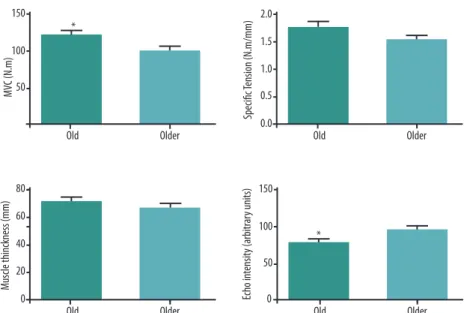
The MIVC was significantly greater (p<0.05) in the Old group (120.3±24.8 N/m) when compared to the Older group (99.2±20.9 N/m). However, when the value was normalized by ST, no signiicant diference (p≥0.05) was observed between the Old and Older group. A greater QEI value was found (p<0.05) in the Older (97.1±17.3 a.u.) compared to the Old group (80.2±14.9 a.u.). In contrast, the QMT did not difer signiicantly between groups (p≥0.05) (Figure 1).
he results of 30SS are presented in Figure 2. he Old group performed a signiicantly more (p<0.05) repetitions (14.1±2.3 repetitions) in the 30SS test than the older group (11.2±1.9 repetitions).
Table 1. Physical characteristics of the participants.
Group Old Older
Age (years) 64.6±0.49* 76.3±0.88
Body mass (kg) 70.15±2.49* 64.83±2.17
Height (m) 1.57±0.13* 1.54±0.23
BMI (Kg/m2) 28.3±1.60* 27.2±0.94
%BF (%) 33±9* 23.2±10.9
Muscle quality of active older women Lopez et al.
Figure 1. Between group comparison of maximal isometric voluntary contraction (MIVC), speciic tension (ST), quadriceps muscle thickness, and quadriceps echo intensity; Old group: aged between 60 and 66 years; Older group: aged between 73 and 83 years; *: Signiicant diferent from Older group (p<0.05).
Figure 2. Total number of repetitions performed in the 30-sit-to-stand test; Old group: aged between 60 and 66 years; Older group: aged between 73 and 83 years; *:Signiicant diferent from Older group (p<0.05).
DISCUSSION
he aim of this study was to compare the maximal strength, muscle mass, muscle quality, and functional performance obtained in two diferent age groups of active elderly women. We observed that although muscle mass has been similar in both groups, the Old group showed better performance in the 30SS compared with the Older group. Besides that, the QEI was higher in the Older, suggesting that active women in the eighth decade of life may present impairments in QEI and functional performance when compared to active women at seventh decade of life, even though they presented similar MT. Furthermore, our results suggest that muscle composition may be more important to muscle functional performance than muscle size in active elderly women.
between, men aged from 60 to 73 years (39.8±6.5 N.m), and older men aged between 76 and 85 years (40.9±6.4 N.m)24. he diferent gender and muscle
group assessed may have contributed to these discrepant results. However, in our study no signiicant diferences were observed when the strength was normalized by muscle thickness. hat was an unexpected result since ST has been shown be higher afected by aging process25. For possible
expla-nation, this age diference may not present changes enough for signiicant alterations in force integrity per muscle thickness between groups.
Regarding muscle quantity, our results did not show signiicant group dif-ferences in QMT. Likewise, Arts et al.16 showed in regression analysis a small
age-related decrease in RF MT and VI MT of women compared to men16. he
later had thicker muscles but presented a larger age-related decline in muscle mass, and strength. An interesting result was the slower decline in age-related muscle thickness in elderly women, suggesting gender diferences in muscle quantity16. hese results indicate that the QMT may sufer little deleterious
efects from sixth to ninth decade of life on women16. However, the MT may
not be representative by exclusively contractile tissue6, and hence careful
should be taken when the muscle size is used to compare diferent populations. Previous investigations reported signiicant associations between MT of knee extensors and maximal knee extension strength6,8-10. Signiicant
diferences between groups were observed regarding muscle strength, it was expected in the current study that the older group would exhibit a lower QMT when compared to the old group. However, both groups showed similar muscle thickness. hese diferences between Arts et al.16 results
and ours may be due to two factors: (1) diferent to Arts et al.16, in which
QMT was measured by the sum of RF and VI16, whereas in the current
study the sum of four quadriceps femoris portions was utilized to deine QMT, what is believed to better represent the knee extensor muscles; and (2) Arts et al.16 study had 6 participants in each age groups, whereas at the
present investigation counted with 12 and 15 participants in age groups. hese diferences and results may suggest that some neural component or changes in intramuscular composition may be more important for deter-mination of the strength capacity than muscle size in active elderly women.
Some previous studies have reported that EI is a predictor of strength capacity6-8,26 and functionality9,10 in elderly population. he inverse
re-lationship between QEI and functional capacity observed in previous studies9,10,17 suggest that smaller EI values may indicate reduced deposition
of non-contractile material on skeletal muscles and, thus, increase the strength capacity for the same muscle volume. In agreement with these indings, we found lower QEI and higher number of repetitions performed in the 30SS test in the Old group. Likewise, Wilhelm et al.10 found a
Muscle quality of active older women Lopez et al.
with the suggestion that non-contractile muscle composition (in this study estimated by QEI) may be more representative of the muscle functionality than muscle size9,10.
It has been reported that muscle size and muscle quality are deter-minant for muscle strength6. Fukumoto et al.6 reported that MT and EI
act independently on isometric knee extension in women of diferent ages (51-87 years). hese results may point out that both muscle quantity and muscle quality contribute to muscle strength. Diferently, our results did not show signiicant diferences in QMT between groups, however a signiicantly higher QEI and lower MIVC were found in the Older group. As a possible explanation, Fukumoto et al.6 suggested that substitutions
of contractile tissue per adipose and ibrous tissue are larger than muscle size decrease and this could inluence QMT values. Moreover, the cur-rent indings suggest that for elderly women in sixth and seventh decade of life the quality of muscles may be more relevant to strength capacity than muscle size. Similarities may be found when the ST is carried out by muscle thickness.
In agreement with previous studies6,16, we found that the QEI was
greater in the Older group when compared with Old group, following a signiicant lower number of repetitions in the 30SS test. In recent studies from our research group9,10 it was observed that the muscle quality
(esti-mated by QEI) may be more representative of the muscle functionality than muscle size9,10. In current study, Older group performed about ~20% less
repetitions on the 30SS compared to Old group. his inding corroborates with Rech et al.9 that found a signiicant negative correlation (r=-0.493;
p<0.01) between QEI and the number of repetitions performed on 30SS test. In additional, they did not found association between maximal isometric knee extension and functional tests (30SS and usual gait speed) (r=0.247, p>0.05), suggesting that EI may be a relevant predictor of functional capac-ity more than maximal strength9,10. However, more studies are necessary
to investigate if QEI is more representative of functionality than muscle strength in both elderly men and women.
CONCLUSION
Cientíico e Tecnológico) and CAPES (Coordenação de Aperfeiçoamento de Pessoal de Nível Superior) for their inancial support.
REFERENCES
1. Metter EJ, Conwit R, Tobin J, Fozard JL. Age-associated loss of power and strength in the upper extremities in women and men. J Gerontol A Biol Sci Med Sci 1997;52(5):B267-76.
2. Doherty TJ. Invited review: Aging and sarcopenia. J Appl Physiol 2003;95(4):1717-27. 3. Tzankof SP, Norris AH. Longitudinal changes in basal metabolism in man. J Appl
Physiol Respir Environ Exerc Physiol 1978;45(4):536-9.
4. Evans JG, Bond J. he challenges of age research. Age Ageing 1997;26(Suppl 4):43-6. 5. Candow DG, Chilibeck PD. Efect of creatine supplementation during resistance
training on muscle accretion in the elderly. J Nutr Health Aging 2007;11(2):185-8. 6. Fukumoto Y, Ikezoe T, Yamada Y, Tsukagoshi R, Nakamura M, Mori N, et al.
Skeletal muscle quality assessed from echo intensity is associated with muscle strength of middle-aged and elderly persons. Eur J Appl Physiol 2012;112(4):1519-25. 7. Cadore EL, Izquierdo M, Conceicao M, Radaelli R, Pinto RS, Baroni BM, et al. Echo
intensity is associated with skeletal muscle power and cardiovascular performance in elderly men. Exp Gerontol 2012;47(6):473-8.
8. Watanabe Y, Yamada Y, Fukumoto Y, Ishihara T, Yokoyama K, Yoshida T, et al. Echo intensity obtained from ultrasonography images relecting muscle strength in elderly men. Clin Interv Aging 2013;8(1):993-8.
9. Rech A, Radaelli R, Goltz FR, da Rosa LH, Schneider CD, Pinto RS. Echo intensity is negatively associated with functional capacity in older women. Age (Dordr) 2014;36(5):9708.
10. Wilhelm EN, Rech A, Minozzo F, Radaelli R, Botton CE, Pinto RS. Relationship between quadriceps femoris echo intensity, muscle power, and functional capacity of older men. Age (Dordr) 2014;36(3):9625.
11. Goodpaster BH, Carlson CL, Visser M, Kelley DE, Scherzinger A, Harris TB, et al. Attenuation of skeletal muscle and strength in the elderly: he Health ABC Study. J Appl Physiol 2001;90(6):2157-65.
12. Delmonico MJ, Harris TB, Visser M, Park SW, Conroy MB, Velasquez-Mieyer P, et al. Longitudinal study of muscle strength, quality, and adipose tissue iniltration. Am J Clin Nutr 2009;90(6):1579-85.
13. Clark BC, Fernhall B, Ploutz-Snyder LL. Adaptations in human neuromuscular function following prolonged unweighting: I. Skeletal muscle contractile properties and applied ischemia eicacy. J Appl Physiol 2006;101(1):256-63.
14. Clark BC, Manini TM, Bolanowski SJ, Ploutz-Snyder LL. Adaptations in human neuromuscular function following prolonged unweighting: II. Neurological proper-ties and motor imagery eicacy. J Appl Physiol 2006;101(1):264-72.
15. Pillen S, Tak RO, Zwarts MJ, Lammens MM, Verrijp KN, Arts IM, et al. Skeletal muscle ultrasound: correlation between ibrous tissue and echo intensity. Ultra-sound Med Biol 2009;35(3):443-6.
16. Arts IM, Pillen S, Schelhaas HJ, Overeem S, Zwarts MJ. Normal values for quantita-tive muscle ultrasonography in adults. Muscle Nerve 2010;41(1):32-41.
17. Nishihara K, Kawai H, Hayashi H, Naruse H, Kimura A, Gomi T, Hoshi F. Frequency analysis of ultrasonic echo intensities of the skeletal muscle in elderly and young individuals. Clin Interv Aging 2014;9(1):1471-8.
In-Muscle quality of active older women Lopez et al.
Corresponding author
Pedro Lopez da Cruz Exercise Research Laboratory (LAPEX), Federal University of Rio Grande do Sul (UFRGS), Rua Felizardo, 750 – Bairro Jardim Botânico, CEP: 90690-200 Porto Alegre, RS, Brazil. Email: [email protected]
19. Craig CL, Marshall AL, Sjostrom M, Bauman AE, Booth ML, Ainsworth BE, et al. International physical activity questionnaire: 12-country reliability and validity. Med Sci Sports Exerc 2003;35(8):1381-95.
20. Berg HE, Tedner B, Tesch PA. Changes in lower limb muscle cross-sectional area and tissue luid volume ater transition from standing to supine. Acta Physiol Scand 1993;148(4):379-85.
21. Korhonen MT, Mero AA, Alen M, Sipila S, Hakkinen K, Liikavainio T, et al. Bio-mechanical and skeletal muscle determinants of maximum running speed with aging. Med Sci Sports Exerc 2009;41(4):844–56.
22. Pinto RS, Correa CS, Radaelli R, Cadore EL, Brown LE, Bottaro M. Short-term strength training improves muscle quality and functional capacity of elderly women. Age (Dordr) 2014;36(1):365-72.
23. Jones CJ, Rikli RE, Beam WC. A 30-s chair-stand test as a measure of lower body strength in community-residing older adults. Res Q Exerc Sport 1999;70(2):113-9. 24. Power GA, Allen MD, Booth WJ, hompson RT, Marsh GD, Rice CL. he inluence
on sarcopenia of muscle quality and quantity derived from magnetic resonance imaging and neuromuscular properties. Age (Dordr) 2014; 36(3):9642.
25. Tracy BL, Ivey FM, Hurlbut D, Martel GF, Lemmer JT, Siegel EL, et al. Muscle quality. II. Efects of strength training in 65-to 75-yr-old men and women. J Appl Physiol 2009; 86(1):195-201.