TERMINAL ILEUM SUBMUCOUS PLEXUS
Study of the VIP-ergic neurons of
diabetic rats treated with ascorbic acid
Jacqueline Nelisis Zanoni
1, Luzmarina Hernandes
1,
Roberto Barbosa Bazotte
2, Marcílio Hubner de Miranda Neto
1ABSTRACT - The aim of this study was to evaluate the effect of the ascorbic acid (AA) supplementation on the neurons that produce the vasoactive intestinal peptide (VIP) in the submucous plexus of the ileum of rat, four months after the induction of experimental diabetes mellitus with streptozotocin. Three groups of rats were used: C - control, D - diabetic, DA - diabetic receiving AA. We have measured the immunoreactivity and area of 80 cellular bodies of VIP-ergic neurons from each studied group. In the diabetic animals, we have observed hyperphagia, polydipsia, and an increase of glycemia and glycated hemoglobin. The VIP-ergic neurons have presented an increase of their immunoreactivity and the highest profiles when compared to the other groups. In the diabetic animals supplemented with AA it has been observed a small reduction in the glycemia and the water and food intake. We have also noticed smaller immunoreactivity in their VIP-ergic neurons, similar to what we have observed in the control group animals (group C).
KEY WORDS: ascorbic acid, diabetes mellitus, streptozotocin, ileum, vasoactive intestinal peptide, submucous plexus, rats.
Plexo submucoso de íleo terminal: estudo dos neurônios VIP Plexo submucoso de íleo terminal: estudo dos neurônios VIP Plexo submucoso de íleo terminal: estudo dos neurônios VIP Plexo submucoso de íleo terminal: estudo dos neurônios VIP
Plexo submucoso de íleo terminal: estudo dos neurônios VIP-érgicos de ratos diabéticos tratados com-érgicos de ratos diabéticos tratados com-érgicos de ratos diabéticos tratados com-érgicos de ratos diabéticos tratados com-érgicos de ratos diabéticos tratados com ácido ascórbico
ácido ascórbico ácido ascórbico ácido ascórbico ácido ascórbico
RESUMO - O objetivo deste estudo foi avaliar o efeito da suplementação com ácido ascórbico (AA) sobre os neurônios que expressam o peptídeo intestinal vasoativo (VIP) no plexo submucoso do íleo de ratos, quatro meses após a indução do diabetes mellitus experimental com estreptozootocina. Três grupos de ratos foram usados: C- controles, D- diabéticos, DA- diabéticos recebendo AA. Foram avaliadas a imunoreatividade e a área de 80 corpos celulares de neurônios VIP-érgicos de cada grupo estudado. Nos animais diabéticos ocorreram hirperfagia, polidipsia, elevação da glicemia e hemoglobina glicada. Os neurônios VIP-érgicos apresentaram aumento da imunorreatividade e os maiores perfis, quando comparados aos demais grupos. Nos animais diabéticos suplementados com AA observou-se pequena redução na glicemia, ingesta de água e de alimento, verificando-se também menor imunorreatividade nos neurônios VIP-érgicos, o que foi semelhante ao observado nos animais do grupo controle (grupo C).
PALAVRAS-CHAVE: acido ascórbico, diabetes mellitus, estreptozootocina, íleo, peptídeo intestinal vasoativo, plexo submucoso, ratos.
Universidade Estadual de Maringá, Maringá PR, Brasil: 1Professor, Department of Morphophysiological Sciences; 2Professor, Department
of Pharmacology. This work was supported by funds from CAPES. Received 23 July 2001. Accepted 15 October 2001.
Dra. Jacqueline Nelisis Zanoni –Departamento de Ciências Morfofisiológicas, Universidade Estadual de Maringá – Av. Colombo, 5790 / Bloco H79– 87020-900 Maringá PR, Brasil. FAX: 44 261 4340. E-mail: [email protected]
The neurological manifestations of diabetes mel-litus (DM) occur in the peripheral nervous system and particularly in the enteric nervous system. It is ob-served in the digestive system a dilatation of the sto-mach, small and large intestine1,2. One of its more
relevant clinical problems is the diabetic diarrhea and constipation3. The etiology of these disturbs are not
completely known. However, degenerative changes in the enteric nervous system are related to the
de-velopment of the diabetic neuropathy. In a more accurate assessment of the DM effects on the ente-ric nerves, we have observed in previous studies, de-veloped in our laboratories, a reduction of the num-ber of myenteric neurons in several intestinal seg-ments2,4. This pathology also promotes changes in
the contents of the enteric neuropeptides such as the vasoactive intestinal peptide5 (VIP), whose
dia-betic rat6. It is believed that changes in the VIP-ergic
neurons, which represent 50% of the overall neu-rons present in the submucous plexus7, may imply
in gastrointestinal disorders6,8 such as the diabetic
diarrhea syndrom9.
Several studies have considered some factors as the responsible for the appearance of degenerative changes. Among the possible causes are: a) periphe-ral nerves lesions, due microcirculation changes (vasa nervorum); b) oxidative stress, which is intensified in DM; c) increased in the sorbitol level10,11. The
oxi-dative stress produce free radicals, which appear due to the increase of non-enzymatic glycation, increase of self-oxidation and increase of metabolic stress10.
Free radicals are usually neutralized by antioxidants agents such as the ascorbic acid (AA)12. The
concen-tration of AA in diabetic people is reduced, since is used to neutralize the free radicals and also because its transportation is inhibited due to the appearance of hyperglycemia as well as its renal absortion13.
The oxidative stress may also result from changes in the sorbitol formation stages. Sorbitol is produced by the glucose reduction, in the reaction catalyzed by the aldose reductase enzyme11. The increase of
the sorbitol level promotes an increase of the intra-cellular osmolarity, with formation of edema, neu-ronal lesions and a consequent reduction of nerve conduction velocity14. Drugs that improve the
con-trol of the oxidative stress and decrease the sorbitol presence through the inhibition of the aldose reduc-tase enzyme may have a relevant role in the treat-ment of diabetes neurological problems. The AA is one of these substances and it has been studied in the treatment of this pathology. The supplementa-tion with AA shows little effects on the blood glyce-mia concentration15,16, but it reduces the capillarity
fra-gility and also the cellular sorbitol concentration12,17,18,
suggesting a neural protector role for this substance. In this paper we have studied the neural protec-tor effect of the ascorbic acid on the VIP-ergic en-teric neurons of the submucous plexus in the ileum of diabetic rat.
METHODS
Animal procedures
Male Wistar rats (Rattus norvegicus rats) weighing 300-400g, aged around 13 weeks were employed. To induce diabetes, rats were starved for 14 hours and then strepto-zotocin (35 mg/Kg b.w., Sigma, USA) was injected i.v.. Non-diabetic rats were employed as control group. Streptozo-tocin injection resulted in a diabetic syndrome with rapid weight loss, polyuria and glycosuria. Rats were divided in 3 groups: ascorbic acid-treated diabetes (DA group), un-treated diabetes (D group) and unun-treated control (C group).
Ascorbic acid was given for 16 weeks from the onset of the diabetes by adding ascorbic acid (Sigma, USA) to drink-ing water (1 g/L prepared fresh each day)16. The animals
were kept in individual metabolic cages in a room with a maintained photoperiod (6:00 a.m. – 6:00 p.m.) and room temperature (RT) (24º ± 2ºC). Water was given ad libitum and Nuvital® lab chow served as the diet.
Thus, 16 weeks after streptozotocin (D and DA groups) or control group, the rats were anesthetized intraperito-neally with thiopental (40 mg/kg-body wt.). Blood was collected by cardiac puncture for the measurement of glycated hemoglobin19, glucose20 and ascorbic acid21 level.
The rats were observed during 4 months. Water consump-tion, food intake and urine elimination were monitored.
Immunohistochemistry and morphological analysis After abdominal incision the ileal segments were col-lected, rinsed in 0.01M phosphate buffer saline (PBS), pH 7.4, and fixed in Zamboni’s liquid for 18 hours22 at 4º C,
the segments were processed according to the immuno-histochemistry technique for whole-mount preparation23
in order to detect the presence of VIP in the submucous plexus.
Briefly, segments were opened along the mesenteric border, washed and dehydrated, diaphanized in xilol and rehydrated. Afterwards were place in 0.01M PBS pH 7.4. Samples were reduced with the aid of a circular sectioner and the mucosa and muscle layers were dissected under stereomicroscope. The submucous layer isolated was in-cubated with polyclonal rabbit anti-VIP (Penninsula Labs, USA) overnight at RT at 1:200. The samples were washed in PBS and then incubated in sequence with the second-ary FITC-conjugated antibody (Penninsula Labs, USA) for 1h at 1:100 (RT) under shaking. In control samples, the primary antibody was substituted by goat serum. The whole-mounts were placed in glycerol-coated slides.
The immunofluorescence was analyzed on a trinocu-lar biological optic microscope, 40X lens, equipped with immunofluorescence filters (FITC) and a kit to capture images IPPWIN-DCAM. The images were taken by a high-resolution camera, transmitted to personal computer and then recorded in a compact disc.
The area (µm2) of 80 cellular bodies of immunoreac-tive VIP-ergic neurons (VIP-IR) from each group studied was measured through the image analysis software Im-age-Pro-Plus 3.0.1.
Statistical analysis
The data were analyzed by the minimum squares method, through the variance analyzes and the Tukey’s test to compare the averages.
We have employed the methodology and General Lin-ear Model (GLM)24 and the Student‘s t-test to compare
Table 1. Body weight, in grams/aged 90 days (BW/90) and body weight, in grams/aged 210 days (BW/210), daily water consumption (DWC), daily food intake (DFI), daily urine elimination (DUE) in untreated controls (C), untreated diabetes (D) and ascorbic acid-treated diabetes (DA) groups.
BW 90/ g BW210/g DWC/ml DFI/ g DUE/ ml
C 338.9±6.8 (10)a 466.0±6.2 (10)a 64.9±5.9 (5)a 30.7±0.9 (5)a 3.3±0.4 (5)a
D 335.7±6.0 (10)a 318.9±4.6 (10)b 158.6±8.4 (5)b 46.9±3.3 (5)b 59.3±7.1 (5)b
DA 339.4±8.8 (10)a 315.6±2.9(10)b 133.6±5.4 (5)c 39.6±3.1 (5)b 72.4±4.0 (5)b
Means followed by different letters in the same column are different by Tukey test (p < 0.05). All results were expressed as mean ± SE. (n) = number of rats.
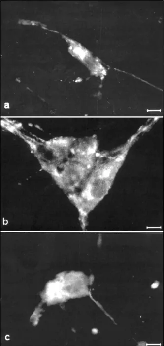
Fig 1. Immunofluorescence micrographs showing VIP immu-noreactivity in the submucous plexus of ileum from untreated controls (a), untreated diabetes (b) and ascorbic acid-treated diabetes (c) groups. Calibration bars = 10 µm.
RESULTS
Streptozotocin promoted a diabetic syndrome, since hyperphagia, polydipsia, polyuria and loss of body weight were observed (Table 1). However, the results for D and DA groups were similar.
Diabetes control and plasmatic ascorbic acid concentration level
The acid ascorbic supplementation reduced the glucose blood level in rats from group DA (p < 0.05). However, we have not noticed differences in the gly-cated hemoglobin level among the groups of dia-betic rats. (Table 2)
The supplementation increased the plasmatic ascorbic acid level in the DA group (p < 0.05) when compared to group D (Table 2).
Immunohistochemistry and morphological analyses
VIP-IR cell bodies were observed in the submu-cous plexus of the ileum of rats in the control group (Fig. 1a). We have showed an increase in the fluo-rescence intensity in nervous fibers and cellular bod-ies of the submucous plexus of the diabetic rats (Fig. 1b). There was no increase in the fluorescence in-tensity of the VIP-IR cell bodies (Fig. 1c) in the rats treated with ascorbic acid (DA group).
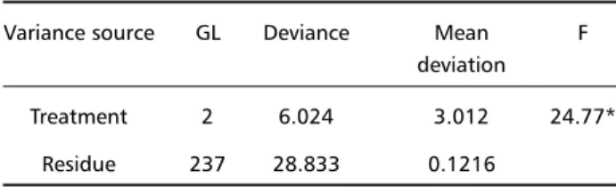
Deviance analyses showed that the VIP-IR cell bodies area average was different in the groups stud-ied (Table 3). The measured areas ranged from 233.7 µm2 (C group) to 1612.8 µm2 (D group).
DISCUSSION
Our studies have shown that the diabetes melli-tus induced by streptozotocin caused changes in the area and in the immunoreactivity of VIP-ergic neu-rons from the submucous plexus of the ileum of rats. Several studies have shown that the diabetes present a differentiated effect, depending on the intestinal region and on the kind of neurotransmitter conside-red. The most evident changes were showed on the VIP neurotransmitters, noradrenaline and serotonin, with no recorded changes for the substance P6,25,26,27.
We have been able to verify that rats with a 16-week-old-diabetes showed an increase in the cell body area of VIP-ergic neurons in the submucous plexus when compared to the control animals. This increase may be related to the synthesis of neuropep-tides in the cell since we have observed an increase in the immunoreactivity of neurons cell bodies in these animals. The immunoreactivity of neurons in
the DA group was similar to that in the control group, showing that the VIP levels in the cells of DA group increased proportionally to the increase of the cellu-lar profile. There was an intensification of the VIP levels in the D group.
Belai & Burnstock26 also observed the increase of
VIP-IR in the submucous plexus of 16-week-old dia-betic rats. Biochemical evaluation, besides the mor-phological evaluation, proved that the VIP levels in-crease in the presence of diabetes5,6. This supports
the relation between the levels of this neurotrans-mitter and the increase of immunoreactivity observed in the light microscopy. It has also been suggested that the increase on the area of cellular body of VIP-ergic submucous neurons may be related to the re-duction on the number of neurons in the myenteric submucous since that, although the enteric plexus are spatially separated, the connection between them suggest they form an integrative unit28. See et al.29
observed an increase on the cellular body volume of submucous VIP-ergic neurons after denervation my-enteric. The authors speculate that in normal condi-tions the myenteric plexus would have an indirect inhibitory action over the submucous plexus to which it is connected. The removal of the inhibitory im-pulse for the submucous neurons could result in a VIP production increase, leading to an increase in the area of the submucous VIP-ergic neurons.
It has been verified, through quantitative analy-ses carried out with the material from the animals employed in this experiment, a reduction in the num-ber of neurons of the myenteric plexus in groups D and DA through the Myosin-V technique (personal communication). This has also been observed previ-ously in the small2 and large4 intestine of rats.
Tak-ing in consideration the results presented by See et al.29, we can infer that the loss of myenteric neurons
itself could already lead to a functional overcharge in the submucous neurons with consequences to their morphology. It may also be possible there was a specific loss of submucous VIP-ergic neurons due to the diabetes; this way, the remaining submucous and myenteric neurons would have to increase their activity to compensate for the lost neurons. This in-crease in their activity would be responsible for an increase in the synthesis processes, thus, the intrac-ellular machinery would become more and more developed making the cell gain volume. Another factor to be considered (and that could also contri-bute to the increase of VIP synthesis by the submu-cous neurons) is the increase of food intake observed in diabetic animals since the liberation of this neu-rotransmitter may be promoted by the mucosa me-chanic stimulation7.
Table 2. Glycemia (GYL), glycated hemoglobin (GHb) and plasma ascorbic acid (AA) in untreated controls (C), untreated diabetes (D) and ascorbic acid-treated diabetes (DA) groups.
GYL/mg.dl-1 GHb/% AA/µg.ml-1
C 144.4 ± 6.7a 4.1 ± 0.3a 26.7 ± 2.7a
D 506.0 ± 8.0b 8.1 ± 0.2b 19.1 ± 3.5ab
DA 452.3 ± 10.6c 7.9 ± 0.5b 32.4 ± 1.8ac
Means followed by different letters in the same column are different by Tukey test (p < 0.05). All results are expressed as mean ± SE. n= 10 rats, for group.
Table 3. Deviance analysis of means from VIP-IR neurons cell bod-ies area in the three studied groups.
Variance source GL Deviance Mean F deviation
Treatment 2 6.024 3.012 24.77*
Residue 237 28.833 0.1216
*Significant to the level of 5 % of probability.
Table 4. Means and standard errors of VIP-IR neurons cell bodies areas in untreated controls (C), untreated diabetes (D) and ascor-bic acid-treated diabetes (DA) groups.
Groups Means ± SE C 551.8 ± 20.4a
D 858.1 ± 33.3b
DA 791.6 ± 30.2b
The increase in the neuronal profile area in dia-betic rats could also be related to the intracellular edema provoked by the sorbitol accrual14. However,
the edema contribution is not the primordial factor and we should not bestow it a higher importance than other factors: these cells presented in the DA group immunoreactivity levels similar to the C group while in the D group the higher immunoreactivity is a sign of intense synthesis activity and/or neurotrans-mitter accrual in the cellular body. If the primary cause of the cellular body increase of these neurons was the edema, the cytoplasmic components would be more scattered and their immunoreactivity would be less than the observed in the control animals.
It has been shown that the diabetes leads to a reduction of the circulatory levels of AA13. As the AA
is a co-factor for the noradrenaline synthesis30 it is
possible that this neurotransmitter may not be syn-thesized with the same efficiency. On the other hand, the peripheral neuropathy related to the sorbitol accrual would compromise the integrity of the sym-pathetic neurons cell bodies and nervous fibers. This would imply in a sympathetic nervous system inhibi-tory action deficit in the submucous collaborating to overcharge the VIP-ergic neurons. This hypoth-esis would explain why although the DA group neu-rons have presented an increase in their cell pro-files, they have expressed a lower immunoreactivity for VIP than the rats’ neurons from D group. This suggested us that, in the DA group, the action of the sympathetic neurons may be more efficient, as much for the higher capacity of noradrenaline syn-thesis as for the better morphological conditions of the sympathetic nervous fibers that connect with the VIP-ergic submucous neurons. Since the AA is an inhibitory substance of the aldose redutase enzyme12, 17,18, which contributes to a decrease in the sorbitol
production, the main agent of peripheral neuropa-thies in diabetic people.
Our result with the use of AA was similar to the one where ponalrestat, an aldose reductase inhibi-tor, was given to rats with chronic diabetes31. This
drug, similarly to AA, prevented the increase of im-munoreactivity of VIP-ergic neurons31. The plasmatic
concentration of ascorbic acid was reduced 27.9% in the animals from the diabetic group when com-pared to the animals from the control group. This reduction has been related to a higher exposition of the diabetic animals to oxidative stress16, higher
urine excretion and to the hyperglycemia, which inhibits the AA transportation to the interior of some cells12.
In human beings suffering from diabetes melli-tus, the reduction of blood AA is of about 30%12.
Per-haps, this reduction in rats happens to be smaller, for unlike human beings, these animals are able to synthesize AA12. The supplementation with AA in the
diabetic animals (DA group) raised the plasmatic level of this substance in 41% when compared to the dia-betic animals that did not receive this supplementa-tion (D group) and in 17.6% when compared to the C group. There were also changes in the glycemic level and in the glycated hemoglobin in the diabetic animals receiving the supplementation, respectively 10.6% and 2.5% less than in the non-supplemented diabetic animals.
Summing up, we have verified the following in the non-supplemented diabetic animals: a great in-crease in the water and food intake; a great rise in the glycemia and glycated hemoglobin; an increase in the immunoreactivity and larger cell profile areas of the VIP-ergic submucous neurons when compared to the other groups. On the other hand, when com-paring the diabetic animals supplemented with AA to the non-supplemented diabetic animals we have observed a smaller reduction in the glycemia, water and food intake and also a smaller immunoreactiv-ity in the VIP-ergic neurons, which was similar to the observed in the animals of the control group (C group).
Acknowedgements - Acknowedgements - Acknowedgements - Acknowedgements -
Acknowedgements - We specially thank Sérgio Piva and Elisabeth Eyko Aoki for their excellent technical assis-tance.
REFERENCES
1. Diani AR, Grogan DM, Yates ME, Risinger DL, Gerritsen GC. Radio-logic abnormalities and autonomic neuropathology of the digestive tract of the Ketonuric Diabetic Chinese Hamster. Diabetologia 1979; 17:33-40.
2. Zanoni JN, Miranda-Neto MH, Bazotte RB, Souza RR. Morphological and quantitative analysis of the neurons of the myenteric plexus of the cecum of streptozotocin diabetic rats. Arq Neuropsquiatr 1997;55:696-702. 3. Clements RS JR, Bell DSH. Diabetic neuropathy: peripheral and
auto-nomic syndromes. Diabetic Neuropathy 1982;71:50-67.
4. Hernandes L, Bazotte RB, Gama P, Miranda-Neto MH. Streptozotocin– induced diabetes duration is important to determine changes in the number and basophily of myenteric neurons. Arq Neuropsiquiatr 2000;58:1035-1039.
5. Ballmann M, Conlon JM. Changes in the somatostatin, substance P and vasoative intestinal polypeptide content of the gastrointestinal tract following streptozotocin-induced diabetes in the rat. Diabetologia 1985;28:355-358.
6. Belai A, Linconl J, Milner P, Crown A, Loesch A, Burnstock G. Enteric nerves in diabetic rats: increase in vasoative intestinal polypeptide but not Substance P. Gastroenterology 1985;89:967-976.
7. Dockray GJ. Physiology of the gastrointestinal tract. 2 Ed. New York: Raven Press, 1994:41-59.
10. Baynes JW. Role of oxidative stress in development of complications in diabetes. Diabetes 1991;40:405-412.
11. Vinson JA, Staretz MA, Bose P, Kassm HM. In vitro and in vivo reduc-tion of erythrocyte sorbitol by ascorbic acid. Diabetes 1989;38:1036-1041. 12. Will JC, Byers T. Does diabetes mellitus increase the requirement for
vitamin C ? Nutr Rev 1996;54:193-202.
13. Cunningam JJ. The glucose/insulin system and vitamin C: implica-tions in insulin-dependent diabetes mellitus. J Am Coll Nutr 1998; 17:105-108.
14. Hosking DJ, Bennett T, Hampton DM. Diabetic autonomic neurophaty. Diabetes 1978;27:1043-1055.
15. Som S, Basu S, Mukherjee D, et al. Ascorbic acid metabolism in diabe-tes mellitus. Metabolism 1981;30:572-577.
16. Young IS, Torney JJ, Trimble ER. The effect of ascorbate supplementa-tion on oxidative stress in the streptozotocin diabetic rat. Free Radic Biol Med 1992;13:41-46.
17. Yue DK, Mclennan S, Fisher E, et al. Ascorbic acid metabolism and polyol pathway in diabetes. Diabetes 1989;38:257-261.
18. Cunningham JJ, Mearkle PL, Brown G. Vitamin C: an aldose reductase inhibitor that normalizes erythrocyte sorbitol in insulin-dependent dia-betes mellitus. J Am Col Nut 1994;13:344-350.
19. Koenig RJ, Peterson CM, Jones RL, Saudek C, Lehrman M, Cerami A. Correlation of glucose regulation and hemoglobin Alc in diabetes mel-litus. N Engl J Med 1976;295:417-420.
20. Bergmeyer HU, Bernet E. Determination of glucose with glucose-oxi-dase and peroxiglucose-oxi-dase. Methods of enzymatic analysis. New York: Verlag Chemie-Academic Press, 1974.
21. Henry RJ, Cannon DC, Winkilman JW. Química cliníca: bases e técnicas. Editorial JIMS, 1980.
22. Stefanini M, Demartino C, Zamboni C. Fixation of ejaculated sperma-tozoa for electron microscopy. Nature 1967;216:173-174.
23. Costa M, Buffa R, Furness JB, Solcia E. Immunohistochemical localiza-tion of polypeptides in pheripheral autonomic nerves using whole mount preparations. Histochemistry 1980;65:157-165.
24. Nelder J, Weddenburn RWM. Generalized linear models. London: Chapman and Hall, 1989.
25. Belai A, Linconl P, Milner P, Burnstock G. Progressive changes in adr-energic, serotonergic and peptidergic nerves in proximal colon of streptozotocin-diabetic rats. Gastroenterology 1988;95:1234-1241. 26. Belai A, Burnstock G. Changes in adrenergic and peptidergic nerves in
the submucous plexus of streptozotocin-diabetic rat ileum. Gastroen-terology 1990;98:1427-1436.
27. Belai A, Linconl J, Milner P, Burnstock G. Differential effect of strep-tozotocin-induced diabetes on the innervation of the ileum and distal colon. Gastroenterology 1991;100:1024-1032.
28. Furness J.B, Costa AM. The enteric nervous system. New York: Churchil Livingstone, 1987.
29. See NA, Epstein ML, Dahl JL, Bass P. The myenteric plexus regulates cell growth in rat jejunum. J Auton Nerv Syst 1990;31:219-229. 30. Levine M. New concepts in the biology and biochemistry of ascorbic
acid. N Engl J Med 1986;314:892-902.