EX
PE
R
IM
EN
TA
L
G
A
ST
R
O
EN
TE
R
O
LO
G
Y
INTRODUCTION
Aging has been defined as a series of changes taking place during life, which reduces the capability of an organism survival. It is associated to metabolic, neuroendocrine, genetic and immunological changes that may contribute to the process of cellular death by
apoptosis or necrosis(2)
The nature of the aging process has been reason to considerable speculation such as: the aging of DNA codification, progressive deterioration in the synthesis of proteins and also of other macro-molecules, attacks
to the immune system and the action of free radicals(15).
Free radicals are molecules that have an extra unpaired electron, which does not have a pair in its external orbit
and usually derives from oxygen. They originate in the
mitochondria, through energy production from glucose
and O2 and are immediately neutralized by the enzymes
within the mitochondria(13, 18). Free radicals play a role
in the aging process since they harm directly, constantly
cells and tissues and also have a cumulative action(20).
The consequence of the free radicals action is called cellular oxidation, and is considered the initial stage of
several diseases. This process can only be neutralized
by the presence of vitamins considered antioxidants (12).
Ascorbic acid is one of these vitamins used to fight off free radicals. In most species, the hepatic metabolism of glucose includes the ascorbic acid synthesis. Human beings, however, do not synthesize it due to the absence of the gulonolactone oxidase enzyme in the metabolic stage. Therefore, it is necessary the daily intake of ascorbic acid
to prevent illnesses such as the scorbutus(14).
Changes in the gastrointestinal neuromuscular func- tions due to aging process have been seen in animal models as well as in human beings, with the evidence of dysphagia and constipation(10). The enteric nervous system (ENS) (responsible
for the control of gastrointestinal functions) is affected by the aging process with a reduction in the number of myenteric neurons in several intestinal segments being observed(8, 16, 19).
Some pathological cases (such as diabetes mellitus) accelerate the aging process, as it can be seen by the alteration in the number and size of enteric neurons(6, 25, 26, 27).
EFFECTS OF ASCORBIC ACID
ON THE VASOACTIVE INTESTINAL
PEPTIDE SYNTHESIS IN THE
ILEUM SUBMUCOUS PLEXUS OF
NORMAL RATS
Jacqueline Nelisis
ZANONI
and Priscila de
FREITAS
ABSTRACT - Background - The aging process is a deteriorating process that attacks the gastrointestinal tract, causing changes in the number and size of neurons from the enteric nervous system. The activity of free radicals on enteric neurons is helped by the significant reduction of antioxidants. Aim - Evaluate the effect of the ascorbic acid supplementation on the neurons that produce the vasoactive intestinal peptide (VIP) in the submucous plexus of the ileum of normal rats for a period of 120 days.
Methods - Fifteen rats were divided in three groups: untreated control with 90 days, untreated control with 210 days and ascorbic acid-treated rats with 210 days. Ascorbic acid was given for 16 weeks from the 90th day of age by adding it to drinking water (1
g/L prepared fresh each day). The ileums were processed according to the immunohistochemistry technique for whole-mount preparation in order to detect the presence of VIP immunoreactive in the cellular bodies and nervous fibers in the neurons of the submucous plexus. We have verified their immunoreactivity and measured the cellular profile of 80 cellular bodies of VIP-ergic neurons from each studied group. Results - The ascorbic acid supplementation did not alter physiological parameters such as water intake and food consumption of the three studied groups. We observed a significant increase of the cellular profile of VIP-ergic neurons in untreated control with 210 days when compared to untreated control with 90 days. The cellular profile of VIP-ergic neurons in ascorbic acid-treated rats with 210 days was bigger than those observed in others groups. Conclusion - The ascorbic acid had a neurotrophic effect on VIP-ergic neurons on the ileum after period 120 days of supplementation. HEADINGS – Ascorbic acid. Vasoactive intestinal peptide. Ileum. Submucous plexus. Rats.
Department of Morphophysiological Sciences, State University of Maringá, Maringá, PR, Brazil.
The majority of researches are carried out with myenteric neurons and few are those with submucous neurons. There are two large groups of submucous neurons: - cholinergic neurons (55%) and those containing the vasoactive intestinal peptide
(VIP) and dynorphin, the non-cholinergic neurons (45%)(7).
Our objective was to study the VIP-ergic submucous neurons of the ileum of normal rats with and without the ascorbic acid supplementation for a period of 120 days.
MATERIAL AND METHODS
The experimental protocols used in this study are in accordance with the ethical principles in animal research prescribed by the Brazilian College of Animal Experimentation (COBEA).
Fifteen male Wistar rats (Rattus norvegicus), weighing
about 300 g, were used. The rats were divided in three groups: untreated control with 90 days (C), untreated control with 210 days (C2) and ascorbic acid-treated rats with 210 days (CA). Ascorbic acid was given for 16 weeks from the 90th day of
age by adding it to drinking water (1 g/L prepared fresh each
day)(24). The rats were kept in individual metabolic cages in a
room with a maintained photoperiod (6:00 a.m.–6:00 p.m.) and at room temperature (RT) (24º ± 2ºC) with water and food
(Nuvital lab chow) ad libitum.
On the sacrifice day, all rats were anesthetized intraperitoneally with thiopental (40 mg/kg-body wt). Blood from groups C2 and CA was collected by cardiac puncture to measure the blood levels
of ascorbic acid(11). Rats from groups C2 e CA were monitored
during the four months of the experiment.
Immunohistochemistry and morphological analysis After an abdominal incision, the ileum segments were collected, rinsed in 0.01M phosphate buffer saline (PBS), pH 7.4, and fixed
in Zamboni’s liquid for 18 hours(23) at 4º C. The segments were
processed according to the immunohistochemistry technique for
whole-mount preparation(4) in order to detect the presence of VIP
immunoreactive (VIP-IR) in the submucous plexus.
Soon after, the segments were opened along the mesenteric border, washed and dehydrated, diaphanized in xylene and rehydrated. Afterwards, they were placed in 0.01 M PBS pH 7.4. Samples were reduced with the aid of a circular sectioner and the mucosa and muscle layers were dissected under stereomicroscope. The isolated submucous layer was incubated with polyclonal rabbit anti-VIP (Penninsula Labs, USA) overnight at RT at 1:200 under shaking. The samples were washed in PBS and then incubated in sequence with the secondary FITC-conjugated antibody (Penninsula Labs, USA) for 1 h at 1:100 (RT) under shaking. In the control samples, the primary antibody was substituted by goat serum. The whole-mounts were placed in glycerol-coated slides.
The immunofluorescence was analyzed on a trinocular bio- logical optic microscope, 40X lens, equipped with immunofluores-cence filters (FITC) and a kit to capture images IPPWIN-DCAM. The images were taken by a high-resolution camera, transmitted to a personal computer and then recorded in a compact disc.
The area (µm2) of 80 cellular bodies of immunoreactive
VIP-ergic neurons (VIP-IR) from each studied group was
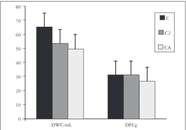
FIGURE 1 - Daily water consumption/mL (DWC), daily food intake/g (DFI) of animals from: untreated control with 90 days (C), untreated control with 210 days (C2) and ascorbic acid-treated rats (CA). All groups, when compared, are not significantly different (P >0.05). (n) = 5 rats per group.
80
70
60
50
40
30
20
10
0
DWC/mL DFI/g
C
C2
CA
measured through the image analysis software
Image-Pro-Plus 3.0.1. Neurons were classified in intervals of 100 µm2
classes and the percentage of each group was calculated. The
values obtained were analyzed by Student’s t test (PRISMA
software). Significance level was set at P < 0.05. Data are reported as means (M) ± standard error (SE) for the indicated number of observations (n).
RESULTS
The plasmatic concentration of ascorbic acid in group C2 was 26.7 ± 2.7 and 48.8 ± 3.8 in group CA, a significant difference
(P < 0.05). There were no difference between the water intake
and the food consumption when we compared the three studied
groups (P > 0.05). The results are shown in Figure 1.
Cellular bodies and VIP-IR neurons fibers from the submucous plexus were found in all studied groups (Figure 2). The lowest immunoreactivity was observed in neurons from group C. We also observed that group C presented neurons with the smallest
cell profile, 108 µm2. Group CA presented neurons with the
largest cell profile 1171.9 µm2.
Figure 3 shows the areas of VIP-IR neurons of the three studied groups. The 4-months aging period (group C2) caused an increase of 50.8 % in the area of VIP-IR neurons when compared
to those observed at 90 days of age (group C) (P < 0.001). The
cellular profile of VIP-IR neurons for group CA was 59.2 % and 17 % larger than those found in groups C and C2 respectively (P < 0.001).
Most VIP-IR neurons in group C had cellular body areas
distributed in the following ranges: from 101-400 µm2 (86.2%),
from 401-600 µm2 (12.5%) and larger than 601 µm2 (1.3%) (Figure
4). We found a reduction of 38.8 % in the area of VIP-IR neurons
results obtained for group C (Figure 4). An increase of 18.8% and
20% in the VIP-IR neurons areas in the 401–600 µm2 and >601
µm2 ranges respectively, were also found when we compared
group C2 to group C; 62.6% of neurons from group CA had a
cell profile above the 601 µm2 range (Figure 4).
FIGURE 2 - Immunostaining of VIP-positive neurons in the submucous plexus from untreated control with 90 days (A), untreated control with 210 days (B) and ascorbic acid-treated rats (C). Bars calibrations: 10 µm
FIGURE 3 - Means and standard errors of cell body areas from VIP-IR neurons from: untreated control with 90 days (C), untreated control with 210 days (C2) and ascorbic acid-treated rats (CA). All groups, when compared, are significantly different (P <0.001). (n) = 5 rats per group
900
800
700
600
500
400
300
200
100
0
C C2 CA
groups
area
Intervals of 100 µm2
FIGURE 4 - Size distribution of VIP-IR submucous neurons in intervals of 100 µm2. Key: untreated control with 90 days (white bar),
untreated control with 210 days (C2) (gray bar) and ascorbic acid-treated rats (CA) (black bar). n= 5 rats per group 40
35
30
25
20
15
10
5
0
101-200 201-300 301-400 401-500 501-600 601-700 701-800 >801
%
DISCUSSION
Zanoni JN, Freitas P. Efeitos do ácido ascórbico sobre a síntese de peptídio intestinal vasoativo do plexo submucoso do íleo de ratos normais. Arq Gastroenterol 2005;42(4)186-90.
RESUMO – Racional - O envelhecimento é um processo deteriorativo que acomete o trato gastrointestinal, provocando alterações no número e tamanho dos neurônios do sistema nervoso entérico. A ação dos radicais livres nos neurônios entéricos é favorecida pela diminuição significativa de antioxidantes. Objetivo - Avaliar o efeito da suplementação com ácido ascórbico sobre os neurônios submucosos do íleo de ratos normais que produzem o peptídio intestinal vasoativo (VIP) por um período de 120 dias. Métodos - Quinze ratos foram divididos em três grupos: controles com 90 dias, controles com 210 dias e tratados com ácido ascórbico com 210 dias. O ácido ascórbico foi administrado durante 16 semanas a partir de 90 dias de idade pela adição em água (1 g/L/dia). O íleo foi processado para obtenção de preparados totais empregados na realização de técnica imunoistoquímica para detectar a presença de corpos celulares e fibras VIP imunoreativas nos neurônios do plexo submucoso. O perfil celular e a imunoreatividade de 80 corpos celulares de neurônios VIP-érgicos de cada grupo estudado foi verificada. Resultados - A suplementação com ácido ascórbico não alterou parâmetros fisiológicos tais como a água ingerida e alimento consumido nos três grupos estudados. Observou-se aumento significativo do perfil celular dos neurônios VIP-érgicos dos animais controles com 210 dias, quando comparados com os controles com 90 dias. O perfil celular dos neurônios VIP-érgicos no grupo de animais tratados com ácido ascórbico foi maior do que aqueles observados nos grupos controles. Conclusão - O ácido ascórbico teve efeito neurotrófico sobre os neurônios VIP-érgicos do íleo após 120 dias de suplementação.
DESCRITORES – Ácido ascórbico. Peptídio intestinal vasoativo. Íleo. Plexo submucoso. Ratos.
number of submucous VIP-ergic neurons in the aging process. However, we do believe that VIP-ergic neurons are also lost in this condition, similar to what occurs to myenteric neurons(16, 19).
SANTER and BAKER(19) evaluated the neuronal density in the
jejunum, ileum, colon and rectum. Using the technique NADH-diaphorase, they observed a 40%-reduction in the myenteric neurons of the jejunum, colon and rectum of rats aged 24 months, when compared to 6-months-old animals. The loss observed in
the colon was still more accentuated, reaching 60%. GABELLA(8)
observed a reduction of 50% in the number of intestinal neurons due to the aging process, together with structural changes and the reorganization of the remaining nervous cells. SOUZA et
al.(22) using the Giemsa method of observed the loss of 34% of
neurons in all parts of the small intestine of old human beings,
especially of the duodenum (38%). According to GOMES et al(9),
the total number of myenteric neurons of the ganglia decreased 37% in human beings over 60-years of age.
CHA et al.(3) studied the changes that take place in
neurons containing neuropeptides in the cortex of old rats, and noticed a reduction in the number of VIP-IR neurons and also those expressing to the neuropeptide Y. The cell profile increase observed in age-related VIP-ergic neurons might be a compensatory effect caused by the eventual reduction in the number of these neurons. The increase in the cell profile was accompanied by the increase in the immunoreactivity, being this an indicator that the cell increased its synthesis process of the VIP neurotransmitter. Another hypothesis for the increase in the profile of submucous VIP-ergic is that
it might be related to the loss of myenteric neurons taking place during aging process. Although the enteric plexus are spatially separated, the connection between them suggests they form an integrative unit(7). SEE et al.(21) observed an
increase on the cellular body volume of submucous VIP-ergic neurons after myenteric denervation. The authors speculate that in normal conditions the myenteric plexus would have an indirect inhibitory action over the submucous plexus to which it is connected. The removal of the inhibitory impulse for the submucous neurons could result in a VIP production increase, leading to an increase in the area of the cell bodies submucous VIP-ergic neurons.
Our study showed an increase of 17.02% in the area of the submucous VIP-ergic neurons supplemented with ascorbic acid when compared to controls without supplementation at the same age (group C2). It also showed an increase of 59.20% when compared to control at 90 days of age (group C). The ascorbic acid may have had a neurotrophic effect, generating an increase in the cellular profile. This increase may be positive, since it is believed that this neurotransmitter works directly in the intestinal mucosa, causing an increase in the intestinal
secretion(5). As for the absorptive function in the rat intestine,
there is considerable evidence for reduction due to age in the
water, sugar, and amino acids transportation(1).
REFERENCES
1. Amenta F, editor. Aging of the autonomic nervous system. Boca Raton: CRC Press; c1993. 354 p.
2. Camilleri M, Lee JS, Viramontes B, Bharucha AE, Tangalos EG. Insights into the pathophysiology and mechanisms of constipation, irritable bowel syndrome, and diverticulosis in older people. J Am Geriatr Soc 2000; 48:1142-50.
3. Cha CI, Lee YI, Lee EY, Park KH, Baik SH. Age-related changes of VIP, NPY and somatostatin-immunoreactive neurons in the cerebral cortex of aged rats. Brain Res 1997;753:235-44.
4. Costa M, Buffa R, Furness JB, Solcia E. Immunohistochemical localization of polypeptides in peripheral autonomic nerves using whole mount preparations. Histochemistry 1980;65:157-65.
5. Dockray GJ. Physiology of enteric neuropeptides. In: Johnson LR, Alpers DH, Christensen J, Jacobson ED, Walsh JH, editors. Physiology of the gastrointestinal tract. 2nd Edition. New York: Raven Press; 1994. p169-210.
6. Fregonesi CE, Miranda-Neto MH, Molinari SL, Zanoni JN. Quantitative study of the myenteric plexus of the stomach of rats with streptozotocin-induced diabetes. Arq Neuropsiquiatr 2001;59:50-3.
7. Furness JB, Costa AM. The enteric nervous system. Edinburgh/New York: Churchill Livingstone; 1987. 290 p.
8. Gabella G. Fall in the number of myenteric neurons in aging guinea pigs. Gastroenterology 1989;96:1487-93.
9. Gomes O, Souza RR, Liberti EA. A preliminary investigation of the effects of aging on the nerve cell number in the myenteric ganglia of the human colon. Gerontology 1997;43:210-7.
10. Hall KE. Aging and neural control of the GI tract. II. Neural control of the aging gut: can an old dog learn new tricks? Am J Physiol Gastrointest Liver Physiol 2002;283: G827-G32.
11. Henry RJ, Cannon DC, Wilkelman JW. Quimica clinica: principios y tecnicas. Barcelona: JIMS; 1980. 2v.
12. Joseph JA, Denisova NA, Bielinski D, Fisher DR, Shukitt-Hale B. Oxidative stress protection and vulnerability in aging: putative nutritional implications for intervention. Mech Ageing Dev 2000;116:141-53.
13. Kuyvenhoven JP, Meinders AE. Oxidative stress and diabetes mellitus-Pathogenesis of long-term complications. Eur J Inter Med 1999;10:9-19.
14. Levine M. New concepts in the biology and biochemistry of ascorbic acid. N Engl J Med 1986;314:892-902.
15. Majumdar APN, Jaszewski R, Dubick MA. Effect of aging on the gastrointestinal tract and the pancreas. Proc Soc Exp Biol Med 1997;215:134-44.
16. Meciano Filho J, Carvalho VC, Souza RR. Nerve cell loss in the myenteric plexus of the human esophagus in relation to age: a preliminary investigation. Gerontology 1995;41:18-21.
17. O’Donnell E, Lynch MA. Dietary antioxidant supplementation reverses age-related neuronal changes. Neurobiol Aging 1998;19:461-7.
18. Parthiban A, Vijayalingam S, Shanmugasundaram KR, Mohan R. Oxidative stress and the development of diabetic complications-antioxidants and lipid peroxidation in erythrocytes and cell membrane. Cell Biol Int 1995;19:987-93.
19. Santer RM, Baker DM. Enteric neuron numbers and sizes in Auerbach’s plexus in the small and large intestine of adult and aged rats. J Auton Nerv Syst 1988;25:59-67. 20. Schöneich C. Reactive oxygen species and biological aging: a mechanistic approach.
Exp Gerontol 1999;34:19-34.
21. See NA, Epstein ML, Dahl JL, Bass P. The myenteric plexus regulates cell growth in rat jejunum. J Auton Nerv Syst 1990;31:219-29.
22. Souza RR, Morateli HB, Borges N, Liberti EA. Age-induced nerve cell loss in the myenteric plexus of the small intestine in man. Gerontology 1993;39:183-8. 23. Stefanini M, De Martino C, Zamboni L. Fixation of ejaculated spermatozoa for electron
microscopy. Nature 1967;216:173-4.
24. Young IS, Torney JJ, Trimble ER. The effect of ascorbate supplementation on oxidative stress in the streptozotocin diabetic rat. Free Radic Biol Med 1992;13:41-6. 25. Zanoni JN, Miranda-Neto MH, Bazotte RB, Souza RR. Morphological and quantitative
analysis of the neurons of the myenteric plexus of the cecum of streptozotocin-induced diabetics rats. Arq Neuropsiquiatr 1997;55:696-702.
26. Zanoni JN, Hernandes L, Bazotte RB, Miranda-Neto MH. Teminal ileum submucous plexus: study of the diabetes rats treated with ascorbic acid. Arq Neuropsiquiatr 2002;60:32-7.
27. Zanoni JN, Buttow NC, Bazotte RB, Miranda-Neto MH. Evaluation of the population of NADPH-diaphorase-stained and myosin-V myenteric neurons in the ileum of chronically streptozotocin-diabetic rats treated with ascorbic acid. Auton Neurosci 2003;104:32-8.