Inluence of early mobilization on respiratory and
peripheral muscle strength in critically ill patients
Influência da mobilização precoce na força muscular periférica e
respiratória em pacientes críticos
INTRODUCTION
Advances in intensive care and mechanical ventilation (MV) in the past two decades have increased critically ill patient survival. However, some patients require prolonged MV (PMV) and are deconditioned due to respi-ratory insuiciency caused by underlying disease, adverse efects of
medica-tions, and prolonged immobilization.(1-3)
Patients in the intensive care unit (ICU) are often confined to their beds, which results in inactivity, immobility, and severe
osteomyoarticular system dysfunction.(4,5) These changes are
predisposing factors for polyneuropathy and/or myopathy in critically ill patients and lead to a 2- to 5-fold increase in MV duration and time
for ventilatory weaning.(6)
Immobility disproportionately affects respiratory muscles because the mechanical ventilator performs a large amount of the respiratory effort, decreasing the spontaneous ventilation effort. This results in the partial or complete absence of neural activation and muscle Camila Moura Dantas1, Priscila Figueiredo
dos Santos Silva1, Fabio Henrique Tavares de Siqueira2, Rodrigo Marinho Falcão Pinto2, Simone Matias2, Caroline Maciel3, Marcia Correa de Oliveira5 Cláudio Gonçalves de Albuquerque3,4, Flávio Maciel Dias Andrade1,2, Francimar Ferrari Ramos2,5,Eduardo Eriko Tenório França1,5
1. Universidade Católica de Pernambuco – UNICAP - Recife (PE), Brazil.
2. Faculdade Integrada do Recife – FIR - Recife (PE), Brazil.
3. Faculdade Mauricio de Nassau – FMN - Recife (PE), Brazil.
4. Faculdade Pernambucana em Saúde – FPS - Recife (PE), Brazil.
5. Hospital Agamenon Magalhães – HAM - Recife (PE), Brazil.
ABSTRACT
Objective: To evaluate the efects of an early mobilization protocol on respi-ratory and peripheral muscles in criti-cally ill patients.
Methods: A randomized controlled clinical trial was conducted with 59 male and female patients on mechanical ventilation. he patients were divided into a conventional physical therapy group (control group, n=14) that received the sector’s standard physical therapy program and an early mobilization group (n=14) that received a systematic early mobilization protocol. Peripheral muscle strength was assessed with the Medical Research Council score, and respiratory muscle strength (determined by the maximal inspiratory and expiratory
pressures) was measured using a vacuum manometer with a unidirectional valve. Systematic early mobilization was performed on ive levels.
Results: Signiicant increases were observed for values for maximal inspiratory pressure and the Medical Research Council score in the early mobilization group. However, no statistically signiicant improvement was observed for maximal expiratory pressure or MV duration (days), length of stay in the intensive care unit (days), and length of hospital stay (days).
Conclusion: he early mobilization group showed gains in inspiratory and peripheral muscle strength.
Keywords: Breathing exercises; Respiratory muscles; Intensive care units
This study was conducted at the Hospital Agamenon Magalhães - HAM - Recife (PE), Brazil.
Conflict of interest: None. Submitted on August 21, 2011 Accepted on May 2, 2012
Corresponding author:
mechanics, thereby reducing the diaphragm’s
ability to generate strength.(5) This atrophy is more
pronounced in the respiratory muscles than in the
peripheral muscles, even though both are inactive.(2)
Respiratory muscle function impairment contributes to exercise intolerance, dyspnea, and hypercapnia; however, it can be improved with adequate physical training.(7)
Electrophysiological studies have demonstrated diffuse neuromuscular abnormalities in limbs of 50% of ICU patients after 5 to 7 days of MV, and the main clinical symptom is physical deconditioning due to
muscle weakness.(3)
Mobilizing critically ill patients confined in the ICU and positioning them in order to prevent joint contractures is an early rehabilitative mechanism that has significant effects on oxygen transportation, maintenance of muscle strength and joint mobility, and lung function and respiratory system performance. All of these factors can facilitate MV weaning, which reduces the length of ICU and overall hospital stays and improves quality of life
after hospital discharge.(8-11)
Rehabilitating patients with respiratory diseases is a well-established way to alleviate symptoms and
optimize function, regardless of disease stage.(8,12)
However, there are currently no standard protocols to guide physical therapy in critically ill patients. This lack is important because these patients have high rates of morbidity and mortality, impaired respiratory function, consequent reduction of
quality of life, and high health care costs.(13)
The aim of the present study was to assess the effects of a mobilization protocol on the peripheral and respiratory muscles of critically ill patients in order to assist in treatment guideline designs.
METHODS
A randomized controlled clinical trial with conce-aled allocation was conducted in the general ICU of the Hospital Agamenon Magalhães - HAM between February 2009 and February 2011. The study was approved by the HAM Research Ethics Committee under protocol number CAE 0039.0.236.000-08.
Patient recruitment was performed by a researcher who made daily visits to the HAM ICU and selected patients who fit the population profile intended for the study using a checklist. When the researcher identified a patient who met the eligibility criteria,
their legal guardian was informed of the aims of the study and an invitation to participate was extended. All volunteers and/or patient guardians included in the study were informed about the purpose of the project and signed an Informed Consent Form (ICF) according to Resolution 196/96 of the Brazilian Ministry of Health.
The study included male and female individuals on MV who met the following inclusion criteria: adequate cardiovascular reserve, demonstrated by
<50% variation in resting heart rate (HR)(14) and
systolic blood pressure (SBP) <200 mmHg or >90 mmHg; adequate respiratory reserve, demonstrated
by peripheral oxygen saturation (SpO2) >90% with
fraction of inspired oxygen (FiO2) <60%, no signs
of respiratory distress and respiratory rate (RR) <25 breaths per minute; and no physical exercise
program prior to study enrollment.(15)
Patients with signs of intracranial hypertension, inability to walk without assistance before acute illness in the ICU, cognitive impairment before being admitted to the ICU, neuromuscular disease, stroke, body mass index (BMI) >40, unconsolidated fracture, MV for more than 7 days, or postoperative recurrence or cancer therapy in the previous 6 months were excluded from the study.
The volunteers were randomly divided into two groups. Conventional physical therapy group (CPTG) patients received passive mobilization of the four limbs five times a week and active-assisted exercises according to patient improvement and cooperation. The early mobilization group (EMG) patients received a systematic early mobilization protocol, twice a day, every day of the week, as depicted in figure 1.
Patient data collection was initiated after these procedures. Recruited patients’ clinical records, demographic information, clinical history, diagnosis, BMI, blood gas data, and Acute Physiology and Chronic Health Evaluation Classification System II (APACHE II) scores were compiled and compared.
and maximal expiratory pressure (PEmax) at the
time of ICU discharge (TICU) were considered as the
final measurements.(13)
PImax and PEmax were assessed based on residual volume and total lung capacity, respectively, in stable patients who were being weaned from MV
using a vacuum manometer (Comercial Médica®,
São Paulo, Brazil) with a unidirectional valve for
40 seconds.(16)
The early mobilization protocol was interrupted in patients with tachycardia or bradycardia; signs of respiratory distress, as evidenced by accessory muscle use, flaring of nostrils (FN), or an increase
in RR >25 breaths per minute; changes in SpO2 to
<90%; and an increase or decrease in mean arterial pressure (MAP) by 20 mmHg.
The Kolmogorov-Smirnov test was used to verify the normal distribution of the study variables. Fisher’s exact tests and chi-squared tests were used to assess differences between proportions, and independent and paired sample Student’s t tests were used to analyze continuous variables. Correlation analyses were performed using Spearman’s correlation test. All conclusions were based on a 5% level of significance, and Microsoft Office Excel 2007 (Manaus, Brazil) and GraphPad Prism, version 4.0 (La Jolla, USA) were used.
RESULTS
Four hundred and thirty-one patients were admit-ted to the ICU during the study period. Among the-se, 372 patients were ineligible based on the inclusion and exclusion criteria. he 59 remaining patients were randomly divided into the CPTG (n=33) and EMG (n=26). Nineteen and 12 deaths occurred in the CPTG and the EMG after the study protocol was initiated, respectively, resulting in a inal sample of 14 patients in each group.
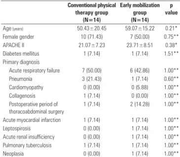
Table 1 shows the overall characteristics of the study sample regarding age, gender, APACHE II score, prevalence of diabetes mellitus, and primary clinical diagnosis. There were no significant differences between the studied groups. None of the patients received neuromuscular blocking agents or corticosteroids following study allocation.
Table 1 - Patient characteristics
Conventional physical therapy group
(N=14)
Early mobilization group (N=14)
p value
Age (years) 50.43±20.45 59.07±15.22 0.21*
Female gender 10 (71.43) 7 (50.00) 0.75**
APACHE II 21.07±7.23 23.71±8.51 0.38*
Diabetes mellitus 1 (7.14) 1 (7.14) 1.51**
Primary diagnosis
Acute respiratory failure 7 (50.00) 6 (42.86) 1.00**
Pneumonia 3 (21.43) 1 (7.14) 0.60**
Cardiomyopathy 0 (0.00) 0 (5.88) 1.00**
Collagenosis 1 (7.14) 0 (0.00) 1.00**
Postoperative period of thoracoabdominal surgery
1 (7.14) 2 (14.28) 1.00**
Acute myocardial infarction 1 (7.14) 1 (7.14) 1.00**
Leptospirosis 0 (0.00) 1 (7.14) 1.00**
Acute renal insufficiency 0 (0.00) 1 (7.14) 1.00**
Pulmonary tuberculosis 1 (7.14) 1 (7.14) 1.00**
Neoplasia 0 (0.00) 1 (7.14) 1.00**
APACHE II - Acute Physiology and Chronic Health Evaluation Classification System II. Age and APACHE II parameters are expressed as the mean ± standard deviation, while the other parameters are shown as absolute values (%). * Student’s t test for independent samples; ** Fisher’s exact test.
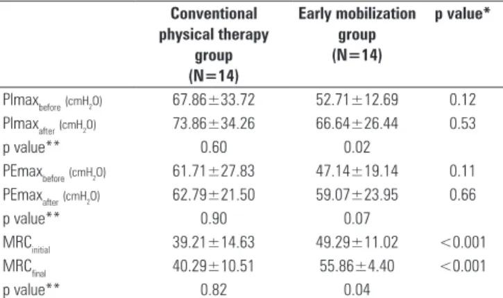
Table 2 shows the PImax, PEmax, and MRC values and variations obtained before and after study protocol implementation. We observed a signiicant increase in PImax after the study period in the EMG (52.71±12.69 before versus 66.64±26.44 after; p=0.02). his phenomenon was not observed for patients in the CPTG (67.86±33.72 before versus 73.86±34.26 after; p=0.60). When analyzing expiratory muscle strength, no signiicant gains were observed for PEmax values in either group. No signiicant increase
Figure 1 – Early mobilization protocol for critically ill patients on mechanical ventilation. ICU - intensive care unit; PS - passive stretching; 4L - four limbs; PM – passive mobilization; PJ - positioning of the joint; UL - upper limbs; AAE – active-assisted exercise; TLtS – transfer from lying to sitting position; MRC – Medical Research Council; ARE – active-resistive exercise; LL - lower limbs; Cycle LL – cycle ergometry for lower limbs; TStC - transfer from sitting to chair; OP – orthostatic posture; CRE - counter-resistance exercise. Source: Adapted from Morris PE, Goad A, Thompson C, Taylor K, Harry B, Passmore L, et al. Early intensive care unit mobility therapy in the treatment of acute respiratory failure. Crit Care Med. 2008;36(8):2238-43.(29)
Stage 1 (Unconscious)
Stage 2 (Conscious)
Stage 3 (Conscious)
Stage 4 (Conscious)
Stage 5 (Conscious)
PS of the 4L
PM of the joints of the
4L (10x)
PJ
Admission into ICU
Discharge from ICU
PS of the 4L
Flexion/extension AAE on the 4L
(10x)
TLtS on the bed for at least 20’
PS of the 4L
ARE on UL (against gravity with help of weight)
TLtS on the edge of the bed
Cycle LL – 3’, 5’, and 10’ with Borg scale between
12 and 13
PS of the 4L
ARE on UL (against gravity and help of
weight)
Cycle LL – 3’, 5’, and 10’ with Borg scale
between 12 and 13
TStC
OP
PS of the 4L
CRE on UL (with help of
weight)
Cycle LL – 3’, 5’, and 10’ with Borg scale
between 12 and 13
Balance training
Walking
Open eyes, direct gaze, protract the tongue
and have grade II muscle strength
in UL
Strength > III in the UL (MRC)
Strength quadriceps ≥ III
in peripheral muscle strength was observed after the study period in the CPTG (39.21±14.63 before versus 40.29±10.51 after; p=0.82); however, a signiicant gain in peripheral muscle strength was observed for the EMG (49.29±11.02 before versus 55.86±4.40 after; p=0.04). When comparing both groups, MRC values before (49.29±11.02 versus 39.21±14.63; p=0.00) and after (55.86±4.40 versus 40.29±10.51; p=0.00) were signiicantly higher in the EMG patients. No signiicant
diferences were observed for total MV duration (TMV,
p=0.60), ICU stay length (TICU, p=0.77), or and hospital
stay length (THosp, p=0.25) between the two groups.
Table 2 – Assessment of muscle strength variables in the conventional physical therapy and early mobilization groups
Conventional physical therapy
group (N=14)
Early mobilization group (N=14)
p value*
PImaxbefore(cmH2O) 67.86±33.72 52.71±12.69 0.12
PImaxafter(cmH2O) 73.86±34.26 66.64±26.44 0.53
p value** 0.60 0.02
PEmaxbefore(cmH2O) 61.71±27.83 47.14±19.14 0.11
PEmaxafter(cmH2O) 62.79±21.50 59.07±23.95 0.66
p value** 0.90 0.07
MRCinitial 39.21±14.63 49.29±11.02 <0.001
MRCfinal 40.29±10.51 55.86±4.40 <0.001
p value** 0.82 0.04
PImax – maximal inspiratory pressure; PEmax - maximal expiratory pressure; MRC - Medical Research Council. The results are expressed as the mean ± standard deviation. Values of PImax are expressed in module. *Student’s t-test for independent samples; **Student’s t-test for paired samples.
DISCUSSION
he present study showed that the EMG exhibited sig-niicant increases in PImax and MRC when compared to
the CPTG, although no diferences were observed in TMV
(days), TICU (days), and THosp (days).
Immobility, physical deconditioning, and muscle weakness are problems often found in patients on MV. These complications inherent to prolonged ventilation are multifactorial, and age, female gender and chronic diseases, such as congestive heart failure, diabetes mellitus, and chronic obstructive pulmonary disease, are independent predictors of ICU-acquired weakness.(17,18)
These complications may lead to delayed weaning from MV and pressure ulcer development with consequent reduction in quality of life after ICU
discharge, progressing to physical deconditioning.(19)
Immobility results in functional muscle loss, which is roughly a 1.3 to 3% loss in muscle strength per
day and up to 10% over 1 week of inactivity.(4)
Epidemiological data show that over 5 million people are admitted to ICUs each year, and many develop complications associated with prolonged bed rest, which significantly affects morbidity, mortality, and
hospitalization costs.(20)
The data in table 1 list the characteristics of the studied sample and their primary clinical diagnosis. There were no differences in the distribution, demonstrating the homogeneity of both groups at the beginning of the study. Similarities were also observed
when the sample was assessed for TICU, TMV, and THosp,
as shown in table 2.
In a randomized study conducted in 458 patients with pneumonia treated at 17 hospitals, Mundy et
al.(21) used a protocol in which patients were transferred
from the bed to a chair or urged to walk for at least 20 minutes during the first hours of admission.
They found that early mobilization reduced the TICU
without complications from the primary disease. Atrophy resulting from disuse and innervation loss in some diseases promotes decreased muscle mass, afecting the musculoskeletal system through myosin iber changes (primarily caused by oxidative stress), decreased protein synthesis, and increased proteolysis. Muscle activity is anti-inlammatory, which plays an increasingly beneicial role in severe diseases, such as acute respiratory dysfunction syndrome (ARDS) and sepsis. In contrast, just 5 days of bed rest can be suicient for the development of increased insulin resistance and vascular dysfunction in healthy individuals. All of these factors contribute to increased risks and complications,
resulting in prolonged TICU.(19,22) By biopsying the human
diaphragm, Levine et al.(23) showed that 18 to 69 hours of
controlled MV is suicient to reduce the cross-sectional area of types I and II muscle ibers, resulting in increased diaphragmatic proteolysis during inactivity, which leads to muscle iber atrophy and increased risk for muscle fatigue. hese processes also delay MV weaning.
gain in expiratory strength was observed in the EMG when compared to the CPTG. This may have been due to the small size of the sample, as indicated by the p-value. Our findings are consistent with the gain in PImax but differ from the gain in PEmax observed
by Chiang et al.(1) in a study that evaluated physical
training effects on 32 patients on prolonged MV who were subjected to strength and resistance exercises, transfers from lying to sitting and sitting to standing, and diaphragmatic exercises. These patients showed significant improvements in the PImax, PEmax, and limb strength on the third and sixth weeks of the study period. These results suggest that physical training could actually attenuate and partially reverse the effects of immobilization.
Peripheral muscle dysfunction, often observed in patients on prolonged MV that is due to immobilization on the bed combined with other factors leads to ICU-acquired weakness, which is defined by an MRC score <48. This functional marker allows for the prognosis of a longer hospital stay and increased risk of mortality
after hospital discharge.(3)
Dysfunction was evident in the studied population at the first evaluation; the CPTG had a mean MRC value of 39.21, whereas the EMG value was 49.29 (p<0.001). After the first assessment, a significant increase in MRC scores was observed in the EMG, with a mean gain of 6.57 (p=0.04), and this significant increase did not occur in the CPTG, for which the gain in MRC was only 1.08. The study by Martin et
al.(13) showed that exercising the muscles inserted in
the rib cage, such as the pectoralis major, results in the improvement of ventilatory mechanics, inspiration, and expiration. The authors assessed 49 patients subjected to an early mobilization program directed at the ULs and found a correlation between peripheral muscle strength score and weaning time.
In the present study, we were able to observe a gain in peripheral muscle strength in the EMG as assessed by the MRC score. his result airms that mobilization in the ICU is feasible and safe when applied early and systematically. In addition, this procedure reduces the efects of immobilization, maintains functional capacity, and reduces the loss of muscle ibers that deteriorate with immobility. Our indings corroborate those reported
by Burtin et al.(24), who showed gains in capacity and
functional status, as well as quadriceps strength, in a randomized controlled study conducted with 90 patients on MV for more than 7 days who were subjected to early exercises through passive ergometry of the lower limbs
(LL) for 20 minutes. Schweickert et al.(25) conducted
a randomized controlled study in which one group of patients was subjected to passive, active-assisted, and free active exercises, including transfer from lying to sitting on the bed, transfer to the chair, balance training, and walking. We found that 59% of the patients returned to functional independence upon hospital discharge, compared to only 35% in the control group. Accordingly, patients in the intervention group had 2.4 fewer days of ventilatory support when compared to the control group. In this study, the systematic mobilization program did not inluence TMV, TICU, or THOSP.
Some authors have demonstrated that early mobilization is an important component in the care of critically ill patients who require prolonged MV; it facilitates improved lung and muscle function,
accelerates the recovery process, and decreases TMV
and TICU,.(1,7,22,26-30)
The present study has several limitations. First, there was a difference between the initial values of PImax between the CPTG and the EMG. However, these groups were selected at random, and the difference was not significant. Another limitation of the study was the small number of patients, which is reflected in the low power of the current study. Sample size calculation performed after the assessment of the initial data indicated that 50 patients per group would have been necessary to achieve a 95% confidence level
and a type II error (b) of 80%.
CONCLUSION
We were able to verify that compared to patients who underwent the standard mobilization program, patients subjected to a systematic early mobilization protocol showed gains in inspiratory muscle strength and peri-pheral muscle strength.
RESUMO
Objetivo: Avaliar os efeitos de um protocolo de mobiliza-ção precoce na musculatura periférica e respiratória de pacientes críticos.
muscu-lar periférica foi avaliada por meio do Medical Research Council
e a força muscular respiratória (dada por pressão inspiratória máxima e pressão expiratória máxima) foi mensurada pelo ma-novacuômetro com uma válvula unidirecional. A mobilização precoce sistemática foi realizada em cinco níveis.
Resultados: Para os valores de pressão inspiratória máxima
e do Medical Research Council, foram encontrados ganhos
sig-niicativos no grupo mobilização precoce. Entretanto, a pressão expiratória máxima e o tempo de ventilação mecânica (dias),
tempo de internamento na unidade de terapia intensiva (dias), e tempo de internamento hospitalar (dias) não apresentaram sig-niicância estatística.
Conclusão: Houve ganho da força muscular inspiratória e periférica para a população estudada quando submetida a um protocolo de mobilização precoce e sistematizado.
Descritores: Exercícios respiratórios; Músculos respiratórios; Unidades de terapia intensiva
REFERENCES
1. Chiang LL, Wang LY, Wu CP, Wu HD, Wu YT. Effects of physical training on functional status in patients with prolonged mechanical ventilation. Phys Ther. 2006;86(9):1271-81.
2. Vassilakopoulos T, Petrof BJ. Ventilator-induced diaphragmatic dysfunction. Am J Respir Crit Care Med. 2004;169(3):336-41.
3. De Jonghe B, Bastuji-Garin S, Durand MC, Malissin I, Rodrigues P, Cerf C, Outin H, Sharshar T; Groupe de Réflexion et d’Etude des Neuromyopathies en Réanimation. Respiratory weakness is associated with limb weakness and delayed weaning in critical illness. Crit Care Med. 2007;35(9):2007-15.
4. Hodgin KE, Nordon-Craft A, McFann KK, Mealer ML, Moss M. Physical therapy utilization in intensive care units: results from a national survey. Crit Care Med. 2009;37(2):561-6; quiz 566-8.
5. Sassoon CS, Zhu E, Caiozzo VJ. Assist-control mechanical ventilation attenuates ventilator-induced diaphragmatic dysfunction. Am J Respir Crit Care Med. 2004;170(6):626-32.
6. Latronico N, Rasulo FA. Presentation and management of ICU myopathy and neuropathy. Curr Opin Crit Care. 2010;16(2):123-7.
7. Forgiarini Junior LA, Rubleski A, Garcia D, Tieppo J, Vercelino R, Dal Bosco A, et al. Avaliação da força muscular respiratória e da função pulmonar em pacientes com insuficiência cardíaca. Arq Bras Cardiol. 2007;89(1):36-41. 8. Perme CS, Southard RE, Joyce DL, Noon GP, Loebe M. Early mobilization of LVAD recipients who require prolonged mechanical ventilation. Tex Heart Inst J. 2006;33(2):130-3.
9. Bourdin G, Barbier J, Burle JF, Durante G, Passant S, Vincent B, et al. The feasibility of early physical activity in intensive care unit patients: a prospective observational one-center study. Respir Care. 2010;55(4):400-7. 10. Stiller K. Phisioterapy in intensive care: towards an evidence-based
practice. Chest. 2000;118(6):1801-13. Review.
11. Gooselink R, Bott J, Johnson M, Dean E, Nava S, Norrenberg M, et al. Physioterapy for adult patients with critical illness: recommendations of the European Respiratory Society and European Society of Intensive Care Medicine Task Force on Physioterapy for Critically ill Patients. Intensive Care Med. 2008;34(7):1188-99.
12. Porta R, Vitacca M, Gilè LS, Clini E, Bianchi L, Zanotti E, et al. Supported arm training in patients recently weaned from mechanical ventilation. Chest. 2006;128(4):2511-20.
13. Martin UJ, Hincapie L, Nimchuk M, Gaughan J, Criner GJ. Impact of whole-body rehabilitation in patients receiving chronic mechanical ventilation. Crit Care Med. 2005;33(10):2259-65.
14. Guidi RM, Paschoal MA. Estudo comparativo da variabilidade da frequência cardíaca, capacidade funcional cardiorrespiratória e lípides no sangue de crianças obesas mórbidas. Anais do XIII Encontro de Iniciação Científica da PUC-Campinas, 21 e 22 de outubro de 2008. Campinas; 2008.
15. Stiller K. Safety issues that should be considered when mobilizing critically ill patients. Crit Care Clin. 2007;23(1):35-53.
16. Dall’Ago P, Chiappa GR, Guths H, Stein R, Ribeiro JP. Inspiratory muscle training in patients with heart failure and inspiratory muscle weakness: a randomized trial. J Am Coll Cardiol. 2006;47(4):757-63.
17. Denehy L, Berney S. Physiotherapy in the intensive care unit. Phys Ther Rev. 2006;11:49-56.
18. Herridge MS, Cheung AM, Tansey CM, Matte-Martyn A, Diaz-Granados N, Al-Saidi F, Cooper AB, Guest CB, Mazer CD, Mehta S, Stewart TE, Barr A, Cook D, Slutsky AS; Canadian Critical Care Trials Group. One year outcomes in survivors of the acute respiratory distress syndrome. N Engl J Med. 2003;348(8):683-93.
19. Vollman MK. Progressive mobility in the critically ill. Crit Care Nurse. 2010;30(2 Suppl):S3-5.
20. Graf J, Koch M, Dujardin R, Kersten A, Janssens U. Health-related quality of life before, 1 month after, and 9 months after intensive care in medical cardiovascular and pulmonary patients. Crit Care Med. 2003;31(8):2163-9. 21. Mundy LM, Leet TL, Darst K, Schnitzler MA, Dunagan WC. Early
mobilization of patients hospitalized with community-acquired pneumonia. Chest. 2003;124(3):883-9.
22. Needham DM. Mobilizing patients in the intensive care unit: improving neuromuscular weakness and physical function. JAMA. 2008;300(14):1685-90.
23. Levine S, Nguyen T, Taylor N, Friscia ME, Budak MT, Rothenberg P, et al. Rapid disuse atrophy of diaphragm fibers in mechanically ventilated humans. New Engl J Med. 2008;358(13):1327-35.
24. Burtin C, Clerckx B, Robbeets C, Ferdinande P, Langer D, Troosters T, et al. Early exercise in critically ill patients enhances short-term functional recovery. Crit Care Med. 2009;37(9):2499-505.
25. Schweickert WD, Pohlman MC, Pohlman AS, Nigos C, Pawlik AJ, Esbrook CL, et al. Early physical and occupational therapy in mechanically ventilated, critically ill patients: a randomised controlled trial. Lancet. 2009;373(9678):1874-82.
26. Caruso P, Carnieli DS, Kagohara KH, Anciães A, Segarra JS, Deheinzelin D. Trend of maximal inspiratory pressure in mechanically ventilated patients: predictors. Clinics (Sao Paulo). 2008;63(1):33-8.
27. Clin E, Ambrosino N. Early physiotherapy in the respiratory intensive care unit. Respir Med. 2005;99(9):1096-104.
28. Chang AT, Boots RJ, Brown MG, Paratz J, Hodges PW. Reduced inspiratory muscle endurance following successful weaning from prolonged mechanical ventilation. Chest. 2005;128(2):553-9.
29. Morris PE, Goad A, Thompson C, Taylor K, Harry B, Passmore L, et al. Early intensive care unit mobility therapy in the treatment of acute respiratory failure. Crit Care Med. 2008;36(8):2238-43.