MAGNETIC RESONANCE SPECTROSCOPY IN
THE DIAGNOSIS AND ETIOLOGICAL DEFINITION
OF BRAIN BACTERIAL ABSCESSES
Lívia Tavares Morais
1, Verônica de Araújo Zanardi
2, Andréia Vasconcellos Faria
2ABSTRACT - We report two patients with bacterial brain abscesses whose etiological diagnosis was correct-ly proposed by association of diffusion weighted images (DWI) and magnetic resonance spectroscopy (MRS) with conventional MRI. Both patients presented ring enhancing lesions with evidences of restricted diffu-sion. On MRS, the abscess caused by aerobic bacteria presented lactate and aminoacids peaks and the ab-scess caused by anaerobic facultative bacteria showed also acetate and succinate peaks. These results are in agreement with an unique previous study that related MRS pattern with bacterial etiology. Convention-al MRI, associated with DWI and MRS is effective in diagnosing bacteriConvention-al abscess and promising in explor-ing its etiology.
KEY WORDS: pyogenic bacterial abscess, spectroscopy, diffusion, magnetic resonance.
Espectroscopia por ressonância magnética no diagnóstico e definição etiológica dos abscessos bacteria-nos cerebrais
RESUMO - Apresentamos dois pacientes com abscessos bacterianos cerebrais cujos diagnósticos etiológicos foram corretamente auxiliados pela associação de difusão e espectroscopia à ressonância magnética con-vencional. Ambos apresentavam lesões com captação anelar de contraste e evidências de restrição à difu-são de moléculas de água. Na espectroscopia, o abscesso causado por bactéria aeróbia apresentou picos de lactato e aminoácidos, enquanto o abscesso causado por bactéria anaeróbia facultativa mostrou, além des-tes, picos de acetato e succinato. Tais resultados concordam com um único estudo prévio que relacionou o padrão de espectroscopia nos abscessos com sua etiologia bacteriana. A ressonância magnética convencio-nal, associada à difusão e à espectroscopia é uma técnica eficiente no diagnóstico de abscessos bacterianos e promissora em explorar suas etiologias.
PALAVRAS-CHAVE: abscesso cerebral piogênico, espectroscopia, difusão, ressonância magnética.
Department of Radiology, Faculty of Medical Sciences, State University of Campinas (UNICAMP), Campinas SP, Brazil: 1
Undergradu-ate student; 2Assistant Professor.
Received 25 April 2007, received in fi nal form 15 August 2007. Accepted 15 September 2007.
Dra. Andréia Vasconcellos Faria - Dept. Radiologia / Faculdade de Ciências Médicas - UNICAMP - Caixa Postal 6111 - 13083-970 Campi-nas SP - Brasil. E-mail: [email protected]
Abscesses can be defi ned as focal suppurative pro-cesses within the brain parenchyma and have been reported to account for 1%-2% and up to 8% of all intracranial space-occupying lesions in patients in de-veloped and developing countries, respectively1.
In-stead of the fact that general abscess are usually di-agnosed by conventional magnetic resonance images (MRI), some bacterial abscesses may have a nonspecif-ic clinnonspecif-ical and morpholognonspecif-ical presentation, simulating those of cystic ring-enhancing mass lesions of varying etiologies1. So, bacterial abscess may be
misdiagnosti-cated as infl ammation caused by other etiologies, as fungical, granulomatous or even as necrotic tumors2.
However, some particular details in conventional MRI may suggest the diagnosis and coupling diffusion weighted images (DWI) with magnetic resonance
spec-troscopy (MRS) has been explored by other authors to improve diagnostic specifi city in focal brain lesions by enabling better lesion characterization1. Besides,
it may be possible to explore the etiologies of bac-terial brain abscesses, differentiating anaerobic from other pyogenic abscess (ie, aerobic or sterile) on the basis of metabolite patterns seen with in vivo MRS1.
Herein, we demonstrate that MRS coupled with DWI and structural MRI is helpful on characterize the etiology of bacterial abscess once the MRS pat-tern shows the metabolic products of specifi c bacte-rial groups.
METHOD
weeks before. He had a previous head trauma and subse-quent cerebrospinal fl uid (CSF) fi stula with spontaneous re-gression. MRI showed a left frontal lesion.
Patient 2 is a 58 years old male patient with diagnosis of Osler-Weber-Rendu disease since he was seven. He presented with 10 days headache. MRI showed a right temporal lesion.
Procedures – MRIs were performed in a 2T scanner (El-scint Prestige®, Haifa, Israel), with T1 and T2 acquisitions in three orthogonal planes, including T1-weighted “spin echo” (SE) gadolinium enhanced images. MRI acquisition param-eters were: sagittal T1 SE, 6 mm thick, fl ip angle=180o; rep-etition time (TR)=430, echo time (TE)=12, matrix 200x350, fi eld of view (FOV)=25x25 cm; T2 and proton density “fast spin echo” (FSE), 6 mm thick, flip angle=160o; TR=4800, TE=108/18, matrix 256x256, FOV=22x22 cm and T2- fl uid-at-tenuated inversion recovery (FLAIR),TR=8500 and 2000 or 100 and 2200, TE=72 or 90, matrix of 256x296 and FOV of 22x22 cm. T1 SE gadolinium enhanced images were obtained in the 3 orthogonal planes. DWIs were aqquired with TR/ TE=500/107 ms, fl ip angle=180°, 6 mm thick, gap=20%, ma-trix 128x128, FOV= 37.3x23.1 cm, 1 nex, with 3 orthogonal gradients, B=0-700 s/mm2.
Single voxel hydrogen magnetic resonance spectrosco-py (MRS) was acquired using PRESS sequence (TR/TE=500/135 ms, number of excitation ((NEX)=200) with regions of inter-est (ROIs) of 8 cm3, centered in the lesions. Prior to the
ac-quisition, a localized shimming at the ROI was performed, followed by water suppression adjustment. Localized shim-ming was repeated to ensure good fi eld homogeneity and until the 1H signal from water within the ROI became as nar-row as possible. The spectra were post-processed using soft-ware supplied by the machine manufacturer (Elscint Pres-tige 2T, Haifa, Israel).
RESULTS
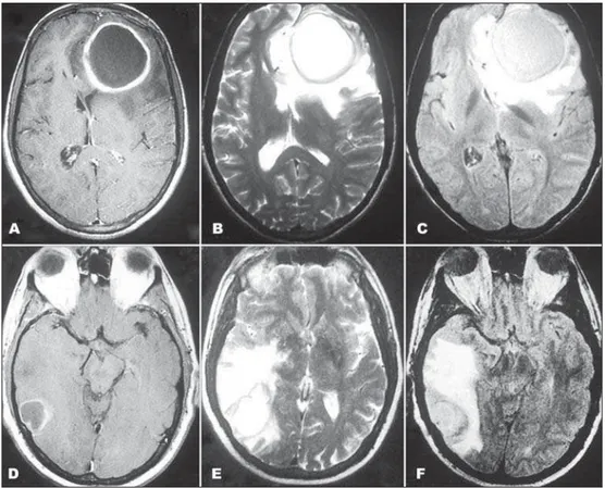
Both lesions were hyperintense in T2 weighted im-ages (WI) and FLAIR and hypointense in T1WI with a ring of hyperintense signal on T1WI and hypointense signal on T2WI, with contrast peripheral enhance-ment and extensive surrounding edema (Fig 1).
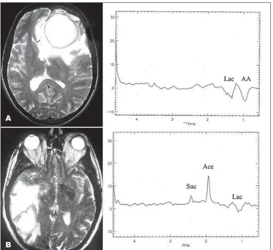
DWI showed hyperintensity in both with a low sig-nal in apparent coeffi cient diffusion (ADC) map (Fig 2). MRS showed many differences between the le-sions. Patient 1 had aminoacid peak (0.9 ppm) and el-evated lactate signal intensity (1.3 ppm) while patient 2 had a succinate (1.9 ppm) and acetate (2.4 ppm) peaks and also elevated lactate signal intensity (1.3 ppm) (Fig 3).
The lesions were surgically removed and culture revealed infection by Streptococcus pneumoniae,
Fig 2. Patient 1 (A and B) and patient 2 (C and D) show lesions with restricted diffusion on DWI (A and C) and ADC map (B and D).
Fig 3. Proton MRS. Patient 1 (A) shows aminoacid peak (AA) and elevated lactate signal intensity (Lac), inverted in TE of 135 ms which makes it easy to differentiate from lipids signal. Patient 2 (B) had suc-cinate (Suc) and acetate (Ace) peaks and also elevated lactate and aminoacid signal intensity. Both MRS performed in necrotic area display absence of characteristic peaks of brain tissue.
obe bacteria, in patient 1 and Streptococcus milleri, facultative anaerobe bacteria, in patient 2.
DISCUSSION
Abscesses usually are hyperintense in T2WI and hypointense in T1WI (as the most part of brain le-sions) but bacterial abscesses have a particular
con-tinuous peripheral ring hypointense on T2WI and and hyperintense on T1WI, a feature that could relate to coagulative necrosis, increased accumulation of hem-orrhagic products and paramagnetic materials such as iron, magnesium and manganese3, and production of
free radicals, secondary to bacterial metabolism4.
granulomatous abscesses (as occur in tuberculosis)5,6
present hemorrhagic areas producing the same alter-ation in peripheral signal intensity, making this fi nd-ing suggestive of bacterial etiology, but nonspecifi c. In addition to conventional MRI, DWI appears to be almost characteristic of bacterial abscesses. Pus is a viscous fl uid content of infl ammatory cells, mucoid protein, bacteria and necrotic tissue. Microscopic dif-fusion movement of water molecules are expected to be decreased in the cavity containing pus, whereas in necrotic tumor or cystic metastases, the cystic contents are less viscous, containing less infl ammatory cells and more serous fl uid7,8. So, bacterial abscess produce high
signal on DWI and low ADC values, ranged from 0.45 to 0.89. This variation might be related to a difference
in the concentration of infl ammatory cells, and differ-ences in necrotic tissue debris, bacteria, and viscosity of abscess fl uid8-10. Other in
fl ammatory processes as fungical disease, toxoplasmosis and necrotic portions of the tumors usually show lower signal intensity on DWI and higher ADCs11.
However, recent studies have shown that restrict-ed diffusion value is an important sign but is not spe-cifi c for cerebral abscess. Restricted diffusion has been found in metastatic squamous cell carcinoma and ra-diation necrosis. The underlying pathophysiology is not known but probably due to sterile liquefaction necrosis. In sterile liquefaction necrosis, it may contain creamy pus like material with polymorphonuclear leu-kocytes12.
Not enough, MRS also may corroborate the diag-nosis of bacterial abscess and progress in etiological diagnosis. MRS is a noninvasive method to access, in vivo, the biochemical environment of a tissue produc-ing direct or indirect information about the mecha-nisms of the disease progression and /or the host reac-tion to the aggression factor. Clinically, it may help to defi ne the nature of different types of brain lesions by the metabolic profi le. In some cases, it helps the differentiation among infections from other etiolo-gies when conventional MR images are incomplete or inconclusive13.
Bacterial abscesses are completely necrotic le-sions and so, all metabolites that characterize the SNC tissue are absent if the region of interest (ROI) is full located in the necrotic center. In fact, in both patients reported, peaks that characterize brain, as N-acetyl aspartate (NAA), considered a neuronal and axonal marker; choline, a constituent metabolite of cell membrane and myelin; and creatine, which plays a major role in energy metabolism, are absent14.
Lactate, an end product of anaerobic glycolysis and a considered marker of hypoxia, is elevated in both patients (identifi ed as a reversed J coupled peak in 1.3 ppm in this applied TE (135 ms), that avoid lipid overlap). However, its elevation is nonspecifi c. Large lactate peaks have been commonly identifi ed in ne-crotizing high-grade neoplasms, infarcts, demyelinat-ing disease, encephalitis, cystic gliomas and in no pyo-genic abscesses including toxoplasmosis15.
Other peaks, indicative of abnormal tissue were identifi ed. Both patients showed amino-acid signals at 0.9 ppm, seen in many brain abscesses and as-signed to valine, leucine, and isoleucine, considered representing accumulated end-products of proteolysis caused by proteolytic enzymes secreted by microor-ganisms or polymorphonuclear leukocytes in pus, or both16-22. Patient 2, infected by an anaerobe
faculta-tive bacteria, presented acetate and succinate, consid-ered being the end products of homolactic and het-erolactic fermentation and key markers of bacterial infection23-25. Other infectious diseases as cysticercosis
may also display succinate elevation26.
Garg et al.1, in an in vivo andin vitro study of 75
patients with brain abscess, also demonstrated dif-ferences in MRS according to bacterial etiology. Suc-cinate and/or acetate were present in anaerobic in-fection. Latter, Lai et al.27 showed the same
fi ndings in MRS performed in low and high magnetic fi elds28.
These metabolite resonances are probably the result of enhanced glycolysis and the fermentative path-ways for energy generation4.
transforms to the next intermediate compound. This phenomenon could result in the absence of succinate in the spectra of aerobe29. Succinate was also earlier
described as a marker for anaerobic infections in clini-cal samples at gas liquid chromatography25.
Beyond MRS represents a potential tool for nonin-vasive diagnosis of brain abscesses, it might be useful for evaluating changes after treatment. With effec-tiveness of antibiotic therapy, a decreasing of these abnormal peaks, produced by infectious organism and intense host infl ammatory reaction, and the lac-tate signal persists30, besides the changes in signal and
retraction of lesion at conventional MRI.
In conclusion, MRS coupled with structural MRI and DWI and MRS is effective and promising not only to diagnosing but also to explore the etiology of bac-terial abscesses. In addition, it also may be a powerful tool to control the response to the treatment.
REFERENCES
1. Garg M, Gupta RK, Husain M, et al. Brain abscesses: etiologic categoriza-tion with in vivo proton MR spectroscopy. Radiology 2004;230:519-527. 2. Faria AV, Reis F, Zanardi VA, Menezes JR, Cendes F. The paĴ ern of pro-ton magnetic resonance spectroscopy in non-neoplastic encephalic le-sions. Arq Neuropsiquiatr 2004;62:429-436.
3. Siegal JA, Cacayorin ED, Nassif AS, et al. Cerebral mucormycosis: proton MR spectroscopy and MR imaging. Magn Reson Imaging 2000;18:915-920. 4. Sudhakar KV, Agrawal S, Rashid MR, Hussain N, Hussain M, Grupta
RK. MRI demonstration of hemorrhage in the wall of a brain abscess: possible implications for diagnosis and management. Neuroradiology 2001;43:218-222.
5. Meyerand ME, Pipas JM, Mamourian A, Tosteson TD, Dunn JF. Classi-fi cation of biopsy-confi rmed brain tumors using single-voxel MR spec-troscopy. Am J Neuroradiol 1999;20:117-123.
6. Kaminogo M, Ishimaru H, Morikawa M, Suzuki Y, Shibata S. Proton MR spectroscopy and diě usion-weighted MR imaging for the diagnosis of intracranial tuberculomas: report of two cases. Neurol Res 2002;24:537-543.
7. Noguchi K, Watanabe N, Nagaoshi T, et al. Role of diě usion-weighted echo-planar MRI in distinguishing between brain abscess and tumour: a preliminary report. Neuroradiology 1999;41:171-174.
8. Kim YJ, Chang KH, Song IC, et al. Brain abscess and necrotic or cystic brain tumor: discrimination with signal intensity on diě usion-weight-ed MR imaging. Am J Roentgenol 1998;171:1487-1490.
9. Lai PH, Ho JT, Chen WL, et al. Brain abscess and necrotic brain tumor: discrimination with proton MR spectroscopy and diě usion-weighted imaging. Am J Neuroradiol 2002;23:1369-1377.
10. Desprechins B, Stadnik T, Koerts G, Shabana W, Breucq C, Osteaux M. Use of diě usion weighted NMR imaging in diě erential diagnosis be-tween intracerebral necrotic tumors and cerebral abscesses. Am J
Neu-roradiol 1999;20:1252-1257.
11. Mishra AM, Gupta RK, Jaggi RS, et al. Role of diě usion-weighted im-aging and in vivo proton magnetic resonance spectroscopy in the dif-ferential diagnosis of ring-enhancing intracranial cystic mass lesions. J Comput Assist Tomogr 2004;28:540-547.
12. Tsui EYK, Chan JH, Cheung YK, Lai D KF, Fong SHN. Evaluation of ce-rebral abscesses by diě usion-weighted MR imaging and MR spectros-copy. Comput Med Imag Graph 2002;26:347-351.
13. Faria AV, Dabus GC, Zanardi VA, Cendes F. Proton magnetic reso-nance spectroscopy and magnetic resoreso-nance imaging fi ndings in a pa-tient with central nervous system paracoccidioidomycosis. J Neuroimag 2004;14:377-379.
14. Castillo M, Kwock L, Mukherji SK. Clinical applications of proton MR spectroscopy. Am J Neuroradiol 1996;17:1-15.
15. Chang L, Cornford ME, Chiang FL, Ernst TM, Sun NCJ, Miller BL. Ra-diologic-pathologic correlation cerebral toxoplasmosis and lymphoma in AIDS. Am J Neuroradiol 1995;16:1653-1663.
16. Poptani H, Gupta RK, Jain VK, Roy R, Pandey R. Cystic intracranial mass lesions: possible role of in vivo MR spectroscopy in its diě erential diagnosis. Magn Reson Imaging 1995;13:1019-1029.
17. Martinez-Perez I, Moreno A, Alonso J, et al. Diagnosis of brain abscess by magnetic resonance spectroscopy: report of two cases. J Neurosurg 1997;86:708-713.
18. Rémy C, Grand S, Laý ES, et al. 1H MRS of human brain abscesses in vivo and in vitro. Magn Reson Med 1995;34:508-514.
19. May GL, Sztelma K, Sorrell TC, Mountford CE. Comparison of human polymorphonuclear leukocytes from peripheral blood and purulent ex-udates by high resolution 1H MRS. Magn Reson Med 1991;19:191-198. 20. Burtscher IM, Holta S. Case report in vivo proton MR spectroscopy of un-treated and un-treated brain abscesses. Am J Neuroradiol 1999;20:1049-1053. 21. Soto-Hernandez JL, Moreno-Andrade T, Góngora-Rivera F, Ramírez-Crescencio MA. Case report: Nocardia abscess during treatment of brain toxoplasmosis in a patient with aids, utility of proton MR spectroscopy and diě usion-weighted imaging in diagnosis. Clin Neurol Neurosurg 2006;108:493-498.
22. Himmelreich U, Accurso R, Malik R, et al. Identifi cation of Staphylococ-cus aureus brain abscesses: rat and human studies with 1H MR spectros-copy. Radiology 2005;236:261-270.
23. Harada M, Tanouchi M, Miyoshi H, Nishitani H, Kannuki S. Brain ab-scess observed by localized proton magnetic resonance spectroscopy. Magn Reson Imaging 1994;12:1269-1274.
24. Phillips KD, Tearle PV, Willis AT. Rapid diagnosis of anaerobic infec-tions by gas-liquid chromatography of clinical material. J Clin Pathol 1976;29:428-432.
25. Gorbach SL, Mayhew JW, BartleĴ JG, Thadepalli H, Onderdonk AB. Rapid diagnosis of anaerobic infection by direct gas-liquid chromatog-raphy of clinical specimens. J Clin Invest 1976;57:478-484.
26. Pandit S, Lin A, Gahbauner H, Libertin CR, Erdogan B. MR spectrosco-py in neurocysticercosis. J Comput Assist Tomogr 2001;25:950-952. 27. Lai PH, Li KT, Hsu SS, et al. Pyogenic brain abscess: fi ndings from in
vivo1.5-T and 11.7-T in vitro proton MR spectroscopy. Am J Neuroradi-ol 2005;26:279-288.
28. Kim SH, Chang KH, Song IC, et al. Brain abscess and brain tumor: discrim-ination with in vivo H-1 MR spectroscopy. Radiology 1997;204:239-245. 29. Stryer L. Citric acid cycle. In Stryer L (Ed). Biochemistry, 4.Ed. New York,
NY: Freeman 1995:509-514.