STUDY OF THE POSITIVITY OF SPONTANEOUS
AND DIRECTED DIAGNOSIS OF LEUKOARAIOSIS
IN THE ELDERLY BY CRANIAL COMPUTERIZED
TOMOGRAPHY, AND ITS CORRELATION WITH
COGNITIVE DEFICIT AND CARDIOVASCULAR
RISK FACTORS
Mauricio de Miranda Ventura
1, Antonio Carlos de Paiva Melo
2,
Henrique Carrete Jr
3, Ricardo Vieira Botelho
4ABSTRACT - Objective:To evaluate the prevalence of leukoaraiosis (LA) and its correlation with vascular risk factors and the cognitive performance of elderly patients. Method:78 patients were randomly selected and submitted to clinic-laboratorial evaluation for vascular risk factors. Two cognitive tests were performed. All patients were submitted to cranial computerized tomography (CT), which was analyzed in two situations: the spontaneous and the directed way. Results:There was no statistically significant difference between the prevalence of spontaneous and the directed diagnoses of LA (20.5 and 18%, respectively). The presence of LA was not significantly correlated with cognitive impairment. Only age was positively associated with LA. Conclusion:Cranial CT is a trustworthy method for the diagnosis of LA. Only age showed a positive as-sociation. No clinical implications were found, concerning the cognitive performance of the patients.
KEY WORDS: leukoaraiosis, cognition disorders, risk factors.
Estudo da positividade do diagnóstico espontâneo e dirigido da leucoaraiose em idosos pela tomografia computadorizada de crânio e sua correlação com déficits cognitivos e fatores de risco cardiovascular
RESUMO:Objetivo:Avaliar a prevalência da leucoaraiose (LA) em idosos e correlacioná-los com fatores de risco vasculares e seu desempenho cognitivo. Método:78 pacientes foram selecionados aleatoriamente e submetidos a avaliação clinico-laboratorial para fatores de risco vascular. Dois testes cognitivos foram reali-zados. Todos os pacientes foram submetidos a tomografia computadorizada (TC) de crânio, que foi analisa-da em duas situações: de forma espontânea e dirigianalisa-da. Resultados:Não houve diferença estatisticamente significativa entre as formas de diagnóstico espontâneo e dirigido da LA (20,5 and 18%, respectivamente). Com relação ao desempenho cognitivo, a presença da LA não determinou prejuízo significativo. Somente a idade mostrou associação positiva com a LA. Conclusão:A TC de crânio é um método confiável para o diagnóstico de LA. A idade foi o único fator de risco que mostrou associação positiva. Não houve prejuízo quanto ao desempenho cognitivo.
PALAVRAS-CHAVE: leucoaraiose, transtornos cognitivos, fatores de risco.
Hospital do Servidor Público Estadual “Francisco Morato de Oliveira”, São Paulo SP, Brasil: 1Médico do Serviço de Geriatria; 2Diretor do Serviço de Neurologia; 3Médico do Serviço de Radiologia; 4Médico do Serviço de Neurocirurgia.
Received 6 June 2007, received in fi nal form 11 September 2007. Accepted 8 October 2007.
Dr. Ricardo Vieira Botelho - Rua Dr. Haberbeck Brandão 68 / 92 - 04027-040 São Paulo SP - Brasil. E-mail: [email protected]
The cranial computerized tomography (CT) is a di-agnostic method which is widely used in the areas of Neurology and Geriatrics. The detection of leukoarai-osis (LA) in the CT of the encephalon is frequent. This term was introduced by Hachinski et al.1, who
de-scribed a radiological alteration characterized by hy-poattenuations around the cerebral ventricles in im-ages obtained by CT. With the advent of magnetic
resonance (MR), the corresponding detection is the al-teration of intensity of the signal (hypersignal) in the acquisition in T2. Due to its greater sensitivity, MR has been considered the best diagnostic method2.
Never-theless, Lopez et al.3 found stronger
its lower costs, frequently precedes the MR in the in-vestigations of pathologic processe of the central ner-vous system (CNS), and it may be the only diagnostic method available for countless populations.
The clinical significance of the LA is uncertain. And, although it is considered part of natural aging associated with brain-vascular risk factors, it could ei-ther contribute to the presence of cognitive defi cits in healthy people, or to worsen defi cits as well as vas-cular dementia in sufferers of Alzheimer’s disease4.
Nevertheless, the real clinical implications of LA re-main unclear.
We have studied the prevalence of LA in an elder-ly population who showed no evidence of cognitive alterations, the effect of previously directed diagnosis for the research of LA and its prevalence, and we cor-related the cognitive defi cit with the presence of co-morbidities and smoking. Our main objective was to evaluate the prevalence of LA (in studies with CT) in a population of non-suffers of dementia or of brain-vascular diseases. The secondary objectives were: to evaluate differences concerning the prevalence of the diagnosis of LA when radiological study was conduct-ed through directconduct-ed method (it was requirconduct-ed, previ-ously, to the radiologist to check the presence of LA and to classify it) and through spontaneous method; to study and compare the cognitive performance of the patients who were suffers of LA and those who were not; to correlate anthropologic data (age and sex), level of education and the presence of chronic-degenerative diseases with LA.
METHOD
With the objective to reduce the selection bias, a list of outpatients (who frequented the Ambulatory of Geriatric Service) were generated and a number was attributed to each one. The candidates were than picked up by a closed envelope lottery. Those who did not bear dementia, brain-vascular diseases or any other disease which was severe enough to cause cognitive defi cit or to prevent them from going through CT, were selected. Patients showing senso-rial defi cit (auditive and/or visual) were excluded to make sure the results of the cognitive tests would not be affected. The research project was approved by the Research Ethics Committee of the Instituto de Assistência Médica ao Servidor Público Estadual, and the patients agreed with the terms of postinformed consentiment.
All patients were submitted to general clinical evalua-tion concerning their cognitive capacity.
The clinic evaluation consisted of medical history, use of medications and physical and neurological exams. We were particulary interested in the presence of the following chronic-degenerative diseases: hypertension (HT), diabetes mellitus (DM), dislipidemy, obesity and smoking. We
identi-fi ed and correlated them with the presence of LA.
The cognitive evaluation was done through Structured Interview for the Diagnosis of Dementia (SIDAM) - validat-ed for the diagnosis of dementia5, for the diagnosis of mild
cognitive impairment6, whose version in Portuguese was
validated by Ventura and Bottino7 and through the Mini
Mental State Exam (MMSE)8.
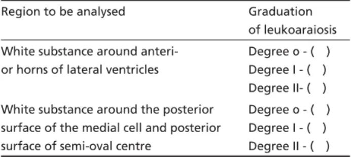
The analysis of CT was done in two moments: in the fi rst phase, a report was issued by the neuroradiologist who did not know the diagnosis of the patient or the objective of the study (spontaneous diagnosis); in the second phase, the neuroradiologist was requested to determine specifi cally the presence or absence of LA, and, in affi rmative case, to quantify the intensity of LA (directed diagnosis), according to the method of van Swieten et al.9 (Table 1).
Statistical analysis – The variables were described by means and standard deviation. The difference between the distribution of the spontaneous and the directed to-mographic diagnosis was evaluated through test of
McNe-mar10. Concerning age, level of education, MMSE and
SI-DAM, the comparison between the groups with or without LA, according to the two methods used, was done through Mann-Whitney test11. For the study of the differences of
dis-tribution of qualitative variables (HT; DM; dislipidemy; obe-sity and smoking) between the groups with and without LA, the exact Fisher test or qui-square test were used. The lev-el of signifi cance was defi ned as p≤0.05. The statistical
pro-gram used was the Statistic 6.0.
RESULTS
Initially, the 78 patients were submitted to the protocol of the study, by accomplishing clinical, neu-rological and cognitive evaluation. Their CTs were
Table 1. Graduation of the lesions of the white substance in the cranial computerized tomography.
Region to be analysed Graduation of leukoaraiosis White substance around
anteri-or hanteri-orns of lateral ventricles
Degree 0 - ( ) Degree I - ( ) Degree II- ( ) White substance around the posterior
surface of the medial cell and posterior surface of semi-oval centre
Degree 0 - ( ) Degree I - ( ) Degree II - ( )
Table 2. Prevalence of chronic-degenerative diseases and their relation with LA.
Without LA With LA p
HT 38 (48.718%) 11(14.103%) 0.5844
DM 11(14.103%) 3 (3.84%) *
submitted to analysis through spontaneous diagnosis. Five patients, or members of their families, declined to take part in the study, and 73 exams, among the initial 78, were submitted to directed diagnosis.
Patients’ average age was 75.1 years (sd: 6.47); 71.8% (56 patients) were female and 28.2% (22 pa-tients) were male.
Concerning their educational background, 55.1% of the patients had low educational level (up to 4 years); 12.8% had medium educational level (4 to 8 years); 32.1% had a high educational level (more than 8 years).
In relation to the chronic-degenerative diseases: 62.82% were hypertense; 64.10% were obese; 50% had dislipidemy; 29.48% were tabagist, and 17.94% had diabetes mellitus. Table 2 shows the prevalence of comorbidies and smoking, as well as the p-value of the association with LA.
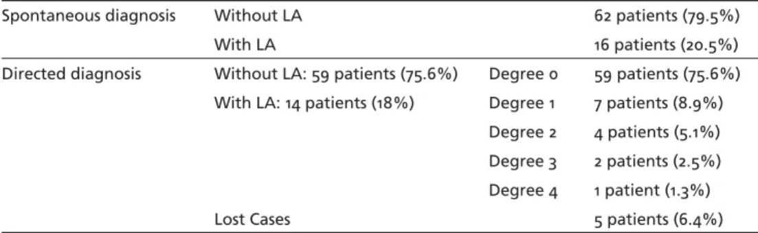
The prevalence of LA perceived was 20.5%, accord-ing to spontaneous diagnosis, and of 18%, accordaccord-ing to directed diagnosis (p=0.727; McNemar). Only in 8 cases the diagnoses were different, depending on the approach used (Table 3). There were no signifi cant differences between the directed and the spontane-ous diagnoses. Based on the lack of difference be-tween the spontaneous and directed methods, we used only the former up to discussion.
In Table 4 we observe that: the age of the individ-uals with LA was signifi cantly higher than that of in-dividuals without LA, according to spontaneous diag-noses (p=0.01); the distribution of patients with and
without LA was not different when compared to their level of education (p=0.630); there were differences, close to the level of signifi cance, concerning the cog-nitive performance of the patients with LA (MMSE: p=0.059; SIDAM: p=0.067) (Table 4).
DISCUSSION
Since its initial description, there has been a lot of discussion in the literature concerning the causes and the clinical correlation of LA. There may even be some confusion relating to previous brain alterations and loss of involutive and nonspecifi c white substance, which occurs as age advances. There has also been discussion concerning the possibility of the existence of LA only as an incidental radiologic diagnosis (with-out clinical correspondence), on assymptomatic pa-tients. There are different neurologic fi ndings which may contribute to the presence of periventricular hy-podensities: perivascular demielinization and gliosis; dilatation of Virchow-Robin spaces; small gaps; loss of axons and glial cells (predominantly oligodendrocytes); rarefaction of myelin associated with spongiosis; pres-ence of multiple gaps and numerous sclerotic plates. Nevertheless, as mentioned by Hachinski et al.1, a
simple infi ltration of the spinal-brain liquid into the periventricular brain parenchyma would be enough to cause radiologic alteration, typical of LA.
According to directed and spontaneous diagnoses, 18 to 20.5% of the elderly patients without evident cognitive defi cit presented tomographic images of LA. There were no signifi cant differences between the two forms of diagnostic evaluation. The spon-taneous diagnosis, done by radiologist without the tendency imposed by the objective of the research must be considered trustworthy.
Patients with LA showed different cognitive per-formance when compared to those without LA. Al-though the difference did not reach the defi ned level of signifi cance, in case we accepted the level of signif-icance of ten per cent (p=0.10), the difference would
Table 3. Prevalence of spontaneous and directed diagnoses of LA.
Spontaneous diagnosis Without LA 62 patients (79.5%)
With LA 16 patients (20.5%)
Directed diagnosis Without LA: 59 patients (75.6%) Degree 0 59 patients (75.6%) With LA: 14 patients (18%) Degree 1 7 patients (8.9%)
Degree 2 4 patients (5.1%) Degree 3 2 patients (2.5%) Degree 4 1 patient (1.3%)
Lost Cases 5 patients (6.4%)
Table 4. Probability associated (p) to differences in the distribu-tion of LA in reladistribu-tion to age, school level and cognitive perfor-mance according to LA’s spontaneous diagnoses (Mann-Whitney).
Variable Without LA With LA p
Age 73.89 79.56 0.001
School level 6.65 5.94 0.630
MMSE 25.28 23.44 0.059
have been determined (p=0.059 MMSE and p=0.067 SIDAM). It is possible that the test used, with lower power of evaluation (non parametric Mann-Whitney), and the size of the sample have contributed to the fi -nal results. Through spontaneous diagnosis, research-ers analyzed only the presence or the absence of LA, regardless of its intensity.
The majority of the patients in our sample were diagnosed as having LA of I and II degrees. It is pos-sible that the prevalence of LA of low intensity has contributed for these results, and that suggests that LA of lower level is less intensely correlated with cog-nitive defi cit. The small number of patients with LA of more advanced levels does not allow us to draw any major conclusions. Our study excluded patients with evident cognitive defi cit.
As far as sensitivity is concerned, the cranial CT is not the best method for the diagnosis of LA, nor is it the best way to qualify it. Nonetheless, because of the high cost of the MR, the greater availability of the CT, the diffi culties for realizing the exam with MR in elderly patients, the CT was considered the most available, economic and functional method. It is worth mentioning that Lopez et al.3 found more
signifi cant clinical-pathological correlation with the CT, specifi cally concerning the appearance of symp-toms of vascular brain disease in probable bearers of Alzheimer’s disease.
In the studies about the correlation between LA and cognitive profi le, Steingart et al.12 studied,
pro-spectively, 105 voluntary normal elderly patients, by CT, psychometric evaluation and neurological exams. In that sample, patients who had LA showed a worse performance in the cognitive tests.
Steingart et al.13 evaluated 113 patients through
the use of the CT (of the skull), neurological exams and psychometric tests. All patients had dementia syn-drome, and 80,5% of them were diagnosed as having Alzheimer’s disease, and 39 were found to have LA. Comparison between these two groups revealed that those patients with LA showed a worse performance in the psychometric tests.
Kluger et al.14 have reported the results of their
study, according to which 6 elderly patients with le-sions of the peri-ventricular white substance in the region of the anterior horn of the lateral ventricle, as their CT (of the skull) revealed, showed sub-clinical defi cits in the motor psychometric tests, when com-pared with other 11 patients who did not present the same lesions. This suggests that theses lesions may affect, primarily, psychomotor activities.
Hogervorst et al.15 compared the CT of healthy
pa-tients to those of papa-tients with Alzheimer’s disease, evaluating the intensity of the LA. The cognitive per-formance of the two groups, according to MMSE (MESM), did not show any statistically significant difference, as far as the normal patients were con-cerned, but in relation to patients with Alzheimer’s disease, the performance, in the MMSE (MESM), was worse among those with LA.
Smith et al.16 studied the in
fl uence of LA in cogni-tive defi cits. The CT of 182 patients with brain hemor-rhage were analyzed, blindly – as far as the clinic his-tory of the patients was concerned – by radiologists, who then quantifi ed the LA. These patients were ac-companied through interviews carried out through the telephone. A positive association was established be-tween cognitive defi cits and the intensity of the LA in patients, after an episode of intra-brain hemorrhage. Hachinski et al.1 note the positive association between
LA and intellectual decay, and discuss its association with Alzheimer’s disease, which is justifi ed by the fact that amyloid angiopathy was found to have the same neuropathological characteristics in both diseases. The studies which compared cognitive perfor-mance in the presence of LA, by means of CT, got to the conclusion that it may lead to a discrete defi cit, with no signifi cant difference. The study carried out by Smith et al.16, which showed a positive
associa-tion with LA, depending on its intensity, evaluated the patients after an episode of severe intra-brain hemorrhage, differently from us who have exclud-ed patients with any sort of brain-vascular problem. Furthermore, differently from our study, there was a post evaluation through telephone interviews about how patients had progressed, and this may have cor-roborated their fi ndings.
Pantoni and Garcia4, in a review article, considered
LA as part of normal aging process. Nevertheless, if it is aggravated by brain-vascular risk factors, it could contribute to the presence of discrete cognitive defi -cits in healthy people, or to worsen cognitive defi cits in patients with Alzheimer’s disease or with vascular dementia, and, eventually, it could increase the risk of brain-vascular problems.
When comorbidities and chronic disease were tak-en as risk factors for LA, our study revealed age as the only variable positively associated with LA. This was observed in both diagnoses, spontaneous and direct-ed, and was corroborated by other studies12,13,15,16.
not conclusive in relation to this possibility: Steingart et al.12, Hogervorst et al.15 and Smith et al.16 did not
fi nd this relationship in their studies, while Steingart et al.13 were the only ones to establish it. In our study,
regardless of the diagnostic approach used, it was not possible to establish a positive association between LA and arterial hypertension. A possible reason is the fact that patients with brain-vascular diseases were excluded, and patients with more intense LA were a minority in our casuistics, although more than 62% of the individuals in the research were hypertense. In other studies which have established this association, the presence of brain-vascular disease was an impor-tant condition for the inclusion of patients. There are doubts concerning the relevance of other vascular risk factors, and it is not probable that dislipidemy, dia-betes mellitus, obesity and smoking play any role in its etiology.
In conclusion, the rate of prevalence of LA in a population who does not have brain-vascular demen-tia or disease was 20.5%. The prevalence of LA did not increase according to the directed nor with the spontaneous mode of research applied. Age was the anthropometric variable which showed the strongest association with the LA. There was an indication of the association of the LA with cognitive defi cit, al-though we have not reached the level of signifi cance determined for this study. Studies with a greater number of individuals, as well as the study of patients with previous cognitive defi cit could make this asso-ciation clearer. No cronic-degenerative diseases, nor smoking, were associated to the presence of LA.
REFERENCES
1. Hachinski WC, Potter P, Mershey H. Leuko-araiosis. Arch Neurol 1987;44:21-23.
2. ErkinjunĴ i T, Ketonen L, Sulkava R, Sipponen J, Vuorialho M, Iivanain-en M. Do white matter changes on MRI and CT differIivanain-entiate vascu-lar dementia from Alzheimer’s disease? J Neurol Neurosurg Psychia-try 1987;50:37-42.
3. Lopez OL, Becker JT, Jungreis CA, et al. Computed tomograpy – but not magnetic resonance imaging – identifi ed periventricular white-mat-ter lesions predict symptomatic cerebrovascular disease in probable Al-zheimer’s disease. Arch Neurol 1995;52:659-664.
4. Pantoni L, Garcia JH. The signifi cance of cerebral white maĴ er abnor-malities 100 years after Binswanger’s report: a review. Stroke 1995; 26:1293-1301.
5. Zaudig M, MiĴ elhammer J, Hiller W, Pauls A, Thora C. SIDAM: a struc-tured interview for the diagnosis of dementia of the Alzheimer type, multi-infarct dementia and dementia of other etiology according to DSM III R and ICD 10. Psychol Med 1991;21:225-236.
6. Zaudig M. A new sistematic method of measurement and diagnosis of mild cognitive impairment and dementia accorging to DSM III R and ICD 10. Int Psychogeriatrics 1992:203-219.
7. Ventura MM, BoĴ ino CMC. Estudo de confi abilidade da versão em por-tuguês de uma entrevista estruturada para o diagnóstico de demência. Rev Ass Med Brasil 2001;47:110-116.
8. Folstein MF, Folstein SE, McHugh. Mini-Mental State. J Psychiatr Res 1975;12:189-198.
9. van Swieten JC, Hħ dra A, Koudstaal PJ, van Għ n J. Grading white mat-ter lesions on CT and MRI: a simple scale. J Neurol Neurosurg Psychi-atry 1990;53:1080-1083.
10. Agresti A. An introduction to categorical data analysis. New York: John Wiley and Sons, Inc, 1996.
11. Conover WJ. Practical nonparametric statistics, 2.Ed. New York: John Wiley & Sons, Inc., 1980.
12. Steingart A, Hachinski VC, Lau C, et al. Cognitive and neurologic fi nd-ings in subjects with diě use white maĴ er lucencies on computed tomo-graphic scan (leuko-araiosis). Arch Neurol 1987;44:32-35.
13. Steingart A, Hachinski VC, Lau C, Fox AJ, Fox H, Lee D, Inzitari D, Mer-skey H. Cognitive and neurologic fi ndings in demented patients with diě use white maĴ er lucencies on computed tomographic scan (leuko-araiosis). Arch Neurol 1987;44:36-39.
14. Kluger A, Glanutsos J, Leon MJ, George AE. Signifi cance of age-related white maĴ er lesions. Stroke 1988;19:1054-1055.
15. Hogervorst E, Ribeiro HM, Molyneux A, Budge M, Smith AD. Plasma homocysteine levels, cerebrovascular risk factors, and cerebral white maĴ er changes (leukoaraiosis) in patients with Alzheimer disease. Arch Neurol 2002;59:787-793.