TRANSORAL APPROACH TO THE
CRANIOVERTEBRAL JUNCTION
José Alberto Landeiro
1, Sávio Boechat
2,
Daniel de Holanda Christoph
2, Mariângela Barbi Gonçalves
2,
Igor de Castro
3, Mario Alberto Lapenta
3, Carlos Henrique Ribeiro
3ABSTRACT - The transoral approach provides a safe exposure to lesions in the midline and the ventral side of the craniovertebral junction. The advantages of the transoral approach are 1) the impinging bony patholo-gy and granulation tissue are accessible only via the ventral route; 2) the head is placed in the extended po-sition, thus decreasing the angulation of the brainstem during the surgery; and 3) surgery is done through the avascular median pharyngeal raphe and clivus. We analyzed the clinical effects of odontoidectomy after treating 38 patients with basilar invagination. The anterior transoral operation to treat irreducible ventral compression in patients with basilar invagination was performed in 38 patients. The patients’ ages ranged from 34 to 67 years. Fourteen patients had associated Chiari malformation and eight had previously under-gone posterior decompressive surgery. The main indication for surgery was significant neurological deteri-oration. Symptoms and signs included neck pain, myelopathy, lower cranial nerve dysfunction, nystagmus and gait disturbance. Extended exposure was performed in 24 patients. The surgery was beneficial to the majority of patients. There was one death within 10 days of surgery, due to pulmonary embolism. Postoper-ative complications included two cases of pneumonia, three cases of oronasal fistula with regurgitation and one cerebrospinal fluid leak. In patients with marked ventral compression, the transoral approach provides direct access to the anterior face of the craniovertebral junction and effective means for odontoidectomy.
KEY WORDS: basilar invagination, odontoidectomy, transoral approach.
Acesso transoral para a junção craniocervical
RESUMO - O acesso transoral é uma via direta e segura às lesões situadas na linha média e na face anterior da junção craniocervical. As vantagens do acesso transoral são as seguintes:1) a compressão óssea e o teci-do de granulação localizam-se anteriormente e são accessíveis pela via anterior; 2) a cabeça teci-do paciente é colocada em extensão, diminuindo a angulação do tronco cerebral durante a cirurgia; e 3) a cirurgia é fei-ta através de um plano avascular na linha média faríngea e clivo. Analisamos os resulfei-tados obtidos após odontoidectomia por via transoral em 38 pacientes portadores de invaginação basilar. Trinta e oito pacien-tes com compressão ventral da junção craniocervical foram submetidos a odontoidectomia por via transo-ral. A idade dos pacientes variou de 34 a 67 anos. Quatorze pacientes apresentavam associação com malfor-mação de Chiari tipo I e 8 já haviam sido submetidos à cirurgia descompressiva por via posterior. A maioria dos pacientes apresentou nucalgia, mielopatia, déficits dos nervos cranianos baixos, nistagmo, e distúrbio da marcha. Em 24 pacientes foi necessário ampliar o acesso transoral através de miotomia do palato mole, ou osteotomia do palato duro ou maxilotomia. A cirurgia proporcionou melhora dos sintomas na maioria dos pacientes. Um paciente faleceu no pós-operatório imediato por causa de embolia pulmonar. Dois pa-cientes tiveram pneumonia, três apresentaram fístula oronasal com regurgitação, e um teve fístula liquóri-ca. Em pacientes com compressão ventral irredutível da junção craniocervical, a via transoral proporcionou uma abordagem direta e ampla ao processo odontoide.
PALAVRAS-CHAVE: invaginação basilar, odontoidectomia, acesso transoral.
1Chefe do Serviço de Neurocirurgia do Hospital da Força Aérea do Galeão, Rio de Janeiro RJ, Brasil, Professor Associado do
Depar-tamento de Cirurgia Geral e Especializada da Faculdade de Medicina da Universidade Federal Fluminense; 2Residente do Serviço
de Neurocirurgia do Hospital da Força Aérea do Galeão, RJ; 3Assistente do Serviço de Neurocirurgia do Hospital da Força Aérea do
Galeão, RJ.
Received 28 February 2007, received in fi nal form 29 August 2007. Accepted 4 September 2007.
Dr. José Alberto Landeiro - Avenida Monsenhor Ascâneo 501 / 204 - 20621-060 Rio de Janeiro RJ - Brasil. E-mail: [email protected]
The transoral approach (TOA) provides the most direct access to pathologies located on the ventral side of the craniovertebral junction (CVJ)1-6. The CVJ
the upper cervical cord. Bone junction abnormalities at the CVJ are likely to compress and affect the local neural and vascular structures, and cerebrospinal fl u-id circulation.
We review the indications and the surgical tech-nique of transoral approachs based on the experience acquired with the surgical treatment of 38 patients with basilar invagination. Stabilization is not empha-sized in this paper.
METHOD
Thirty-eight patients with basilar invagination were op-erated on by the senior author between 1994 and 2004. Patients treated via the transoral approach for other con-ditions (rheumatoid arthritis, tumors and trauma) were ex-cluded from this report.
Preoperative evaluation – Preoperative neuroimaging investigations consisted of plain radiographs followed by dynamic polytomography. The computerized tomography (CT) with tridimensional reconstruction and magnetic reso-nance imaging (MRI) provide a perfect visualization of the physiopathology of the lesion.
Preoperative evaluation also included orthodontal exam-ination, detection of oropharyngeal infection, the nutrition-al condition of the patient and the functionnutrition-al state of lower cranial nerves. Poorly nourished patients were fi rst treat-ed with enteral fetreat-eding. Patients with lower cranial nerve defi cits had tracheostomies during preoperative treatment.
All patients started the neurodiagnostic procedures with thin section computed tomography (CT) scans in coronal, axial and sagittal views followed by MRI scans in the fl exed and extended positions in order to verify stability.
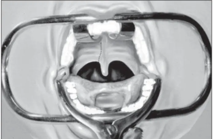
Surgical procedure – The patient was positioned supine with the head slightly extended and secured in the Mayfi eld support with three-point skull fi xation, with the operating table placed in a slight Trendelenburg position. The surgeon stood behind or on the right side of the patient. Usually tra-cheostomy was not necessary; a nasotracheal armored tube provided adequate ventilation. The use of self-retaining re-tractors facilitated the surgery. The tongue and endotrache-al tube were depressed, and the soft pendotrache-alate elevated and compressed backwards, assisted by two nasal tubes fi xed to the palate. High upward migration of the odontoid process may require incision of the soft palate, avoiding the uvula (Fig 1). The use of teeth protectors was useful and helped tofi x the retractor.
The palpated anterior tubercle of the atlas was an im-portant landmark for the incision, because it is usually lo-cated in the center of the lesion. Therefore, an incision of 2.5 cm above and below the atlas tubercle was usually suffi -cient to reach the inferior border of the clivus and the body of the axis. A midline incision in the posterior mucosa and the pharyngeal muscles was done in one single plane up to the anterior longitudinal ligament.
The area of incision was infi ltrated with 1:200000 solu-tion of xylocaine with adrenaline to reduce mucosal
bleed-Fig. 1. Incising the soft palate to one side of the uvula.
Fig 2. After infi ltration, a midline incision is made in the pha-ryngeal mucosa. To allow for retraction, the incision should ex-tend well above and below the lesion.
Fig 3. The ligaments and muscles are dissected from the bone. The switches retain the soft tissue.
Fig 4. The clivus, arch of C1 and the body of C2 are exposed. Half of the anterior arch of C1 has been removed.
Fig 5. Midsagittal T2-weighted image at the craniovertebral junc-tion showing intrusion of the odontoid peg into the foramen magnum and severe cervicomedullary junction compression.
Fig 6. Midsagittal T2-weighted postoperative image, demon-strating removal of the odontoid process.
Fig 7. Postoperative CT scan in the sagittal view after odon-toidectomy. The anterior ring of the atlas has been spared.
The anterior tubercle of the arch of C1 is the surgical key to
the CVJ. The longus colli muscles are attached to it on both sides, and the anterior longitudinal ligament is attached in the midline. Intraoperative fl uoroscopy can help in the lo-calization of the C1 tubercle and C2 body. About 12-15 mm of the anterior arch of atlas was removed with the drill. This maneuver exposes the base of the odontoid process (Figs 2, 3 and 4). In severe invagination, it may be necessary to re-sect the caudal clival bone.
The odontoidectomy was performed with a high-speed drill with cutting bars until the posterior cortical was reached, which was subsequently removed by drilling with diamond burrs. Before drilling the posterior cortical bone, traction of the odontoid peg was performed, followed by section of the alar and apical ligaments, and resection by drilling or with a pituitary rongeur (Figs 5, 6 and 7). In order to achieve adequate decompression, the ligaments and the tectorial membrane were excised. The operation was com-pleted when the pulsatile dura protruded ventrally into the decompression site. The cavity was then irrigated with an-tibiotic solution and the hemostasy was checked. The clo-sure was performed in two layers, muscular and mucosal, with Vicryl 3.0 continuous stitches. In the case of myotomy of the uvula and the soft palate, closure was performed in three layers. The orogastric tube that was placed prior to the surgery was retained. A rigid neck collar was used until the stability of the CVJ was determined.
The patients remained intubated for 48 hours, with the removal of the tube done only after a functional study of
Table 1. Clinical fi ndings.
No. of patients
Neck pain 37
Myelopathy 31
Sensory disturbance 16
Gait disturbance 15
Lower cranial nerve palsies 7
Nystagmus 4
the larynx and pharynx. Tracheostomy was indicated in cas-es of postoperative lower cranial nerve defi cits.
Postoperative care – The cicatrization of the mucosa did not present any problems. However, careful oral and nasal asepsis was crucial. The patient should not take any solids orfl uids orally for approximately six days following surgery. During this period, nutrition and medication should be ad-ministered intravenously or through a feeding tube. Cor-ticosteroids should be administered intravenously during thefi rst three days, until labial, tongue and pharynx ede-ma completely subsides.
RESULTS
Tables 1 and 2 show the clinical features and the postoperative evaluation. Thirty-eight patients were submitted to transoral decompression surgery. Twen-ty patients were male and eighteen female (mean age at surgery, 39.6 yrs; range, 38-67 yrs). A signif-icant number of patients presented with neck pain and motor disturbances. Twenty-two patients were handicapped but still independent with limited activ-ity. Eleven had severe neurological defi cits and were in nursing care. Thirty-one patients had pyramidal signs; 16 had sensory disturbances; 12 had cerebellar signs such as gait disturbance and nystagmus; and seven had lower cranial nerve defi cits. Specifi c signs such as short neck, and cervical hyperlordosis were seen in 27 patients.
All patients had basilar impression, nine had asso-ciated Chiari I malformation and fi ve had associated syringomyelia. Partial or total assimilation of the at-las was observed in 20 patients, and irreducible ven-tral encroachment in 38 patients.
Postoperative results – The dens was removed in 38 patients. Two patients needed to be reoperated on because the odontoid peg had not been totally re-moved and the symptoms remained. Twenty-four pa-tients required extended exposure. Soft palate myoto-my was applied in 20 patients, two required maxillo-tomy and two required hard palate osteomaxillo-tomy. Table 3 shows the mortality, morbidity and complications. One patient died in the immediate postoperative pe-riod because of pulmonary embolism. Three patients had pulmonary infection, one developed CSF leakage, and a leak in the dura mater was detected and treat-ed during the surgery. Dehiscence of soft palate re-quiring resuture occurred in three patients. One de-veloped nasal regurgitation. Signifi cant improvement in neurological defi cits was observed in 14 patients and stabilization of the neurological defi cits in 23.
Stabilization – Of the 38 patients who underwent odontoidectomy in this series, 20 required a stabili-zation procedure. Lateral tomography in fl exed and extended positions of the CVJ was done 10 days af-ter surgery to deaf-termine craniocervical stability. Two patients had previously been operated on and the craniocervical junction was fi xed with Luque/Hartshill rectangles. The other 18 patients were submitted to occiptocervicalfi xation with the inside-outside tech-nique, originally developed by Pait et al.7.
DISCUSSION
The specifi c treatment used on patients with basi-lar invagination depends on whether the bone ab-normality can be reduced to its normal position and also on the direction of the compression. The tran-soral approach should be restricted to the midline in patients with extradural pathologies. Anterior surgi-cal decompression is indicated in patients with irre-ducible ventral compression of the cervicomedullary junction and can be extended by mobilization of the maxilla, incision of the soft palate and osteotomy of the hard palate. The enlarged transoral route allows a better approach to more extensive lesions in this
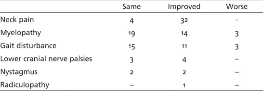
Table 2. Postoperative neurological status.
Same Improved Worse
Neck pain 4 32 –
Myelopathy 19 14 3
Gait disturbance 15 11 3
Lower cranial nerve palsies 3 4 –
Nystagmus 2 2 –
Radiculopathy – 1 –
Table 3. Mortality, morbidity in the 38 patients who underwent a transoral approach.
No. of cases Mortality (respiratory failure) 1 Morbidity (pulmonary infection) 3
CSF leak 1
region and in patients with higher upward migration of the odontoid process8-10.
TOA is now a well-established surgical procedure for anterior decompression at the CVJ1-3,5,6,11,12. This
ap-proach provides direct access to ventral lesions at the CVJ13-17 and has gained great acceptation after
Me-nezes et al.15-18 formulated physiological criteria and
guidelines for treatment of certain disorders of the CVJ. Until recently, most CVJ malformations were treated by foramen magnum decompression and sta-bilization when unstable19,20.
Thirty-eight patients with basilar invagination underwent an odontoidectomy through TOA. Most patients presented with myelopathy and neck pain. The recovery level of patients depended on the mag-nitude of the preoperative defi cits. Patients suffering from light myelopathy who could walk had a bet-ter recovery than those who could not. Patients who were unable to walk had their clinical conditions sta-bilized after surgery. Eleven of 15 patients who had gait disturbance recovered and four of seven who had cranial nerve disfunction improved after surgery. One patient died in the postoperative course because of pulmonary embolism and three developed pulmo-nary infection.
Although some authors mention the removal of intradural lesions through this approach, the major indication for this approach is in the treatment of extradural lesions due to complications originated from treating intradural lesions such as abscesses, meningitis and cerebrospinal fluid fistulas3-5,13,21,22.
One patient in our series developed a cerebrospinal fl uid fi stula that was detected and closed during the surgery. However, problems encountered in these ap-proaches included inadequate exposure of the lateral margins, lack of proximal control of the vertebral ar-teries and the possibility that incisions of the clival dura can lead to excessive hemorrhaging that can be diffi cult to control21.
On strictly technical grounds, many authors dis-agree on various points. For example, preoperative tracheostomy versus oral or nasal intubation, incision of soft palate or retraction, multilayer or single layer closure of the soft palate11,12. We close the posterior
pharyngeal wall in two layers and the soft palate in three layers.
Soft-palate incision provides access to the lower clivus and the high CVJ15-18. This technique provides
superior operative exposure compared to suture of the soft palate and its retraction into the nasal cavity. The enlarged TOA allows a better approach to more
extensive lesions in this region, in cases with higher upward migration of odontoid process in patients in whom there is limited mandibular excursion1,8-10.
However, an incision of the soft palate or osteotomy of hard palate has a high likelihood of complications such as dysphagia, nasal regurgitation and regurgi-tation with speech and swallowing. Enlarged access was necessary in most of our cases. The type of surgi-cal procedure and whether it is necessary to enlarge the access through an incision of the soft palate, os-teotomy of the hard palate, or eventual need for a maxillotomy or mandibular osteotomy, will depend on the analysis of a patient’s pathology and on the study of preoperative imaging. The CT with bone landmarks and tridimensional reconstruction accom-panied by MRI allows a good understanding of the physiopathology of the lesion. The imaging allows one to establish the relations of the odontoid pro-cess with the soft and hard palate, the extension of the vertical migration, the degree of subluxation and the condition of the neural tissue23. Three patients
had soft palate dehiscence and required resuture and long term enteral nutrition.
The importance of tracheostomy should not be overemphasized. It allows prevention of postoperative respiratory complications and is a safeguard against airway obstruction from postoperative lingual ede-ma. Preoperative tracheostomy was not performed in any case in our series. Postoperative tracheostomy was required in 10 cases because of the involvement of the lower cranial nerves and where prolonged in-tubation was required in the postoperative period. An analysis of the instability of the CVJ is manda-tory in the immediate postoperative stage2,5,11,17. The
following situations make the atlantoaxial complex vulnerable to dislocation: turning the anesthetized patient, transferring patients onto the bed or table, waiting for posterior fusion, particularly if traction is released and it is not immediately replaced. There-fore, we recommend extreme care with every patient after TOA. The large majority of patients that submit to an odontoidectomy become unstable and should be stabilized with rigid external braces until the inter-nalfi xation is completed11,16,17,24,25. Menezes et al.16
whom the arch of the atlas was removed, fi xation was mandatory. According to Menezes and Van Gilder17,
when osteoarthritis was present in the atlantoaxial joints, posterior fi xation was often not required. Al-though some authors mention ventral fi xation26–28,
posterior stabilization is more practical and safer7,29.
Of 38 patients, 18 underwent a posterior fi xation us-ing an inside–outside technique as proposed by Pait et al.7 and two were already fi xed with the Ransford
loop technique29. The method of
fi xation with tita-nium plates allows artifact-free imaging with the CT and MRI scans for the postoperative period.
In conclusion, TOA allows direct access to the cli-vus, craniovertebral junction and the anterior aspects of the fi rst three cervical segments. In principle, TOA is meant for ventral compressive lesions. The main in-dications for TOA are irreducible ventral compression, with the most common being basilar invagination.
REFERENCES
1. Appuzo MLJ, Weiss MH, Heiden JS. Transoral exposure of the atlanto-axial region. Neurosurgery 1978;3:201-207.
2. Crockard HA. The transoral approach to the base of the brain and up-per cervical cord. Ann R Coll Surg Engl 1985;67:321-325.
3. Crockard HA, Bradford R. Transoral transclival removal of schwannoma anterior to the craniocervical junction. J Neurosurg 1985;62:293-295. 4. Bonkowski JJA, Gibson RD, Snape L. Foramen magnum meningioma;
transoral resection with a bone baĝ e to prevent CSF leakage: case re-port. J Neurosurg 1990;72:493-496.
5. Crockard HA, Sen CN. The transoral approach for the management of intradural lesions at the craniovertebral junction: review of 7 cases. Neu-rosurgery 1991;28:88-98.
6. Crockard HA, Johnston F. Development of transoral approaches to le-sions of the skull base and craniovertebral junction. Neurosurg Quart 1993;3:61-82.
7. Pait TG, Al-MeĞ y O, Boop FA, Arnautovic KI, Rahman S, Ceola W. Inside-outside technique for posterior occiptocervical spine instrumentation and stabilization: preliminary results. J Neurosurg 1999;90(Suppl): S1-S7. 8. Anand VK, Harkey HL, Al-MeĞ y, O: Open door maxillotomy approach
for lesions of the clivus. Skull Base Surg 1991;1:215-217.
9. Kanamori Y, Miyamoto K, Hosoe H, Fujitsuka H, Tatematsu N, Shin K. Transoral approach using the mandibular osteotomy for atlantoaxi-al verticatlantoaxi-al subluxation in juvenile rheumatoid arthritis associated with mandibular micrognathia. J Spinal Disord Tech 2003;16:221-224. 10. Vishteh AG, Beals SP, Joganic EF, et al. Bilateral sagiĴ al split
mandibu-lar osteotomies as an adjunct to the transoral approach to the anterior
craniovertebral junction: technical note. J Neurosurg (Spine 2) 1999;90: 267-270.
11. Di Lorenzo N: Craniocervical junction malformation treated by transoral approach. A survey of 25 cases with emphasis on postoperative instabil-ity and outcome. Acta Neurochir (Wien) 1992;118:112-116.
12. Goel A, Bhatjiwale M, Desai K. Basilar invagination: a study based on 190 surgically treated patients. J Neurosurg 1998;88:962-968.
13. Drake CG. Management of aneurysms of posterior circulation. In: You-mans JR (Ed). Neurological surgery. Vol 2, Philadelphia, Saunders CO, 1973;787-806.
14. Hadley MN, Spetzler RF, Sonntag VKH. The transoral approach to the superior cervical spine: a review of 53 cases of extradural cervicomed-ullary compression. J Neurosurg 1989;71:16-23.
15. Menezes AH. Developmental and acquired abnormalities of the cranio-vertebral junction. In Van Gilder JC, Menezes AH, Dolan KD (Eds). The craniovertebral junction and its abnormalities. New York: Futura Pub-lishing 1987:109-158.
16. Menezes AH, Van Gilder JC, Graf CJ, McDonnell DE. Craniocervical ab-normalities: a comprehensive surgical approach. J Neurosurg 1980;63: 444-445.
17. Menezes AH, Van Gilder JC. Transoral-transpharyngeal approach to the anterior craniocervical junction: ten-year experience with 72 patients. J Neurosurg 1988;69:895-903.
18. Sawin PD, Menezes AH. Basilar invagination in osteogenesis imperfec-ta and related osteochondrodysplasias: medical and surgical manage-ment. J Neurosurg 1997;86:950-960.
19. Da Silva J. Basilar impression and Arnold-Chiari malformation: surgi-calfi ndings in 209 cases. Neurochirurgia 1992;35:189-195.
20. Zileli M, Cagli S. Combined anterior and posterior approach for man-aging basilar invagination associated with type I Chiari malformation. J Spinal Disord Tech 2002;15:284-289.
21. Sen CN, Sekhar LN. Surgical management of anteriorly placed lesions at the craniocervical junction-An alternative approach. Acta Neurochir (Wien) 1991;108:70-77.
22. Yamaura A, Makino H, Isobe K, Takashima T, Nakamura T, Takemiya S. Repair of cerebrospinal fl uid fi stula following transoral transclival
ap-proach to a basilar aneurysm. J Neurosurg 1979;50:834-836.
23. Pappas CTE, Rekate HL. Role of magnetic resonance imaging and three dimensional computerized tomography in cranio vertebral junction anomalies. Pediatric Neurosurg 1988;14:18-22.
24. Dickman CA, Crawford N, Brantley AGU, Sonntag VKH. Biomechanical eě ects of transoral odontoidectomy. Neurosurgery 1995;36:1146-1153. 25. Dickman CA, Locantro J, Fessler RG. The influence of transoral
od-ontoid resection on stability of craniovertebral junction. J Neurosurg 1992;77:525–530.
26. Andrade JR, MacNab I. Anterior occipito-cervical fusion using an extra-pharyngeal exposure. J Bone Joint Surg (Am) 1969;51:1621-1626. 27. Goel A, Karapurkar AP. Transoral plate screw fi xation of the
craniover-tebral junction: a preliminary report. Br J Neurosurg 1994;8:743-745. 28. Vender JR, Harrison SJ, McDonnell DE. Fusion and
instrumenta-tion at C1-C3 via the high anterior cervical approach. J Neurosurg 2000;92(Suppl):S24-S29.
29. Ransford AO, Crockard HA, Pozo JL, Thomas NP, Nelson IW. Cranio-cervical instability treated by contoured loop fi xation. J Bone Joint Surg