Arq Neuropsiquiatr 2007;65(4-B):1101-1104
1101
HIPPOCAMPAL SCLEROSIS AND STATUS EPILEPTICUS
CAUSE OR CONSEQUENCE ?
A MRI study
Gustavo Wruck Kuster
1, Pedro Braga-Neto
1, Denizart Santos-Neto
1,
Maria Teresa Garcia Santana
1, Antonio Carlos Martins Maia Jr
2,
Orlando Graziani Povoas Barsottini
1ABSTRACT -
Background:
Transient imaging abnormalities, including changes on diffusion-weighted
imag-ing (DWI), may be seen in status epilepticus. These abnormalities can be followed by hippocampal
sclero-sis.
Case report:
We report a 15-year-old lady with focal non convulsive status epilepticus (NCSE) and
fo-cal slowing on EEG. DWI exhibited abnormal hyperintense signals in bilateral temporal and insular cortices.
After 3 weeks, MRI performed a localizated hippocampal atrophy.
Conclusion:
The MRI findings indicated
vasogenic and cytotoxic edema during seizure activity and subsequent loss of brain parenchyma.
KEY WORDS: status epilepticus, magnetic resonance imaging (MRI), diffusion-weighted images, reversible
MRI abnormalities, hippocampal sclerosis.
Esclerose hipocampal e status epilepticus: causa ou conseqüência? Um estudo de RM
RESUMO -
Introdução:
Anormalidades transitórias de imagem, incluindo imagens de ressonância
magné-tica por difusão (DWI), podem ser vistas no status epilepticus. Essas anormalidades podem ser seguidas de
esclerose hipocampal.
Relato de caso:
Nós relatamos uma jovem de 15 anos com status focal não
convul-sivo e lentificação focal no EEG. DWI mostrava sinal hiperintenso em regiões temporais bilaterais e córtex
insular. Após 3 semanas, RM de encéfalo mostrava atrofia localizada do hipocampo.
Conclusão:
Os
acha-dos de RM indicam edema vasogênico e citotóxico durante as crises epilépticas com subseqüente atrofia de
parênquima cerebral.
PALAVRAS-CHAVE: status epilepticus, ressonância nuclear magnética (RNM), sequência difusão, alterações
reversíveis de RNM, esclerose hipocampal.
1Division of General Neurology, Department of Neurology of Federal University of São Paulo, São Paulo SP, Brazil; 2Fleury Institute,
Magnetic Resonance Imaging Unit, São Paulo SP, Brazil.
Received 4 June 2007, received in fi nal form 17 August 2007. Accepted 18 September 2007.
Dr. Gustavo Wruck Kuster - Rua Onze de Junho 643 / apt 164 - 04041-052 São Paulo SP - Brasil. E-mail: [email protected]
Brain magnetic resonance imaging (MRI) is
man-datory as a diagnostic workup for patients with
epi-leptic seizures in order to delineate any structural
ep-ileptogenic lesion. Transient signals abnormalities can
occur and have been attributed to functional
chang-es due to seizure activity
1. Convulsive status
epilepti-cus causes brain MRI changes after generalized and
focal motor seizures
2, although these
fi
ndings in
non-convulsive status epilepticus (NCSE) are exceptional
3,4.
Focal NCSE occurs mostly as complex partial status
epilepticus (CPSE) and several articles have reported
transient MRI abnormalities after such situation
5,6.
We report a case of a young female who
present-ed a unique, long-lasting series of partial seizures
with marked transient MRI abnormalities, followed
by localized brain atrophy in despite of complete
clin-ical recovery.
CASE
A 15 year old right-handed female was admitted in our
Neurological Unit presenting apathy and speech arrest since
5 days before the admission. Her medical records were
unre-markable for perinatal pathological events, febrile seizures,
cardiac diseases, cerebral injuries or infectious diseases.
Psy-chomotor development was normal. Family history was
neg-ative for epilepsy.
At hospital admission, she had no fever and arterial
blood pressure was normal. In neurological examination,
she presented with drowsiness, masticatory movements and
right hand dystonia.
al-Arq Neuropsiquiatr 2007;65(4-B)
1102
Status epilepticus: hippocampal sclerosis Kuster et al.
pha / theta activity of 6-9 Hz over the left central and
mid-dle line regions (Fig 1).
Benzodiazepines, followed by intravenous
phenobar-bital were done with no improvement. She required
intu-bation, mechanical ventilation and continuous infusion of
benzodiazepines in a Intensive Care Unit (ICU).
A CT scan of the brain on the day of admission showed
a left temporal hypodensity with slight contrast
enhance-ment. Cerebrospinal
fl
uid (CSF) analyzed on day 1 was
acel-lular with normal levels of glucose and protein. Further CSF
samples on the day 7 and 14 were also acellular. Routine
haematology and blood biochemistry were normal.
Microbi-ological studies were negative. Human Imunode
fi
ciency
Vi-rus (HIV), Hepatitis B viVi-rus (HBV) and Hepatitis C viVi-rus (HCV)
serologies were negative.
On the seventieth day the patient had an EEG
improve-ment, a partial consciousness recovery and mechanical
ven-tilation was discharged. By this time, the first brain MRI
scan was done.
Three weeks later, after a complete clinical recovery, a
follow-up MRI was performed.
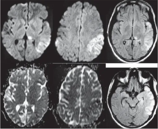
MRI abnormalities –
At the admission, serial MRI scans
(1,5T, Sonata, Siemens, Erlanger, Germany) were performed.
After repeated episodes of NCSE, diffusion-weighted
imag-es and T2-weighted imagimag-es FLAIR exhibited abnormal
hy-perintense signals in bilateral temporal and insular cortices.
These abnormalities appeared more extensive on
diffusion-weighted images than on T2-diffusion-weighted images FLAIR. The
hippocampi appeared abnormal and also presented
bilat-eral abnormal hyperintense signals, without atrophy, more
extensive on the left hippocampus. There was no disruption
of blood-brain barrier (BBB) on T1-weighted images after
in-travenous administration of Gd-DTPA (Fig 2).
After 3 weeks, there were no signal abnormalities on
diffusion-weighted images and T2-weighted images FLAIR.
However, enlargement of the subarachnoid space just near
the left temporal lobe and enlargement of the temporal
horn of the right lateral ventricle were evident, indicating
localized brain atrophy (hippocampal atrophy) (Fig 3).
Fig 1. EEG showed the presence of continuous spikes with al-pha / theta activity of 6-9 Hz over the left central and middle line regions.
Arq Neuropsiquiatr 2007;65(4-B)
1103 Status epilepticus: hippocampal sclerosis Kuster et al.
DISCUSSION
We describe here a case of non-convulsive status
epilepticus characterized by clinical,
electroenceph-alographical and MRI findings. Brain MRI showed
reversible diffusion and T2-weighted images
abnor-malities in bilateral temporal and insular lobes, which
disappeared after three weeks, giving place to
corti-cal atrophy. The patient experienced clinicorti-cal as well
as EEG recovery after this period.
Partial seizures and status epilepticus are
associ-ated with a local change of blood
fl
ow and neuronal
metabolism. This is clearly demonstrated by numerous
PET and SPECT studies which have shown increased
cerebral flow and metabolite consumption in the
area of epileptic focus
7-9. These
fi
ndings have been
con
fi
rmed by functional MRI, with important clinical
implications
10.
The above-mentioned metabolic phenomena are
proportional to the frequency and duration of the
seizures
1,11,12. They determine a transitory alteration of
the BBB, an increased of vascular permeability and the
subsequent appearance of cerebral edema. BBB could
be differently compromised and this could explain the
MRI
fi
ndings reported in literature, which vary from
signal alteration caused by vasogenic and/or cytotoxic
edema to contrast enhancement, as a result of either
damage or breakdown of the BBB, respectively
13,14.
The association of hippocampal abnormalities
with chronic epilepsy and status epilepticus is well
stablished
15-19. The nature of the neuropathological
changes found depends on how long standing they
are. In early cases there are ischemic changes in
neu-rones in vulnerable areas (hippocampus, thalamus
and striatum) with acute astrocytic reation. In the
later cases there is neuronal loss and gliosis in these
areas. It is generally accepted that status epilepticus
directly results in hippocampal sclerosis
20,21, but many
of the reports are of cases with encephalitis, with
se-vere complications of status epilepticus such hypoxia,
hypoglycemia or with other major systemic illness
17.
In our case, no underlying cause was found. The
CSF and serological studies showed no evidence of
encephalitis. The patient did not develop major
sys-temic disturbances such as hypoxia or hypoglycemia
22.
These factors suggest that in this case, the
abnormali-ties detected on MRI are a direct result of the
pa-tient’s status epilepticus. An alternative explanation
would be that the hippocampal changes caused the
status epilepticus, rather then the other way around.
We believe that the clinical picture, the appearance
of new changes in MRI after the onset of the status
epilepticus, make this explanation unlikely.
REFERENCES
1. Jackson GD, Conelly A, Cross JD, Gordon I, Gadian GD. Functional magnetic resonance imaging of focal seizures. Neurology 1994;44:850-856.
2. Kim JA, Chung JI, Yoon PH. Transient MR changes in patients with ton-icoclonic seizures or status epilepticus: periictal diě usion weight
Arq Neuropsiquiatr 2007;65(4-B)
1104
Status epilepticus: hippocampal sclerosis Kuster et al.
ing. Am J Neuroradiol 2000;22:1149-1160.
3. Callahan DJ, Noetzel MJ. Prologed abcense status epilepticus associated with carbamazepine therapy, increased intracranial pressure, and tran-sient MRI abnormalities. Neurology 1992;42:2198-2201.
4. Castro-Costa CM, Vale OC, Leitão V, et al. Epilepsia partialis continua (Ko-shevhikov): a preliminary case report. Arq Neuropsiquiatr 2000;58: 916-918. 5. Chu K, Kang DW, Kim JY, Chang KH, Lee SK. Diě usion-weighted MRI
in nonconvulsive status epilepticus. Arch Neurol 2001;58:993-998. 6. Lansberg MG, O’Brien MW, Norbash AM. MRI abnormalities
associat-ed with partial status epilepticus. Neurology 1999;52:1021-1027. 7. Theodore WH, Newmark ME, Sato S, Brook R. Fluorodeoxyglucose
pos-itron emission tomography in refractory complex partial seizures. Ann Neurol 1983;14:429-437.
8. Lee BI, Markand OM, Welman HN, Krepshaw J. HIPDM-SPECT in pa-tients with medically intractable complex partial seizures. Arch Neurol 1988;45:397-402.
9. Juhasz C, Scheidl E, Szirmai I. Reversible focal MRI abnormalities due to status epilepticus: an EEG, SPECT, transcranial Doppler follow up study. Electroenceph. Clin Neurophysiol 1998;107:402-407.
10. Detre JA, Alsop DC, Aguirre JK. Coupling of cortical and thalamic ic-tal activity in human partial epilepsy: demonstration of functional MRI. Epilepsia 1996;37:657-661.
11. Sammaritano M, Andermann F, Melanson D, Pappius HM. Prolonged fo-cal cerebral edema in partial status epilepticus. Epilepsia 1985:26;334-339. 12. Kramer RE, Luders H, Lesser RP, Weinstein MR, Dinner DS. Transient
focal abnormalities of neuroimaging studies during focal status
epilep-ticus. Epilepsia 1987;28:528-532.
13. Yaě e K, Ferriero D, Barkovic AJ. Reversible MRI abnormalities follow-ing seizures. Neurology 1995;45:104-109.
14. M El- Koussy. Focal status epilepticus: follow up by perfusion and dif-fusion MRI. Eur Radiol 2002;12:568-574.
15. Corsellis JAN, Bruton CJ. Neuropathology in status epilepticus in hu-mans. In Delgado-Escueta AV, Porter RJ (Eds). Advances in Neurology. Vol 34. Status epilepticus. New York, 1983:129-139.
16. Margerison JH, Corsellis JAN. Epilepsy and the temporal lobes: a clini-cal eletroencephalography and neuropathologiclini-cal study of the brain in epilepsy. Brain 1966:84;429-530.
17. Nobuyuki K. Magnetic resonance imaging and PET findins in sta-tus epilepticus following hypoglycemia. Ann Nuclear Med 2006;20: 371-376.
18. Nixon J, Bateman D, Moss T. An MRI and neuropathological study of a case of fatal status epilepticus. Seizure 2001:10;588-591.
19. Keun-Sik H. Diě usion changes sugesting vasogenic oedema during par-tial status epilepticus. Seizure 2004;13:317-321.
20. Parmar H. Acute symptomatic seizures and hippocampus damage: DWI and MRS fi ndings. Neurology 2006;66:1732-1735.
21. Bauer G, Godwald TH, Dodensberg J, et al. Transient and permanent MRI abnormalities aĞ er complex partial status epilepticus. Epilep Be-hav 2006;8:666-671.