DNA damage by ochratoxin A in rat
kidney assessed by the alkaline
comet assay
Institute for Medical Research and Occupational Health, Zagreb, Croatia D. ðeljeñic´,
A.-M. Domijan and M. Peraica
Abstract
There are few studies of ochratoxin A (OTA) genotoxicity in experi-mental animals and the results obtained with cell cultures are inconsis-tent, although the carcinogenic potential of OTA for the kidney of experimental animals has been well established. We studied the genotoxic potential of OTA in the kidney of adult female Wistar rats (5 in each group) treated intraperitoneally with OTA (0.5 mg kg body weight-1 day-1 for 7, 14, and 21 days) measuring DNA mobility on
agarose gel stained with ethidium-bromide using standard alkaline single-cell gel electrophoresis (comet assay). Negative control ani-mals were treated with solvent (Tris buffer, 1.0 mg/kg) and positive control animals were treated with methyl methanesulfonate (40 mg/ kg) according to the same schedule. OTA concentrations in plasma and kidney homogenates in 7-, 14-, and 21-day treated animals were 4.86 ± 0.53, 7.52 ± 3.32, 7.85 ± 2.24 µg/mL, and 0.87 ± 0.09, 0.99 ± 0.06, 1.09 ± 0.15 µg/g, respectively. In all OTA-treated groups, the tail length, tail intensity, and tail moment in kidney tissue were signifi-cantly higher than in controls (P < 0.05). The tail length and tail moment were higher after 14 days than after 7 days of treatment (P < 0.05), and still higher after 21 days (P < 0.05). The highest tail intensity was observed in animals treated for 21 days, and it differed significantly from animals treated for 7 and 14 days (P < 0.05). OTA concentrations in plasma and kidney tissue increased steadily and OTA concentration in kidney tissue strongly correlated with tail intensity and tail moment values. These results confirm the genotoxic potential of OTA, and show that the severity of DNA lesions in kidney correlates with OTA concentration.
Correspondence
A.-M. Domijan
Institute for Medical Research and Occupational Health Ksaverska c. 2 10001 Zagreb Croatia
Fax: +385-1-4673-303 E-mail:[email protected]
Research supported by the Mutagenesis Unit, Institute for Medical Research and Occupational Health and by the Ministry of Science, Education and Sports of the Republic of Croatia.
Received September 12, 2005 Accepted August 29, 2006
Key words •Comet assay •DNA adducts •Experimental animals •Mycotoxin
Introduction
Ochratoxin A (OTA) is a mycotoxin pro-duced by some strains of Penicillium and Aspergillus molds that naturally contami-nate food and feed under all climatic condi-tions (1). Humans are exposed to OTA by ingestion of contaminated food of vegetable
other-wise rare urothelial tumors has also been attributed to exposure to OTA. It was proven that OTA increases the incidence of uncom-mon tubular cell adenomas and of tubular cell carcinomas in rats of both sexes in a dose-dependent manner and the incidence and multiplicity of fibroadenomas of the mammary gland in female rats (3). The In-ternational Agency for Research on Cancer classified OTA as a 2B group compound (possibly carcinogenic to humans and with sufficient evidence for carcinogenicity in laboratory animals), although the mechan-ism of its carcinogenicity is not understood (4). It is believed that this assumed OTA carcinogenicity should reflect in DNA changes. The genotoxicity of OTA was tested in various cell lines, but the results were inconclusive (4), probably because routine genotoxicity tests were contrasting, equivo-cal or negative in compounds whose geno-toxic activity depended on tissue-specific mechanisms. Furthermore, the genotoxic activity is organ and tissue specific for com-pounds with distinct targets for carcinoge-nicity (5). The genotoxicity of OTA in labo-ratory animals has been postulated mainly on the basis of the results of 32P-postlabeling experiments, which showed OTA-depend-ent formation of DNA adduct spots (6,7). The single available report on genotoxicity tested by the comet assay in rat kidneys revealed DNA fragmentation, but animals were treated with a single, very high oral OTA dose (10 mg/kg body weight) (8).
The alkaline single-cell gel electropho-resis (comet) assay is a sensitive and power-ful method for determining DNA strand breaks and alkali labile sites (oxidative DNA base damage, and DNA-DNA/DNA-protein cross-linking, DNA adducts) at the cell level and has been widely used for genotoxicity studies. Since the comet assay does not re-quire cell cultivation, the genotoxic poten-tial of a substance of interest can be assessed in the cells of specific target tissue. The aim of the present study was to determine the
time course of OTA-produced DNA damage by assessing alkali-labile sites using the comet assay in rat kidney tissue.
Material and Methods
Chemicals
OTA (99% purity), Na2EDTA, methyl methanesulfonate, and the chemicals needed for the comet assay were purchased from Sigma (St. Louis, MO, USA). Tris (hydroxy-methyl) aminomethane and NaCl were from Kemika (Zagreb, Croatia). Water (Merck, Darmstadt, Germany) and methanol (Ke-mika, Zagreb, Croatia) used for the high-performance liquid chromatography (HPLC) mobile phase were of HPLC grade.
Animal treatment schedule
Adult female Wistar rats weighing 190 g maintained on a 12-h light/dark cycle and at a constant temperature of 24ºC were kept in macrolone cages. Animals were fed a stand-ard diet for laboratory rodents (Pliva, Zagreb, Croatia) and had free access to water. The study was approved by the Ethics Commit-tee of the Institute for Medical Research and Occupational Health, Zagreb, according to current laws of the Republic of Croatia.
The rats were randomly assigned to nine groups of 5 animals each receiving either OTA (0.5 mg/kg body weight) dissolved in Tris buffer or solvent only (1.0 mL/kg, nega-tive control animals) intraperitoneally (ip) every day for up to three weeks. The ip treatment was chosen because it is consid-ered to be a valuable tool for clarifying the mechanism of action of mycotoxins (9). Posi-tive control animals were treated with meth-yl methanesulfonate (40 mg/kg body weight) according to the same schedule.
washed in saline, and kept on ice until the comet assay and OTA analysis.
OTA analysis
OTA concentrations in plasma and 10% kidney homogenate (0.9% sterile solution of NaCl) were determined by HPLC according to methods described by Peraica et al. (10) and Bauer et al. (11), respectively.
OTA was determined by HPLC (Varian, Walnut-Creek, CA, USA) with fluorescence detection. The guard column and analytical column were LiChrospher RP-18 (Merck, Darmstadt, Germany) 4.0 x 4.0 and 125.0 x 4.0 mm, respectively, with 5-µm particles. Chromatography results were processed us-ing Star Chromatography Workstation soft-ware (version 5.0; Varian).
For OTA detection, the mobile phase consisted of methanol, water and acetic acid (70:30:2) with a flow rate of 0.5 mL/min. Detector wavelengths were set at λex 336 and λem 464. OTA was identified by constant retention time and quantified by comparison with peak areas of spiked samples. For the calibration curve, OTA-free plasma and kid-ney homogenates were spiked with a known amount of OTA and were cleaned up as samples. OTA-positive samples were con-firmed by adding a known amount of stand-ard OTA solution.
Preparation of kidney tissue homogenate for the comet assay
Tissue samples were homogenized in chilled homogenization buffer, pH 7.5, con-taining 75 mM NaCl and 24 mM Na2EDTA to obtain a 10% tissue solution. A Potter-type homogenizer was used, and kidney samples were kept on ice during and after homogenization (12).
Alkaline comet assay
We used a modified alkaline comet assay
protocol according to Sasaki et al. (12). Six microliters of kidney homogenate was sus-pended on 0.5% low-melting agarose and sandwiched between a layer of 0.6% nor-mal-melting agarose and a top layer of 0.5% low melting agarose on fully frosted slides. The slides were kept on ice during the poly-merization of each gel layer. After the solidi-fication of the 0.6% agarose layer, the slides were immersed in a lysis solution (1% so-dium sarcosinate, 2.5 M NaCl, 100 mM Na2EDTA, 10 mM Tris-HCl, 1% Triton X-100, and 10% DMSO) at 4oC. After 1 h, the slides were placed in electrophoresis buffer (0.3 M NaOH, 1 mM Na2EDTA, pH 13) for 10 min at 0ºC to allow DNA to unwind. Electrophoresis was performed for 10 min at 300 mA and 1 V/cm. The slides were neu-tralized with Tris-HCl buffer, pH 7.5, and stained with 20 µg/mL ethidium-bromide for 10 min. Each slide was analyzed using the Leitz Orthoplan (Wetzlar, Germany) epi-fluorescence microscope. One hundred cells were analyzed on each slide using the comet assay II automatic digital analysis system (Perceptive Instruments Ltd., Halstead, Suf-folk, UK) to determine tail length and tail intensity. Tail length (µm) is the distance of DNA migration from the center of the body of the nuclear core and is used to evaluate the extent of DNA damage. Tail intensity (%) is the percentage of the genomic DNA that migrates during electrophoresis from the nuclear core to the tail. Both tail length and tail intensity are measured automati-cally by image analysis software. Tail mo-ment was calculated according to the fol-lowing formula: tail moment = tail length x tail intensity/100 (12). According to Robbi-ano et al. (8), data are shown as DNA-damaging potency (comet assay endpoint treated - comet assay endpoint control)/OTA concentration µM).
Statistical analysis
endpoints between the kidney samples from control and treated rats were determined by the non-parametric Kruskal-Wallis test. The same statistical method was used to estimate the significance of differences between dif-ferent treatment durations. Probability val-ues of P < 0.05 were considered to be statis-tically significant.
The correlation between doses of OTA in kidney homogenates and comet assay end-points was tested using the linear regression analysis.
Results
During the experiment there were no
changes in body or kidney weight of OTA-treated rats compared to negative control animals.
The OTA concentrations in plasma (µg/ mL) and kidney homogenate (µg/g tissue) of treated animals are reported as means ± SD (Table 1). During the experiment, OTA con-centrations in plasma and kidney tissue in-creased steadily.
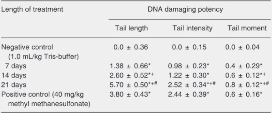
We assessed the time course of OTA genotoxicity in the kidney of treated animals using the comet assay as a highly effective tool for the biomonitoring of DNA integrity. In all OTA-treated groups (0.5 mg kg body weight-1 day-1 for 7, 14, and 21 days), the tail length in kidney tissue was significantly higher than in controls (P < 0.05; Table 2). Tail length in animals treated for 7, 14, and 21 days increased with the length of treat-ment, and the difference between all groups was significant (P < 0.05). Tail intensity in kidney cells of all treated groups was higher than in controls (P < 0.05; Table 2). The highest tail intensity was observed in ani-mals treated for 21 days, and the difference was significant in respect to animals treated for 7 and 14 days (P < 0.05). Tail moment also increased with the length of treatment, with significant differences between all groups (P < 0.05; Table 2).
Discussion
The genotoxic properties of OTA have been previously tested using the comet assay on Madin-Darby canine kidney (MDCK) cells, human fibroblasts, and human-derived hepatoma (HepG2) cells (13-15). OTA in-duced single-strand breaks in MDCK and HepG2 cells in a concentration-dependent manner, while in human fibroblasts it caused DNA breaks in a time- and dose-dependent manner. The increase in all comet assay endpoints observed in the present study agrees with DNA strand breaks detected with the comet assay in the above mentioned in vitro studies.
Table 1. Concentrations of ochratoxin A in plasma and kidney of rats receiving ochratoxin A (ip, 0.5 mg kg body weight-1 day-1) for 7, 14 and 21 days.
Length of treatment (days) Concentration of ochratoxin A
Plasma (µg/mL) Kidney (µg/g)
7 4.86 ± 0.53 0.87 ± 0.09
14 7.52 ± 3.32 0.99 ± 0.06
21 7.85 ± 2.24 1.09 ± 0.15
Data are reported as means ± SD for 5 animals in each group.
Table 2. Tail length, tail intensity (% of total genomic DNA found in the tail of the comets), and tail moment (tail length x tail intensity/100) measured with comet assay in kidney cells of animals treated with ochratoxin A.
Length of treatment DNA damaging potency
Tail length Tail intensity Tail moment
Negative control 0.0 ± 0.36 0.0 ± 0.15 0.0 ± 0.04 (1.0 mL/kg Tris-buffer)
7 days 1.38 ± 0.66* 0.98 ± 0.23* 0.4 ± 0.29* 14 days 2.60 ± 0.52*+ 1.22 ± 0.30* 0.6 ± 0.12*+
21 days 5.70 ± 0.50*+# 2.52 ± 0.34*+# 0.8 ± 0.12*+#
Positive control (40 mg/kg 3.80 ± 0.43* 2.44 ± 0.39* 0.6 ± 0.16* methyl methanesulfonate)
Data are reported for kidney cells from control animals as the mean for all three time points (N = 15) and from animals treated with 0.5 mg ochratoxin A (OTA)/kg body weight for 7, 14 and 21 days (N = 5 in each group). Data are reported as means ± SEM (endpoint value treated animals - endpoint value corresponding control)/OTA, 1.23 µM).
*P < 0.05 compared to control; +P < 0.05 compared to 7-day treatment; #P < 0.05
OTA is undoubtedly a carcinogenic com-pound that causes tumors in rat kidneys (3). Until now, evidence of OTA genotoxicity in vivo was obtained mostly by measuring DNA adducts in the kidneys of mice and rats using the 32P-post-labeling method (6,7). While some investigators claim that OTA binds covalently to DNA, forming OTA-DNA ad-ducts (16), others used a very sensitive method (liquid chromatography/mass spec-trometry/mass spectrometry) to show that DNA binding of OTA in exposed rats cannot be detected and is unlikely to reveal the mechanism of OTA toxicity (17).
Robbiano et al. (8) were the first to report OTA genotoxicity based on tests using alka-line single-cell gel electrophoresis in the rat kidney. The statistically significant increase in the average frequency of DNA breaks and/or alkali labile sites was obtained with a very high single OTA dose (10 mg/kg body weight, that is ½ LD50). In the present study, 0.5 mg OTA kg body weight-1 day-1 for 7 days was sufficient to significantly increase the tail length and intensity in the kidney of experimental animals. Significant differences in tail length, tail intensity and tail moment between 7 and 21 days of treatment indicate that these lesions are time dependent.
When tested using linear regression
anal-ysis, OTA concentration in kidney tissue showed a high positive correlation with tail length, tail intensity and tail moment. OTA concentration was found to correlate more strongly with tail intensity (r2 = 0.9936, P = 0.02) and tail moment (r2 = 0.9874, P = 0.02) than with tail length (r2 = 0.8679, P = 0.24). These results are expected because, during electrophoresis, DNA does not migrate as fragments. The tail of the comet is the result of the migration of relaxed DNA loops that are pulled by the electric field to a limited distance from the core. The length of these loops determines the length of the comet tail. Since tail intensity indicates the number of DNA breaks, it is assumed that beyond some critical amount of damage, what increases is tail intensity rather than tail length (18).
The present study confirms the genotox-icity of OTA reported in earlier studies on cell cultures and on experimental animals. The genotoxicity of OTA found in our study is consistent with its carcinogenic potential in the kidney of experimental animals.
Acknowledgments
We wish to thank Mrs. Jasna Milekovic´, Mrs. Marija Kramaric´, and Mrs. Mirjana Matašin for technical assistance.
References
1. Speijers GJA, van Egmond H. Worldwide ochratoxin A levels in food and feeds. In: Creppy EE, Castegnaro M, Dirheimer G (Editors),
Human ochratoxicosis and its pathologies. Paris: John Libbey Eurotox Ltd.; 1993. p 85-100.
2. Krogh P. Mycotoxin porcine nephropathy: a possible model for Balkan endemic nephropathy. Second International Symposium on Endemic Nephropathy. 1974 March 25-31; Sofia. 1974.
3. Boorman GA. Technical Report on the Toxicology and Carcinogen-esis Studies of Ochratoxin A in F344/N Rats. Durham: Research Triangle Park, National Institute of Health; 1989.
4. IPCS. WHO Food Additives Series. Safety evaluation of certain mycotoxins in food. Geneva: World Health Organization; 2001. 5. Robbiano L, Carrozzino R, Bacigalupo M, Corbu C, Brambilla G.
Correlation between induction of DNA fragmentation in urinary blad-der cells from rats and humans and tissue-specific carcinogenic activity. Toxicology 2002; 179: 115-128.
6. Pfohl-Leszkowicz A, Chakor K, Creppy EE, Dirheimer G. DNA
ad-duct formation in mice treated with ochratoxin A. In: Castegnaro M, Pleština R, Dirheimer G, Chernozemsky IN, Bartsch H (Editors),
Mycotoxins, endemic nephropathy and urinary tract tumours. Lyon: IARC Scientific Publications No. 115. International Agency for Re-search on Cancer; 1991. p 245-253.
7. Petkova-Bocharova T, Stoichev II, Chernozemsky IN, Castegnaro M, Pfohl-Leszkowicz A. Formation of DNA adducts in tissues of mouse progeny through transplacental contamination and/or lacta-tion after administralacta-tion of a single dose of ochratoxin A to the pregnant mother. Environ Mol Mutagen 1998; 32: 155-162. 8. Robbiano L, Baroni D, Carrozzino R, Mereto E, Brambilla G. DNA
damage and micronuclei induced in rat and human kidney cells by six chemicals carcinogenic to the rat kidney. Toxicology 2004; 204: 187-195.
10. Peraica M, Domijan AM, Fuchs R, Lucic A, Radic B. The occurrence of ochratoxin A in blood in general population of Croatia. Toxicol Lett
1999; 110: 105-112.
11. Bauer J, Gareis M, Gedek B. Determination and occurrence of ochratoxin A in slaughtered swine. Berl Munch Tierarztl Wochenschr
1984; 97: 279-283.
12. Sasaki YF, Nishidate E, Izumiyama F, Matsusaka N, Tsuda S. Simple detection of chemical mutagens by the alkaline single-cell gel electrophoresis (Comet) assay in multiple mouse organs (liver, lung, spleen, kidney, and bone marrow). Mutat Res 1997; 391: 215-231.
13. Lebrun S, Follmann W. Detection of ochratoxin A-induced DNA damage in MDCK cells by alkaline single cell gel electrophoresis (comet assay). Arch Toxicol 2002; 75: 734-741.
14. Russo A, La Fauci L, Acquaviva R, Campisi A, Raciti G, Scifo C, et al. Ochratoxin A-induced DNA damage in human fibroblast:
protec-tive effect of cyanidin 3-O-beta-d-glucoside. J Nutr Biochem 2005; 16: 31-37.
15. Ehrlich V, Darroudi F, Uhl M, Steinkellner H, Gann M, Majer BJ, et al. Genotoxic effects of ochratoxin A in human-derived hepatoma (HepG2) cells. Food Chem Toxicol 2002; 40: 1085-1090.
16. Faucet V, Pfohl-Leszkowicz A, Dai J, Castegnaro M, Manderville RA. Evidence for covalent DNA adduction by ochratoxin A following chronic exposure to rat and subacute exposure to pig. Chem Res Toxicol 2004; 17: 1289-1296.
17. Mally A, Zepnik H, Wanek P, Eder E, Dingley K, Ihmels H, et al. Ochratoxin A: lack of formation of covalent DNA adducts. Chem Res Toxicol 2004; 17: 234-242.