Genetic damage induced by trophic doses of lead in the neotropical fish
Hoplias malabaricus
(Characiformes, Erythrinidae) as revealed by the comet
assay and chromosomal aberrations
Marta Margarete Cestari
1, Priscilla Maria M. Lemos
1, Ciro Alberto de Oliveira Ribeiro
2,
João Ricardo M. Alves Costa
2, Emilien Pelletier
3, Marcos V.M. Ferraro
1,6, Mário Sérgio Mantovani
5and Alberto Sergio Fenocchio
4,51
Universidade Federal do Paraná, Departamento de Genética, Curitiba, PR, Brazil.
2Universidade Federal do Paraná, Departamento de Biologia Celular, Curitiba, PR, Brazil.
3Institut des Sciences de la Mer de Rimouski, Allée des Ursulines-Rimouski, Quebec, Canada.
4Universidad Nacional de Misiones, Departamento de Genética, Posadas, Misiones, Argentina.
5Universidade Estadual de Londrina, Departamento de Biologia Geral, Londrina, PR, Brazil.
6Universidade Federal de Santa Catarina, Colégio de Aplicação, Florianópolis, SC, Brazil.
Abstract
The effects of clastogenic or mutagenic agents have rarely been studied in neotropical fish species exposed to contaminated water. In this study, the genetic damage caused by lead in the widely distributed South American fish, Hoplias malabaricus, was assessed using the comet (SCGE) assay and by testing for chromosomal aberrations. Eighteen specimens were acclimatized to laboratory conditions and then chronically exposed to contaminated food by feeding prey (Cyprinus sp.) injected intraperitoneally with doses of inorganic lead adjusted to give a contamination level of 21µg of Pb2+
.g-1
net weight ofH. malabaricus. Three fish were sampled for chromosomal analysis after four doses (18 days) and another three after eight doses (41 days) of lead and the results then compared with three untreated controls kept under lead-free conditions. An additional six treated fish and three controls were sampled for the comet assay after 13 doses (64 days). Exposure to lead significantly increased the frequency of chromosomal aberrations and the frequency of tailed cell nuclei, the latter indicating DNA damage. These results show thatH. malabaricus is a useful biological model for screening the clastogenic effects of lead and possibly other xenobiotics. The genetic damage seen here illustrates the need to investigate the potential effects of heavy metals on fish species in South America.
Key words:chromosomal aberrations, comet assay, genotoxicity,Hoplias malabaricus, lead, mutagenesis.
Received: September 10, 2002; Accepted: October 21, 2003.
Introduction
The pollution of freshwater systems has long been considered a serious problem in Europe and North America (Heath, 1995). In contrast, there have been few studies of the effects of heavy metal and organic pollutants on aquatic organisms in neotropical ecosystems. The presence of heavy metals in industrial and municipal wastewater and in mine tailings has generated concern about the potential mutagenic and carcinogenic actions of these substances in aquatic organisms. In numerous aquatic ecosystems in Bra-zil, native species are often directly threatened by their
ex-posure to water-borne agents or, indirectly, through a con-taminated food chain (Oliveira Ribeiroet al., 2000).
The potential for intoxication by lead and its organo-metallic derivatives in aquatic ecosystems, atmospheric particles, and soil has increased as a result of industrial ac-tivities (Pain, 1995). Investigations of the toxic effects of chemical pollutants on fish cells have demonstrated the im-portance of cytogenetic aberrations as an important indica-tor for environmental moniindica-toring and assessment. Studies in laboratory animals have shown that exposure to lead at levels 10 mg Pb2+.mL-1of blood leads to chromosomal ab-errations (tetraploidy, mitotic anomalies, chromatid breaks), and these effects may be related to inteference with the mechanisms of replication, transcription and DNA re-pair (Goyer and Moore, 1974). Part of this effect on chro-www.sbg.org.br
Send correspondence to Marta Margarete Cestari, Universidade Federal do Paraná, Departamento de Genética, Caixa Postal 19071, 81531-990 Curitiba, PR, Brazil. E-mail: margaces@ufpr.br.
mosomes may be related to the ability of lead to form bridges with structural proteins that can adversely affect the degree of chromosomal condensation
Fish are good indicators for assessing the genotoxic and mutagenic effects of xenobiotics and physical agents (Al-Sabti, 1986). Sister chromatid exchange (SCE) tests have been applied to various fish species (Kligerman, 1979; Vigfussonet al., 1983) and the clastogenic effects of carci-nogenic-mutagenic chemicals on kidney cells ofCyprinius carpiohave been described (Al-Sabti, 1986). The comet as-say is useful for evaluating genetic alterations and has been used as a rapid method to monitor genotoxicity in bullheads and carp (Monteith and Vanstone, 1995) and to detect the effects of carcinogens, such as aflatoxins, in trout and chan-nel catfish (Abd-Allahet al., 1999).
The use of neotropical fish to study the mutagenic ef-fects of chemicals is still relatively uncommon (Matsumoto and Cólus, 2000). Hoplias malabaricus is an important predatory freshwater fish widely distributed throughout South America (Lopez and Fenocchio, 1994) and easily adapts to laboratory conditions. Since this species has only a few (2n = 40 or 2n = 42) biarmed, relatively large chromo-somes, it is easy to handle in chromosomal studies. In this study, we used the comet (SCGE) assay and a test for chro-mosomal aberrations to assess the genetic damage caused by lead fed toH. malabaricus. To our knowledge, this is the first report to examine chromosomal damagein vivo fol-lowing long-term trophic exposure of this species to inor-ganic lead.
Material and Methods
Experimental design
MatureH. malabaricus(average weight = 87.4±8.9 g) were collected from the Canguiri fish farm at the Federal University of Paraná (Curitiba, PR, Brazil). Before being used, the fish were acclimatized individually for 20 days in an aquarium containing well-aerated, dechlorinated water at 21 °C and a constant photoperiod (12 h light/dark cycle). Food (young specimens ofCypriniussp. 5 days) was pro-videdad libitum. TwelveH. malabaricuswere divided into three groups (A, B and D) for treatment with lead and six other fish were used as controls (groups C and E). For in-toxication with lead, the food items were injected intra-peritoneally with a solution of Pb(NO3)2. The volume of
solution injected into the prey fish was adjusted to give a normalized ingested dose of 21µg Pb2+
.g-1wet weight ofH. malabaricus. The control group was fed fish injected with distilled water. Groups A and B were exposed to lead for 18 and 41 days, respectively, and the fish then used for chro-mosomal aberration analysis. Group D fish were exposed for a longer time (64 days, 13 doses) and the genetic effects were evaluated using the comet (SCGE) assay. In this case, group E served as the controls. Fish from each group were anesthetized with 0.02% MS222 (SIGMA (ethyl-ester.3.
aminobenzoic acid). Kidney and blood samples were used for the chromosomal aberrations test and for the comet as-say, respectively.
Chromosomal preparations
Mitotic chromosomes were obtained from short-term cell kidney cultures, as described elsewhere for fish cyto-genetics (Fenocchioet al., 1991). Air-dried chromosome slides were stained with 5% Giemsa in phosphate buffer (pH 6.8). The frequency of chromosomal aberrations was scored by counting approximately 100 metaphase plates per individual. The different types of chromosomal aberra-tions,i.e., gaps, breaks, and fragments, were grouped into a single category: altered metaphase plates.
Comet assay
This assay was done essentially as described by Speit and Hartmann (1999) using blood cells collected via the caudal vein into a 5 mL heparinized syringe. Initially, an aliquot of blood (10µl) was diluted in 1 mL of fetal calf se-rum. The slides for microscopy were prepared using 10µL of this cell suspension and 120µL of low melting point agarose at 37 °C followed by incubation in lysis solution (1 mL of Triton X-100, 10 mL of DMSO and 89 mL of stock lysis solution [2.5 M NaCl, 100 mM EDTA, 10 mM Tris, -adjust to pH 10 with ~0.8% NaOH-, 10 mM Na-lauroyl sarcosine]) for 1 h in the dark at 8 °C.
After lysis, the slides were placed in buffer (0.3 N NaOH, 1 mM EDTA, pH > 13) for 20 min to unravel the DNA. Electrophoresis was done for 20 min at 25 V and 300 mA, after which the slides were neutralized for 15 min in 0.4 M Tris, fixed in absolute ethanol for 10 min and stained with ethidium bromide (0.02µg/mL).
Comet formation was observed with a Nikon epifluo-rescence microscope (filter B-3A; excitation: λ = 420-490 nm; emission:λ= 520 nm) at a magnification of 400X. One hundred cells were analyzed per sample (treated and controls) and were visually classified (Kobayashi et al.,
1995) based on the migration of the fragments (tail size) (class 0 - no visible damage, 1 - a short tail smaller than the diameter of the nucleus, 2 - a tail length 1-2 times the diam-eter of the nucleus, and 3 - a tail length >two times the di-ameter of the nucleus). The total score for 100 comets was obtained by multiplying the number of cells in each class by the damage class, and ranged from 0 (all undamaged) to 300 (all maximally damaged).
Statistical analysis
The mean frequencies of the chromosomal alterations in fish exposed to lead for 18 (A) and 41 (B) days and in the control group (C) were compared by one-way analysis of variance (ANOVA), with a value of p≤0.05 indicating sig-nificance. The comet assay data were scored as classes based on the damage seen and were analyzed by theχ2
in which the total number of comets in each treatment was compared with the controls.
Results and Discussion
Table 1 shows the frequency of chromosomal aberra-tions in metaphase plates ofH. malabaricus. Because of the low frequencies observed, and in order to facilitate compar-isons, all chromosomal alterations in each group were pooled into a single category.
Control fish showed almost no chromosomal abnor-malities (1.1%) when compared to those fed lead-conta-minated food for 18 or 41 days (25.6% and 19.1% of altered plates, respectively). Despite the low number of specimens used, these differences were significant (p < 0.05) because of the low variability among plates. Both treated groups showed an increase in several types of chromosomal abnor-malities, including chromatid gaps and breaks, chromo-somal fragmentation, chromatin decondensation and pericentric inversions (Table 1, < 0.05). Chromatid breaks
were the predominant chromosomal aberrations after treatment.
There was a quantitative relationship between the oc-currence of chromosomal abnormalities and the ingestion of lead. This metal can alter the genetic material of fish (Al-Sabti, 1985) following bioaccumulation in fish organs (not assessed here). The mutagenic or clastogenic activities of lead are related to disturbances in enzyme regulation that probably affect the replication, translation and repair of ge-netic material (Goyer and Moore, 1974).
An unexpected significant difference (Table 1 p < 0.05) was observed between groups A and B, with a greater number of altered metaphase plates after 18 days compared to 41 days. This difference could be related to the activation of appropriate repair mechanisms after a few weeks of exposure, and could counteract the undesired ef-fects of lead on cells. However, this divergence could also reflect the low number of replicates used. A similar finding has been reported using the micronucleus (MN) test. How-ever, in this case, the lower frequency of MN seen in the longest exposed groups was more probably related to the inhibitory effect of lead on the cell cycle than to the induc-tion of cell repair mechanisms (Al-Sabti and Hardig, 1990; Poongothaiet al., 1996). Thus, lead may affect both chro-mosomes and key enzymes involved in metabolic changes in the cell.
To examine the effects of a long-term exposure to lead, fish (group D) were exposed to 13 doses (64 days) and analyzed using the comet assay. Table 2 shows that lead significantly increased the frequency of tailed nuclei in treated fish, thus indicating lead-induced DNA damage. The effects were classified into four classes of damage
Table 1- Frequency of chromosomal aberrations in metaphase plates of Hoplias malabaricustreated with trophic doses of Pb2+.
Normal metaphase
plates
Altered metaphase
plates
Altered plates
(%)
Control (C) 202±4.6 2.3±1.7 1.1
Treatment A (18 days) 122±38.8 42±14.6* 25.6
Treatment B (41 days) 135.6±39.1 31.6±9.1*+ 19.1
The frequencies were calculated based on 1800 counts for each group and are the mean±SD for three fish per group. *p < 0.05 compared to the con-trol group and+p < 0.05 compared to treatment A.
Table 2- DNA damage inHoplias malabaricuswhole blood cells after 64 days of trophic and subchronic exposure to lead.
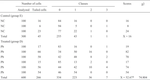
Number of cells Classes Scores χ2
Analyzed Tailed cells 0 1 2 3
Control (group E)
NC 100 16 84 16 0 0 16
NC 100 6 94 5 0 1 8
NC 100 23 77 22 1 0 24
Total 300 45 255 43 1 1 X = 16
Treated (group D)
Pb 100 17 83 16 0 1 19
Pb 100 66 34 50 16 0 82
Pb 100 58 42 48 8 2 70
Pb 100 15 85 13 2 0 17
Pb 100 56 44 42 10 4 74
Pb 100 54 46 54 0 0 54
Total 600 266 334 223 36 7 X = 52.67* 74.804
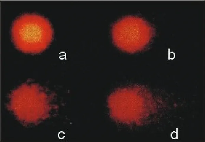
based on the DNA migration (Figure 1). Some interindividual differences were detected, in agreement with other studies (Mitchelmore and Chipman, 1998). Our results also agreed with those of Abd-Allahet al. (1999) showing that the comet assay is an effective, simple and rapid method for assessing genetic damage caused by heavy metals in fish.
The few reports that have examined fish chromo-somal aberrations have been limited to studying the effects of radiation on some cichlid species (Manna and Som, 1982; Som and Manna, 1984). This lack of information may reflect the difficulty in using conventional methods to study species with small chromosomes. A solution to this problem could be the use of certain neotropical cyprino-dontiform fish species that have a small number of rela-tively large chromosomes (Alinket al., 1980). The viability of this approach was confirmed here forH. malabaricus,
which has a low diploid number and relatively large, biarmed chromosomes, but no heteromorphic sex chromo-somes.
In conclusion, this report is one of the first to use the comet assay to evaluate DNA damage following chronic exposure to Pb2+in the diet. The results obtained show that the neotropical fishH. malabaricusmay be a useful natural model for screening the clastogenic effects of lead on fish chromosomes.
Acknowledgements
This work was supported by the Federal University of Paraná Foundation (FUNPAR). A.S.F. and E.P. were sup-ported by the Conselho Nacional de Desenvolvimento Científico e Tecnológico (CNPq, process no. 305218/02-6) and the National Science and Engineering Research Coun-cil of Canada, respectively.
References
Abd-Allah GA, El-Fayoumi RI, Smith MJ, Heckmann RA and O’Neill KL (1999) A comparative evaluation of aflatoxin B1
genotoxicity in fish models using the comet assay. Mutat Res 446:181-188.
Alink GM, Frederix-Wolters EMH, van der Gaaf MA, van de Kerkhoff JFJ and Poels CLM (1980) Induction of sis-ter-chromatid exchanges in fish exposed to Rhine water. Mutat Res 78:369-374.
Al-Sabti K (1985) Frequency of chromosomal aberrations in the rainbow trout,Salmo gairdneriRch., exposed to five pollut-ants. J Fish Biol 26:3-9.
Al-Sabti K (1986) Comparative micronucleated erythrocyte cell induction in three cyprinids by five carcinogenic-mutagenic chemicals. Cytobios 47:47-54.
Al-Sabti K and Hardig J (1990) Micronucelus test in fish for mon-itoring the genotoxic effects of industrial waste products in the Baltic Sea, Sweden. Comp Biochem Physiol 97C:79-82. Baatrup E (1991) Structural and functional effects of heavy metals
on the nervous system, including sense organs, of fish. Comp Biochem Physiol 100C:253-257.
Fenocchio AS, Venere PC, Cesar AC, Dias AL and Bertollo LAC (1991) Short term culture from solid tissues of fishes. Caryologia 44:161-166.
Goyer RA and Moore JF (1974) Cellular effects of lead. Adv Exp Med Biol 48:447-462.
Heath AG (1995) Water Pollution and Fish Physiology. 2nd edi-tion. Lewis Publishers, Boca Raton, pp 1-5.
Kligerman A (1979) Induction of sister chromatid exchanges in the central mudminnow followingin vivoexposure to muta-genic agents. Mutat Res 64:205-217.
Kobayashi H, Sugiyama C, Morikawa Y, Hayashi M and Sofuni T (1995) A comparison between manual microscopic analysis and computerized image analysis in the single cell gel elec-trophoresis. MMS Commun 3:103-115.
Lopez PA and Fenocchio AS (1994) Confirmation of two differ-ent cytotypes for the neotropical fishHoplias malabaricus
(Gill, 1903) (Characiformes). Cytobios 80:217-221. Manna GK and Som RC (1982) Effect of X-rays on the somatic
chromosomes of the exotic fish,Tilapia mossambica. Proc Indian Acad Sci 91:121-133.
Matsumoto FE and Cólus IMS (2000) Micronucleus frequencies inAstyanax bimaculatus(Characidae) treated with cyclo-phosphamide or vinblastine sulfate. Genet Mol Biol 23:489-492.
Mitchelmore CL and Chipman JK (1998) DNA strand breakage in aquatic organisms and the potential value of the comet assay in environmental monitoring. Mutat Res 399:135-147. Monteith DK and Vanstone J (1995) Comparison of the microgel
electrophoresis assay and other assays for genotoxicity in the detection of DNA damage. Mutat Res 345:97-103. Oliveira Ribeiro CA, Pelletier E, Pfeiffer WC and Rouleau C
(2000) Comparative uptake, bioaccumulation and gill dam-ages of inorganic mercury in tropical and nordic freshwater fish.Environ Res 83:286-292.
Pain DJ (1995) Lead in the environment. In: Hoffman DJ, Rattner BA, Burton GA Jr and Cairns J Jr (eds) Handbook of Ecoto-xicology. Lewis Publishers, Boca Raton, pp 356-391. Poongothai K, Shayin S and Usharani MV (1996) Induction of
micronuclei in fish by polluted water and heavy metals. Cytobios 86:17-22.
Som RC and Manna GK (1984) A preliminary study of somatic chromosome aberrations in F1 offprint and their X-irradiated parents ofTilapia(fish). Perspect Cytol Genet 4:331-335.
Speit G and Hartmann A (1999) The comet assay (single cell gel test), a sensitive genotoxicity test for the detection of DNA damage and repair. In: Henderson DS (ed) Methods in
Mo-lecular Biology: DNA Repair Protocols - Eukaryotic Sys-tems. v. 113, Totowa, Humana Press, pp 203-211.
Vigfusson NV, Vyse ER, Pernsteiner CA and Dawson RJ (1983)
In vivoinduction of sister-chromatid exchange inUmbra limi by the insecticides endrin, chlordane, diazinon and guthion. Mutat Res 83:61-68.