Abstract
Occupational exposure to ionizing radiation (IR) can damage DNA. The study evaluated the genotoxic proile and repair indicatives of DNA from peripheral blood lymphocytes of health workers exposed occupationally to IR by adopting comet assay. Biomonitoring was done with ninety individuals; among them 45 were health profes-sionals and the rests were non-profesprofes-sionals. Blood samples were collected after 48 h (2 d; non-exposed) and 168 h (7 d; exposed). The 7 d IR exposed group signiicantly increased in the rates and fre-quency of damage, while 2 d unexposed group exhibited more than 20% of DNA repair as compared to the respective control groups. The DNA damage was observed in more signiicant to the younger workers (18-27 y). However, the hematological abnormalities were not observed, despite of their positive correlation in genotoxic pro-ile. Signiicant and positive correlations were observed in relation to the used medicaments, low consumption of vegetables as well as the type and place of work. In conclusion, biomarkers involved in comet assay can be applied in biomonitoring of genetic instability, including IR induced phenomena.
Genotoxicity and DNA Repair Indicative in Blood Cells
after Occupational Exposure to Ionizing Radiation
ORIGINALRonald Gerard Silva1, Marcus Vinícius Oliveira Barros Alencar2,3, Jadson Silva Teixeira3, Reyca Rodrigues e Silva3, Márcia Fernanda Correia Jardim Paz2,3, João Marcelo de Castro e Sousa4, Rai Pablo Sousa de Aguiar3, Ricardo Melo de Carvalho3, Antonio Luiz Gomes Junior3, Ana Maria Oliveira Ferreira da Mata3, José Victor de Oliveira Santos3, Md. Torequl Islam2,3,5, Paulo Michel Pinheiro Ferreira2,6, Ana Amélia de Carvalho Melo-Cavalcante2,3, Jaqueline Nascimento Picada1
1 Postgraduate Program in Cellular Biology Applied to Health (PPG Bio Saúde), Lutheran University of Brazil, Canoas, Brazil.
2 Northeast Biotechnology Network (RENORBIO), Post-Graduate Program in Biotechnology, Federal University of Piauí, Teresina (Piauí)-64.049-550, Brazil.
3 Laboratory of Genetical Toxicology, Federal University of Piauí, Teresina (Piauí)-64.049-550, Brazil.
4 Department of Biological Sciences, Federal University of Piauí, Picos-64.049-550, Brazil.
5 Department of Pharmacy, Southern University Bangladesh, Mehedibag (Chittagong)-4000, Bangladesh.
6 Department of Biophysics and Physiology, Federal University of Piauí, Teresina (Piauí)-64.049-550, Brazil.
Contact information:
Md. Torequl Islam.
[email protected]Keywords
Occupational Exposure; Ionizing Radiation; Genetic Damage; DNA Repair.
Introduction
metabolites, deamination, alkylations and so on). The increased levels of DNA damage and ineficient repair mechanisms are molecular events of many pathogenesis and diseases such as neurodegene-rative diseases and cancer [1]. Genomic instability leads to the accumulation of mutations, is the initial step in the process of carcinogenesis. Recent data point to an association between genetic changes and cancer [2].
IR causes direct damages to the DNA, with the formation of radicals that generate lesions, such as single and double breaks in DNA strands, as well as change in bases. The overall outcomes are the bad effects on cell cycle, the repair capacity of genetic materials or apoptosis [3], and proceeding on oxi-dative damage to DNA, lipids, proteins and cell es-sential metabolites, with changes in the expression of proteins, metabolites, as well as in epigenetic events [4]. In this context, the biological effects of IR in humans exposed at low doses have being of relevant interests in relation to the effects of genetic instability, circulatory problems and in cancer [5].
The comet assay can be applied in clinical moni-toring studies, an understanding of the pathogene-sis of cancer and degenerative diseases, tumors pre-diction, radiation and chemotherapies and infertility studies, in addition to evaluation of occupational and environmental risks [1], including IR therapies, and in occupational and/or accidental exposure [2]. Its alkaline version has versatility in the detection of various lesions, for example: single and/or double stranded breaks, apurinic sites, oxidative damage and eficacy of repair [6], allowing the detection of damage at low doses, such as 25 cGy, with res-ponse to damage and DNA repair mechanism [7].
By the application of the comet assay in studies of occupational biomonitoring front to damage indu-ced by IR in health professionals, our present study aimed to evaluate the frequency and levels of DNA damage in peripheral blood lymphocytes, along with the DNA repair rates after 7 d of exposure, and 2 d of no exposure. The data were also correlated
with the hematological parameters and to possible interference factors to the genotoxic analysis, inclu-ding age, gender, lifestyle, time and place of work.
Matherials and methods
Ethical aspects
In this study, the independent variables were ma-nipulated, using blood cells and oral mucosal cells of 45 health professionals (radiologists, technolo-gists and technicians) exposed occupationally to IR for genotoxic evaluation and study of repair. The study is following the international standards and guidelines established for research projects involving human subjects, and was approved by the Ethics Committee on Human Research, based at the Lutheran University of Brazil - ULBRA/RS, CAAE: 38570914.8.0000.5349. The participants of this study, after signing the Term of Free and Informed Consent (TFIC), aware of the importance and the individual beneits of the cellular respon-se evaluation in non-neoplastic cells, answered a public health questionnaire, obeying the protocol published by the International Commission for En-vironmental Protection to Mutagens and Carcino-gens (ICEPMC).
Sampling
Comet assay
The alkaline (pH >13) comet assay was performed with slight modiication from the method described byPu et al. [8]. Briely, blood samples were obtained by veni puncturing from the individuals. Then, the samples were immediately processed. In lab, 5 μL of blood (whole blood with heparin) were pipet-ted, which were added to 95 μL of low melting agarose, placed on slides embedded in agarose gel, and covered with cover slips. The slides were then placed in the refrigerator to prevent loss of material following by the removal of the cover slips. Four slides were made for each patient; 2 of those were exposed to 5 µM of hydrogen peroxide (H2O2) for 5 min (before exposure to lysis). After 2 h on lysis so-lution, the slides were immersed in an alkali buffer (pH >13) for 25 min. This process allowed the un-folding of the DNA chains through the breaking of the secondary and tertiary structures present in the cell nucleus. After unfolding, the slides were subjec-ted to an electrical current in electrophoresis unit, which induced the migration of DNA fragments in the direction of the electric current for 25 min at 25 volts and 300 mA. All these steps were carried out under indirect and dark place to avoid further da-mage to the DNA. After electrophoresis, the slides were immediately neutralized with 0.4 M Tris buffer (pH 7.5), then ixed, washed with distilled water and dried overnight. After hydrating with distilled water for 5 min, the slides were stained with silver solution as described by Chavan et al.[9]. The results were expressed in damage index (DI) and frequency of damage (FD). The ID was obtained by evaluating the tail type, classiied from 0 to 4 (50 cells per slide in duplicate) in an optical microscope with the magniication of 100X. Intact nuclei appear round (Class 0 - no damage), while in the damaged cells, the DNA migrates from the nucleus towards the anode during the electrophoresis, showing a "tail" of sedimented fragments, like a comet: Class 1 (mi-nimum damage) to 4 (maximum damage). FD was calculated by subtracting cells with zero damage
from 100, that is, based on the number of cells with damage vs those without damage.
Hematological parameters of IR-exposed health workers
Blood samples were collected for the complete blood count (CBC). The tests were performed ac-cording to the protocols established by LabtestD-iagnóstica AS.
Statistical analysis
Normality was assessed by the Kolmogorov-Smirnov test. The t-Student test was used to test the popu-lation characteristics. To compare the damage ob-served in comet assay, post-test Bonferroni Multiple Comparison Test, while the Mann Whitney test was performed for micronucleus assay. The GraphPad-Prism software (version: 5.0) was used to carry out these analyzes. All statistical analyzes, including Spearman's rho correlation, were considered signi-icant when P <0.05.
Results and discussion
Characterization of the health workers
Socio-economical features and the lifestyle of the health professionals unexposed (control group) and exposed to IR are presented in Table 1. By the appli-cation of analysis of variance statistical test (ANOVA) and t-Students test, no signiicance (P >0.05) was observed for age, smoking, use of medicaments, vegetable consumption, in comparison to the indi-viduals exposed to IR. The data point about the im-portance of risk factors for genetic instability, such as the continued use of medications and, especially, the poor consumption of vegetables.
(MN) [10]. Micronutrients hold DNA protection ca-pabilities and, when in nutritional deiciencies, occur the inducing the MN formation [11]. Breakage and loss of chromosomes involve different cellular dys-functions, such as acentric chromosome fragments resulting from double breaks, MN formation, which
can be affected by the co-factors, for example- magnesium and calcium [12]. However, the indivi-dual response to stress may vary along with various conditions, for instance the particular function and the combination of genes, absorption and meta-bolism, cell death (apoptosis/necrosis, cell cycle control, DNA repair and immune response, and mi-cronutrient deiciencies) [13]. There are reports on the associations between genetic polymorphisms and MN formation, and thus genetic variants may modulate the effects of environmental exposure to genotoxic agents, as well as age, lifestyle charac-teristics (alcohol, tobacco, folates), cardiovascular diseases and cancer [14].
DNA damage profile in peripheral blood lymphocytes of IR exposed individuals
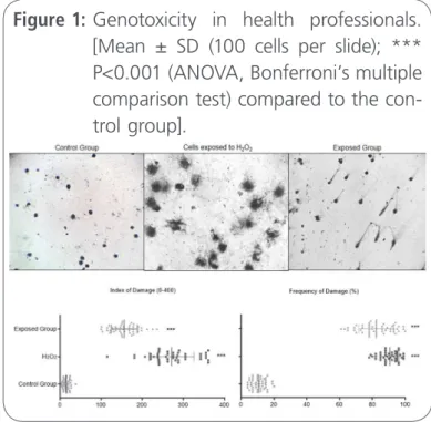
Evaluation of DNA damage can be used as an accu-rate biomarker for quantifying toxicogenetic risks of IR, which are also related to the cancer etiologically [15]. Health workers exposed occupationally to IR showed genotoxicity in blood cells signiicantly (P <0.0001) where an increase in the rates and fre-quencies of DNA damage was observed (Figure 1). To evaluate the susceptibility to oxidative damage,
Figure 1: Genotoxicity in health professionals. [Mean ± SD (100 cells per slide); *** P<0.001 (ANOVA, Bonferroni’s multiple comparison test) compared to the con-trol group].
Table 1. General features of the IR exposed and unexposed groups.
Subjectfeatures Not exposed group (n=45)
Exposed group n = 45)
Age1
31.73 ± 7.44 (18-45)
31.47 ± 7.34 (19 47) Gender2
Male 68.9 (n=31) 66.7 (n=30) Female 31.1 (n=14) 33.3 (n=15) Ethnic2
Caucasian 28.9 4.4
Black 31.1 31.1
White 40.0 64.4
Mulatto 22.0
-Smoking2
Yes 22.2 (n=10) -No 77.8 (n=35) 100 (n=45) Alcohol consumption2
Yes 66.7 (n=30) 44.4 (n=20) No 33.3(n=15) 55.5 (n= 25) Prescribedmedication use2
No 33.3 (n=15) 55.5(n=25) No 44.4(n=20) 66.7(n=30) Notreported 33.4(n=15) 0.00 Vegetable intake2
Yes 20,0 (n=9) 13.3 (n=6) No 80.0 (n=36) 86.7(n=39) PPE use3
Yes - 100(45)
1: Mean ± standard deviation (SD); 2: Percentage; 3: Personal Protective Equipment: coat, mask, apron and boots. P> 0.05 (ANOVA) t-Student test; Professionals reported that consumption of
blood cells from health professionals were exposed to H2O2 and the results showed similarities between the damage seen in cells exposed and unexposed to H2O2.
In an occupational study, the nuclear medicine physicians showed DNA damage in leukocytes with the application of comet assay. Correlation with smoking was not observed in the study [16]. Howe-ver, Khisroon et al. suggested that the radiologists’ professionals are susceptible to the IR-induced ge-notoxicity [17].
The comet assay is a simple, rapid, versatile and sensitive method, which indicates the DNA damage and repair capacity [7]. Thus, this assay has been applied in studies of a wide variety of chemicals, biochemicals and even IR induced genotoxic events in test systems including humans [2]. The alkaline version of comet assay is capable to detect various lesions, such as single and/or double stranded, apu-rinic sites, the oxidative damage in genetic materials [18] even at low IR doses (e.g. - 25 cGy) [19].
IR is evident to cause DNA damage, such as sin-gle and/or double breaks, base modiications, cross-links [20]. The double breaks are more deleterious effects induced by IR and are associated with the acute toxicity and cancer/cancer-induced situations [21], notably, genomic disorganization and phos-phorylation of histone H2AX at serine 130 residues are evident in this kind of breaks [22]. IR also indu-ces DNA damage by mechanisms associated with reactive oxygen species (ROS), which can lead to lipid peroxidation [23]. The hydroxyl radical (●OH) can be produced by UV and IR, which may cause production of other ROS. The ultimate results are the lesions in DNA [24].
The cytogenetic impact of X-rays, even at low doses (5-3 Gy), or disclosed to induce DNA damage, especially in the assessment of chromosomal abnor-malities [25]. The radio-sensitivity can be attributed to the increase of chromosomal aberrations and changes in DNA repair [26].
DNA repair profile in peripheral blood lymphocytes after IR exposure
Humans are continually exposed to IR from natural and anthropogenic sources, with the induction of several biological effects at high and low doses (50-500 mGy), but with individual variations of respon-ses by proteomic differences between individuals [27], due to the gene polymorphism associated with DNA damage, as well as those involved in the re-pair [28]. Health professionals from the clinics, af-ter a week of occupational exposure to IR, showed increased rates and frequency of damage in peri-pheral blood lymphocytes, when compared to non-exposed workers, as shown in Figure 1.
It was also observed that, after 48 h, in the wee-kend, these damage frequencies have been reduced by about 20%, suggesting repair, as noted by signi-icances between the increased frequency of injury observed after one week of exposure and the ones observed after 48 h (without exposure). These data were corroborated with the percentage of DNA re-pair, calculated by considering the damage obser-ved during one week of exposure, which reached, on average of a 20% repair (Figure 2). In its alkaline version, the comet assay estimates the DNA injury
Figure 2: Frequency of Damage (FD) and percen-tage of DNA repair in peripheral blood lymphocytes of health workers expo-sed to ionizing radiation (IR).
and also determines the repair effectiveness with predictive value for clinical studies on IR effects, with responses in individualized cells, once it is a rapid method for detecting DNA damage and repair possibilities in cell lines that decrease the length of tail after 60 minutes application of radiation [29].
The biochemical mechanisms of DNA repair have been well characterized by Aziz Sancar and Paul Modrich[30], but how these mechanisms are regulated, still to be found out [31]. As charac-terized in the previous item, IR induces different DNA lesions, once the double breaks are the most common and lead to genetic instability, cell death and cancer. The double breaks induced by the IR repair by homologous and non-homologous recombination [32], in the G2 phase of the cell cycle [33].
Repair indicative were also observed by the analy-sis of the types of damage (0-4) of the DNA
du-ring the observation period. The data presented in
Figure 3 indicate signiicance (P <0.05) in damages graded by 1, 2, 3 and 4 regarding to the control group. More frequent damages were observed in type 1, 2 and 4.
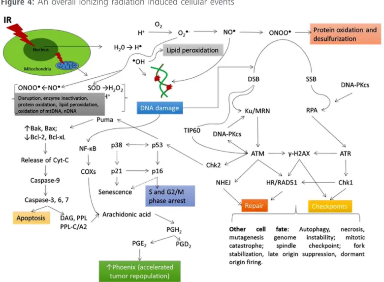
It should be emphasized that failures in these repair mechanisms can lead to DNA damage and apoptosis [34], as well as changes in DNA (mu-tations, chromosomal aberrations, translocations) [35]. However, in tumor cells, the repair is consti-tuted a resistance to radiotherapy [36]. An unders-tanding of cell responses to the DNA damage is important for the comprehension of the molecular mechanisms for post-transcriptional modiication factors involving in responses to the cellular da-mage, primarily related to double strand breaks
(Figure 4). The repair path for double failures can be attributed to the repair by homologous recom-bination (HR) and non-homologous recomrecom-bination (NHR), which occurs in the G2 period of the cell cycle. The phosphorylated RPA replication protein is a marker of response to IR [33]. The double breaks are potentially lethal, since they cannot be repaired, leading to genomic instability. One study indicates that the ATM proteins are the sensor for IR and responsible for HR repair [37].
The responses to DNA damage are important for the maintenance of the genome, such as DNA re-pair, apoptosis and senescence. The apoptosis are often used as cancer risks and are controlled by the p53 protein [38]. Notably, p53, p63 and p73 proteins, and transcription factors involved in the induction of the cell cycle arrest are important, emphasizing that no repair or incorrect repair leads to apoptosis [7]. The repair of nuclear injury induced by IR depends on the mitochondria, as they consu-me energy (ATP), and cells exposed to radiation in-crease oxygen consumption, with a reduction in the kinases phosphorylation (CDK1), which operate in the cell cycle and nuclear DNA repair. These aspects are important for the understanding of genotoxic stress conditions [39].
Figure 3: Possible DNA repair in blood cells of health professionals exposed occupatio-nally to the IR, considering the types of damage (0-4).
Figure 4: An overall ionizing radiation induced cellular events
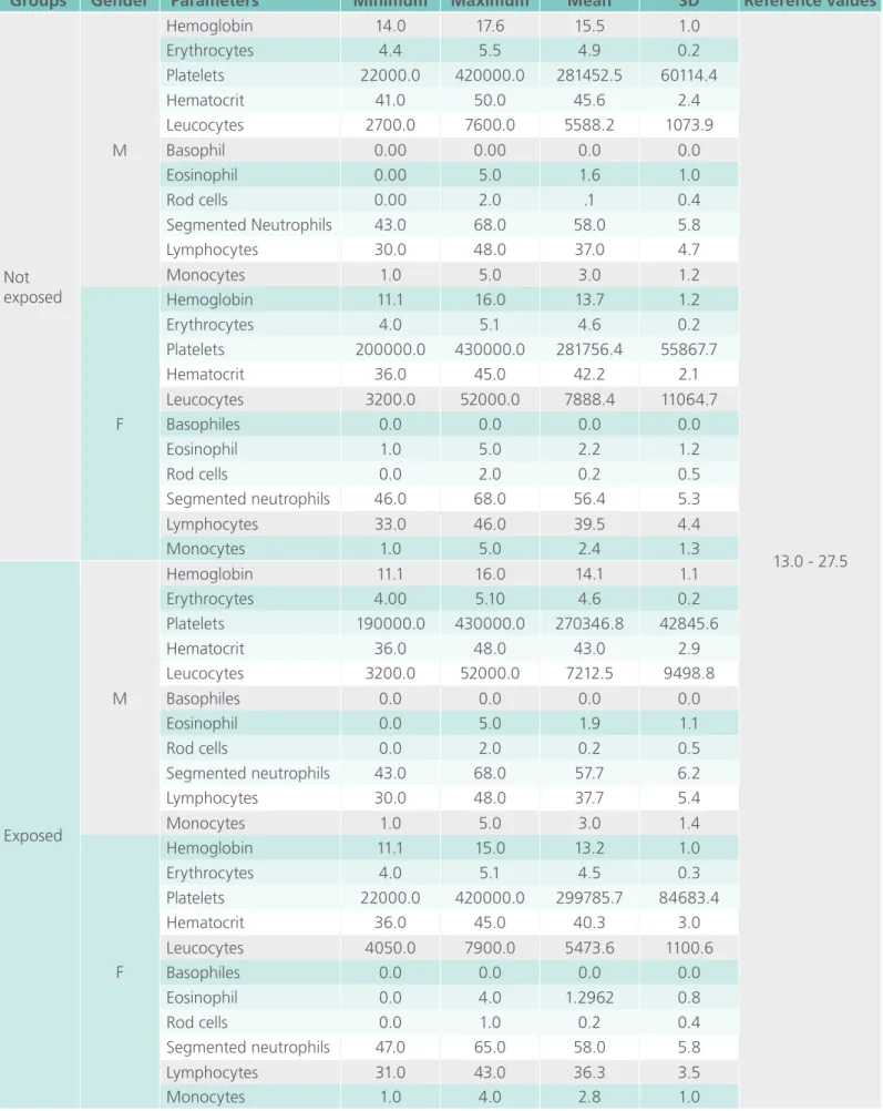
Table 3. Hematological parameters of exposed and unexposed groups to ionizing radiation.
Groups Gender Parameters Minimum Maximum Mean SD Reference values
Not exposed
M
Hemoglobin 14.0 17.6 15.5 1.0
13.0 - 27.5 Erythrocytes 4.4 5.5 4.9 0.2
Platelets 22000.0 420000.0 281452.5 60114.4 Hematocrit 41.0 50.0 45.6 2.4 Leucocytes 2700.0 7600.0 5588.2 1073.9 Basophil 0.00 0.00 0.0 0.0 Eosinophil 0.00 5.0 1.6 1.0
Rod cells 0.00 2.0 .1 0.4
Segmented Neutrophils 43.0 68.0 58.0 5.8 Lymphocytes 30.0 48.0 37.0 4.7
Monocytes 1.0 5.0 3.0 1.2
F
Hemoglobin 11.1 16.0 13.7 1.2 Erythrocytes 4.0 5.1 4.6 0.2 Platelets 200000.0 430000.0 281756.4 55867.7 Hematocrit 36.0 45.0 42.2 2.1 Leucocytes 3200.0 52000.0 7888.4 11064.7 Basophiles 0.0 0.0 0.0 0.0 Eosinophil 1.0 5.0 2.2 1.2
Rod cells 0.0 2.0 0.2 0.5
Segmented neutrophils 46.0 68.0 56.4 5.3 Lymphocytes 33.0 46.0 39.5 4.4
Monocytes 1.0 5.0 2.4 1.3
Exposed
M
Hemoglobin 11.1 16.0 14.1 1.1 Erythrocytes 4.00 5.10 4.6 0.2 Platelets 190000.0 430000.0 270346.8 42845.6 Hematocrit 36.0 48.0 43.0 2.9 Leucocytes 3200.0 52000.0 7212.5 9498.8 Basophiles 0.0 0.0 0.0 0.0 Eosinophil 0.0 5.0 1.9 1.1
Rod cells 0.0 2.0 0.2 0.5
Segmented neutrophils 43.0 68.0 57.7 6.2 Lymphocytes 30.0 48.0 37.7 5.4
Monocytes 1.0 5.0 3.0 1.4
F
Hemoglobin 11.1 15.0 13.2 1.0 Erythrocytes 4.0 5.1 4.5 0.3 Platelets 22000.0 420000.0 299785.7 84683.4 Hematocrit 36.0 45.0 40.3 3.0 Leucocytes 4050.0 7900.0 5473.6 1100.6 Basophiles 0.0 0.0 0.0 0.0 Eosinophil 0.0 4.0 1.2962 0.8
Rod cells 0.0 1.0 0.2 0.4
Segmented neutrophils 47.0 65.0 58.0 5.8 Lymphocytes 31.0 43.0 36.3 3.5
Hematological evaluation of patients exposed to IR
The biological effects of IR are primarily derived from the generation of electrons that generate free radicals and subsequent attack DNA, proteins and lipids. However, this situation can be derived directly by the deposition and energy, generating systemic effects [40]. Studies on doses and effects of IR in relation to the cytopenia (leucopenia, thrombocyto-penia), as hematopoietic cell responses front to the chronic exposure, have been proposed for different types of blood cells to measure the damage [41]. In our study, no changes were observed in hema-tological parameters in exposed and unexposed in-dividuals within the observation period (Table 2). However, a decreased level in hemoglobin content and red blood cells were observed in the exposed group in comparison to the control group (data not shown).
Blood cells reduce oxidative stress in DNA dama-ge from blood mononuclear cells by phosphoryla-tion of histone H2AX, recruit repair, which indicates that a large amount of hemoglobin protects against oxidative damage to DNA [42]. The phosphorylation of histone H2AX is an earlier event to repair DNA breaks induced by IR to maintain the genetic sta-bility [43].
Correlations between the genetic damage in relation to age, gender and lifestyle with genotoxic and hematological biomarkers
Low doses of IR inducing oxidative damage, initia-te responses partially by endogenous antioxidants, with heterogeneity in biological matrices, species, and age. However, there are adaptations to antio-xidant responses [44]. Oxidative damage can cause disruption of biopolymers [45], and may affect pro-teins, protein complexes and lipids [46].
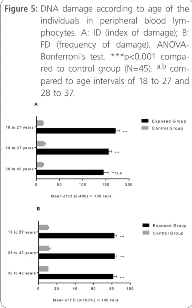
For the analysis of possible differences between the mean ages in relation to genotoxic damage, the ID and FD were used. The data not only points
genotoxicity in workers exposed to X-rays, when compared to unexposed group, but also indicate that workers in mean age of 18-27 y were more sus-ceptible to DNA damage induced by occupational exposure to X-rays (Figure 5A and 5B). Studies show that the environmental, occupational, dietary and lifestyle effects may be important in clinical analysis of risk of DNA damage, which can be evaluated by the comet assay [47]. It is noteworthy that, the repair capacity of single and double strand breaks in peripheral blood decrease with the age of the in-dividuals [7], despite of having a signiicant positive association [48].
Studies show an association between the risk of IR (average of 1.5 Gy) on the induction of
gioma in smokers and nonsmokers as well as in relation to gender. The data indicate signiicant risks for smokers and for women [49].
Although an exposure to IR is relevant for diag-nosis, but the cytotoxic and genotoxic harms may occur with a number of chemical agents, indivi-dually or synergistically with IR [50]. Otherwise, the adaptive responses are the phenomena with IR ex-posure, related to the variability in the expression of genes, endocrine and hormonal factors [51, 52]. In this context, not only diagnostic assays but also the understanding of the inluence of environmen-tal factors, lifestyle and clinical investigations are crucial for factors in bio-genetic monitoring [47].
With the application of statistic of Speraman's rho correlation, positive and signiicant correlations between lifestyle (Table 1) with the genotoxic bio-markers were observed. Corroborating with the hypothesis that occupational exposure to IR has genotoxicity risks, rising indices and frequency of damage (Figure 1), the data showed positive corre-lations between these parameters of genotoxicity with the type and time of work. However, the geno-toxicity observed was also correlated with the use of prescription drugs and with the low consumption of vegetables (Table 3). In our study, a positive and sig-niicant correlation was observed between hema-tological parameters and genotoxicity biomarkers.
Multiple nutrients and its interactions, optimize genomic stability and DNA repair capacity [53]. The increased levels of DNA injury and ineficient repair mechanisms are molecular events of many diseases, including cancer and neurodegenerative diseases [1].
Pharmaceutical drugs can cause genotoxic da-mage and/or carcinogenic effects, and therefore should be considered in the evaluation of the risk/ beneit ratio [54]. Many pharmaceutical formulations are evident to induce toxic phenomena in various organs. Therefore, investigations of the molecular mechanisms are necessary, aiming to the beneits of the therapeutic potential [55]. Hopefully, regulatory agencies, notably in Europe and in US are always
conscious about this matter [56]. Among the other adverse effects, carcinogenic potentials are the pri-me concerns in risk assesspri-ment. Chronic effects are of major concerns in this context, as it may lead varieties of deleterious events including cancer [1].
Conclusion
Occupational exposure to IR, even at low doses in health professionals may cause genotoxicity. In this study, we performed alkaline comet assay to inves-tigate two important biomarkers as damage index and frequency of damage of DNA in peripheral blood cells (lymphocytes) in an exposed group and an unexposed group. Our indings suggest that, the DNA damage was more prominent in younger professionals in relation to the old in the exposed
Table 3. Correlations between the life style and bio-markers.
Parameters Sperman’s
rho p value
Lifestyle versus biomarkers
Time of work vs index of
damage 0.637 0.001** Work place vs index of damage 0.309 0.039*
Work place vs frequency of
damage 0.324 0.030*
Index of damage vs prescribed
medication 0.339 0.000** Frequency of damage vs
prescribed medication 0.688 0.000** Vegetable intake vs index of
damage 0.324 0.000** Vegetable intake vs frequency of
damage 0.161 0.000** Haemoglobin mg/dLvs index of
damage 0.249 0.000** Haemoglobin mg/dLvs frequency
of damage 0.153 0.000** Erythrocyte count vs index of
damage 0.161 0.000** Erythrocyte count vs frequency
group. However, an average of 20% DNA repair was observed, when compared to the frequency of damage after a week of exposure. We founded a correlation between the genotoxic damage and damage type with duration of work, drug as well as micronutrient users. In conclusion, comet assay may be a helpful tool to monitor the genotoxicity induced by IR.
References
1. V. Gunasekarana, G. V. Raj, P. Chand. A Comprehensive Review on Clinical Applications of Comet Assay. Journal of Clinical and Diagnostic Research 2015; 9: GE01-GE05.
2. A. Collins, G. Koppen, V. Valdiglesias, M. Dusinska, M. Kruszewski, P. Møller, et al. The comet assay as a tool for human biomonitoring studies: The ComNet Project. Mutation Research Review: Mutation Research 2014; 759: 27-39.
3. G. Sulli, R. Di Micco, F. d’Adda di Fagagna. Crosstalk between chromatin state and DNAdamage response in cellular senescence and cancer. Nature Review in Cancer 2012; 12: 709-720. 4. A. J. Reisz, N. Bansa, J. Qian, W. Zhao, C. M. Furdui. Effects
of Ionizing Radiation on Biological Molecules-Mechanisms of Damage and Emerging Methods of Detection. Antioxidants & Redox Signaling 2014; 21: 260-292.
5. W. Ru¨hm, G. E. Woloschak, R. E. Shore, T. V. Azizova, B. Grosche, O. Niwa, S. Akiba, T. Ono, K. Suzuki, T. Iwasaki, N. Ban, M. Kai, C. H. Clement, S. Boufler, H. Toma, N. Hamada. Dose and dose-rate effects of ionizing radiation: a discussion in the light of radiological protection. Radiation and EnvironmentalBiophysics 2015; 54: 379-401.
6. E. I. Cortes-Gutierrez, M. I. Davila-Rodriguez, J. L. Fernandez, C. Lopez-Fernandez, A. Gosalbez, J. Gosalvez. New Application of the Comet Assay: Chromosome-Comet Assay. Journal of Histochemistry and Cytochemistry 2011; 59: 655-660.
7. S. Nicolai, A. Rossi, N. Di Daniele, G. Melino, M. Annicchiarico Petruzzelli, G. Raschellà. DNA repair and aging: the impact of the p53 family. Aging 2015; 7: 1050-1065.
8. X. Pu, Z. Wang, J. E. Klaunig. AlkalineComet Assay for Assessing DNA Damage in Individual Cells. Current Protocol in Toxicology 2015; 65:3.12.1-3.12.11.
9. P. Chavan, R. Kumar, R. Kirubagaran, V. P. Venugopalan. Chlorination-induced genotoxicity in the mussel Perna viridis: assessment by single cell gel electrophoresis (comet) assay. Ecotoxicology and Environmental Safety 2016 130: 295-302. 10. P. Thomas, J. Wu, V. Dhillon, M. Fenech. Effect of dietary
intervention on human micronucleus frequency in lymphocytes and buccal cells. Mutagenesis 2011; 26: 69-76.
11. Z. Zhang, X. Wang, J. Li, C. Liu, Q. Zhang. Inhibitory effects of Enteromorpha linza polysaccharide on micronucleus of Allium sativum root cells. International Journal of Biology and Macromolecules 2016; 87: 252-255.
12. F. Decarpentrie, O. A. Ojarikre, M. J. Mitchell, P. S. Burgoyne. Recombination between the mouse Y chromosome short arm and an additional Y short arm-derived chromosomal segment attached distal to the X chromosome PAR. Chromosoma 2016; 125: 177-188.
13. D. Harnicek, E. Kampmann, K. Lauber, R. Hennel, A. S. Cardoso Martins, Y. Guo, C. Belka, S. Mörtl, E. Gallmeier, R. Kanaar, U. Mansmann, T. Hucl, L. H. Lindner, W. Hiddemann, R. D. Issels. Hyperthermia adds to trabectedin effectiveness and thermal enhancement is associated with BRCA2 degradation and impairment of DNA homologous recombination repair. International Journal of Cancer 2016; 139: 467-479.
14. C. Bolognesi, S. Bonassi, S. Knasmueller, M. Fenech, M. Bruzzone, C. Lando, M. Ceppi. Clinical application of micronucleus test in exfoliated buccal cells: A systematic review and metanalysis. Mutation Research Review: Mutation Research 2015; 766: 20-31.
15. Z. Nikitaki, C. E. Hellweg, A. G. Georgakilas, J.- L. Ravanat. Stress-induced DNA damage biomarkers: applications and limitations. Frontiers Chemistry 2015; 3: 35.
16. E. B. Nuechterlein, L. Ni, E. F. Domino, J. K. Zubieta. Nicotine-speciic and non-speciic effects of cigarette smoking on endogenous opioid mechanisms. Prog Neuropsychopharmacology in Biolgical Psychiatry 2016; 69: 69-77.
17. M. Khisroon, A. Khan, M. Naseem, N. Ali, S. K. S. B. Rasheed. Evaluation of DNA Damage in Lymphocytes 1 of Radiology Personnel2 by Comet Assay. Advance Publication. Journal of Occupational Health 2015.
18. L. A. Corcuera, L. Arbillaga, A. Vettorazzi, A. Azqueta, A. López de Cerain. Ochratoxin A reduces alatoxin B1 induced DNA damage detected by the comet assay in Hep G2 cells. Food Chem Toxicol. 2011 Nov;49(11):2883-9.
19. Wilson J, Zuniga MC, Yazzie F, Stearns DM. Synergistic cytotoxicity and DNA strand breaks in cells and plasmid DNA exposed to uranyl acetate and ultraviolet radiation. Journal of Applied Toxicology 2015; 35: 338-349.
20. M. H. Bourguignon, P. A. Gisone, M. R. Perez, et al. Genetic and epigenetic features in radiation sensitivity part I: Cell signalling in radiation response. European Journal of Nuclear MedicineandMolecular Imaging 2005; 32: 229-246.
21. S. Barnard, S. Boufler, K. Rothkam. The shape of the radiation dose response for DNA double-strand break induction and repair Genome Integrity 2013, 4: 1.
23. H. Yu. Typical Cell Signaling Response to Ionizing Radiation: DNA Damage and Extranuclear Damage. Chinese Journal of Cancer Research 2012; 24: 83-89.
24. M. Dizdaroglu, P. Jaruga Mechanisms of free radical–induced damage to DNA. Free Radical Research 2012; 46: 382-419. 25. A. Suetens, K. Konings, M. Moreels, R. Quintens, M. Verslegers,
E. Soors, K. Tabury, V. Grégoire, S. Baatout. Higher Initial DNA Damage and Persistent Cell Cycle Arrest after Carbon Ion Irradiation Compared to X-irradiation in Prostate and Colon Cancer Cells. Frontiers Oncology 2016; 6: 87.
26.H. Mozdarani. Biological Complexities in Radiation Carcinogenesis and Cancer Radiotherapy: Impact of New Biological Paradigms. Genes 2012; 3: 90-114.
27. S. Nishad, A. Ghosh. Dynamic changes in the proteome of human peripheral bloodmononuclear. Mutation Research 2016; 797: 9-20 .
28. S. Gulati, A. Yadav, N. Kumar, Kanupriya, N. K. Aggarwal, R. Kumar, R. Gupta. Effect of GSTM1 and GSTT1 Polymorphisms on GeneticDamage in Humans Populations Exposed to Radiation From Mobile Towers. Archives of Environmental Contamination and Toxicology 2016; 70: 615-625.
29. D. J. Mckenna, B. A. Doherty, C. S. Downes, S. R. Mckeown, V. J. Mckelvey-Martin. Use of the Comet-FISH Assay to Compare DNA Damage and Repair in p53 and hTERT Genes following Ionizing Radiation. PLoS ONE 2012; 7: e49364.
30. D. Constantinescu-Aruxandei, B. Petrovic-Stojanovska, J. C. Penedo, M. F. White, J. H. Naismith. Mechanism of DNA loading by the DNA repair helicase XPD. Nucleic Acids Research 2016; 44: 2806-2815.
31. N. C. Bauer, A. H. Corbett, P. W. Doetsch. The current state of eukaryotic DNA base damage and repair. Nucleic Acids Research 2015; 43: 10083-10101.
32. J. R. Chapman, M. R. Taylor, S. J. Boulton. Playing the end game: DNA double-strand break repairpathway choice. Molecular Cell2012; 47: 497-510.
33. N. I. Nakajima, Y. Hagiwara, T. Oike, R. Okayasu, T. Murakami, T. Nakano, et al. Pre-Exposureto Ionizing Radiation Stimulates DNA Double StrandBreak End Resection, Promoting the Use ofHomologous Recombination Repair. PLoS One 2015; 10: e0122582.
34. D. A. Gewirtz, S. E. Holt, L. W. Elmore. Accelerated senescence: an emerging role in tumor cell response to chemotherapy and radiation. Biochemistry and Pharmacology 2008; 76: 947-957. 35. L. Huang, A. R. Snyder, W. F. Morgan. Radiation-induced genomic
instability and its implications for radiation carcinogenesis. Oncogene 2003; 22: 5848-554.
36. E. Mladenov, S. Magin, A. Soni, G. Iliakis. DNA double-strand break repair as determinant of cellular radiosensitivity to killing and target in radiation therapy. Frontiers in Oncology 2013; 3: 113.
37. P. Jeggo, M. F. Lavin. Cellular radiosensitivity: how much better do we understand it? International Journal of Radiation Biology 2009; 85: 1061-1081.
38. J. A. Lemon, K. Taylor, K. Verdecchia, N. Phan, D. R. Boreham. The inluence of trp53 in the dose response of radiationinduced apoptosis, dna repair and genomic stability in murine haematopoietic cells. Dose-Response 2014; 12: 365-385. 39. L. Qin, M. Fan, D. Candas, G. Jiang, S. Papadopoulos, L. Tian, G.
Woloschak, D. J. Grdina, J. Jian. L CDK1 Enhances Mitochondrial Bioenergetics for Radiation-Induced DNA Repair. Cellular Reports 2015; 13: 2056-2063.
40.R. Radman, E. J. Bland, N. Sangworachat, C. Bucke, T. Keshavarz. Effects of oligosaccharides and polysaccharides on the generation of reactive oxygen species in different biological systems. Biotechnology and Applied Biochemistry 2006; 44: 129-133.
41. I. V. Akushevich, G. A. Veremeyeva, G. P. Dimov, S. V. Ukraintseva, K. G. Arbeev, A. V. Akleyev, A. I. Yashin. Modeling hematopoietic system response caused by chronic exposure to ionizing radiation. Radiation and EnvironmentalBiophysics 2011; 50: 299-311.
42. M. D. Baldissera, M. R. Sagrillo, M. F. de Sá, T. H. Grando, C. F. Souza, G. F. de Brum, S. C. da Luz, S. S. Oliveira, A. L. De Mello, K. Nascimento, E. Tatsch, R. N. Moresco, A. S. da Silva, S. G. Monteiro. Relationship between DNA damage in liver, heart, spleen and total blood cells and disease pathogenesis of infected rats by Trypanosoma evansi. Experimental Parasitology 2016; 161: 12-19.
43. D. Hudson, I. Kovalchuk, I. Koturbash, B. Kolb, O. A. Martin, O. Kovalchuk. Induction and persistence of radiation-induced DNA damage is morepronounced in young animals than in old animals. Aging 2011; 3: 609-620.
44. D. Einor, A. Bonisoli-Alquati, D. Costantini, T. A. Mousseau, A. P. Møller. Ionizing radiation, antioxidant response and oxidative damage: A meta-analysis. Science of the Total Environment 2016; 548-549: 463-471.
45. O. Loseva, E. Shubbar, S. Haghdoost, B. Evers, T. Helleday, M. Harms-Ringdahl. Chronic low dose rate ionizing radiation exposure induces premature senescence in human ibroblasts that correlates with up regulation of proteins involved in protectionagainst oxidative stress. Proteomes 2014; 2: 341-362. 46. W. W. Kam, R. B. Banati. Effects of ionizing radiation on
mitochondria. Free Radical Biology and Medicine 2014; 65: 607-619.
47. A. Azqueta, J. Slyskova, S. A.S. Langie, I. O’ N. Gaivão, A. Collins. Comet assay to measure DNA repair: approach and applications. Frontiers Genetics 2014; 5: 288.
International Archives of Medicine is an open access journal publishing articles encompassing all aspects of medical scien-ce and clinical practiscien-ce. IAM is considered a megajournal with independent sections on all areas of medicine. IAM is a really international journal with authors and board members from all around the world. The journal is widely indexed and classified Q1 in category Medicine.
Publish in International Archives of Medicine 49. P. Flint-Richter, L. Mandelzweig, B. Oberman, S. Sadetzki.
Possible interaction between ionizing radiation, smoking, and gender in the causation of meningioma Neuro-Oncology 2011; 13: 345-352.
50. L. Manti, A. D’Arco. Cooperative biological effects between ionizing radiation and other physical and chemical agents. Mutation Research 2010; 704: 115-122.
51. M. – K. Boss, R. Bristow, M. W. Dewhirst. Linking the History of Radiation Biology to the Hallmarks of Cancer. Radiation Research 2014; 181: 561-577.
52. M. Nenoi, B. Wang, G. Vares. In vivo radioadaptive response: A review of studies relevant to radiation-induced cancer risk. Human and Experimental Toxicology 2015; 34: 272-283. 53.M. F. Fenech. Nutriomes and nutrient arrays - the key
toNpersonalised nutrition for DNA damage prevention and cancer growth control. Genome Integrity 2010, 1: 11.
54. M. Moretti, M. G. Grollino, S. Pavanello, R. Boniglioli, M. Villarini, M. Appolloni, M. Carrieri, L. Sabatini, L. Dominici, L. Stronati, G. Mastrangelo, A. Barbieri, C. Fatigoni, G. B. Bartolucci, E. Ceretti, F. Mussi, S. Monarca. Micronuclei and chromosome aberrations in subjects occupationally exposed to antineoplastic drugs: a multicentric approach. International Archives of Occupational and Environmental Health 2015; 88: 683-695.
55. Z. Hu, J. Wahl, M. Hamburger, A. Vedani. Molecularmechanisms of endocrine and metabolic disruption: An in silico study on antitrypanosomal natural products and some derivatives. Toxicological Letters 2016; 252: 29-41.