O
r
IG
IN
A
l
A
r
T
IC
lE
ABSTRACT CutaneousBcelllymphoma(CBCL)isalymphoproliferativedisorderofneoplasticBcellofthe skinwithawiderangeofclinicalmanifestations.Commonly,theclinicalfeaturesofCBCLare plaques,nodules,orulcerativelesions.Skinisoneofthecommonsitesforextra-nodallympho- masinpatientswithAIDSandBcelltypeislesscommonthanTcelltype.Onlyrecently,theexist-enceofBcelllymphomaspresentingclinicallyintheskinwithoutevidenceofextra-cutaneous involvementhasbeenacceptedasprimaryCBCL.Here,wearepresenting5patientswithcutane- ousinvolvementinthesettingofHIV/AIDSdisease.Twoofthemwereprimarycutaneousnon-Hodgkinlymphomas.AllwereCBCL;3wereimmunoblastic,1wasplasmablastic,andtheother wasaBurkittlymphoma.Weanalyzedtheepidemiological,clinical,virological,andimmunologi-calcharacteristicsofthisgroupofpatients. Keywords:AIDS,HIV,cutaneouslymphoma.[Braz J Infect Dis 2010;14(1):81-85]©ElsevierEditoraLtda.
Authors MarceloCorti1 LuisDeCarolis1 RubénSolari1 MaríaF.Villafañe1 RicardoSchtirbu2 DanielLewi3 MarinaNarbaitz4 1HIV/AIDSDivisionand 2Histopathology Laboratory,Infectious DiseasesF.J.Muñiz Hospital; 3OncologySection,General AcuteHospitalJ.A. Fernández; 4Histopathology Laboratory,National AcademyofMedicine; BuenosAires,Argentina. Submittedon:08/15/2009 Approvedon:10/10/2009 Correspondence to: MarceloCorti DivisionofHIV/AIDS disease Puán381,2ºpiso, C1406CQG BuenosAires,Argentina. E-mail:marcelocorti@ fibertel.com.ar Wedeclarenoconflict ofinterest. INTRODUCTION
Non-Hodgkin´s lymphomas (NHL) are fre-quent malignancies in AIDS patients. The estimated related risk of NHL associated withHIVinfectionis100timesgreaterthan ingeneralpopulation,andtheriskincreases with the progressive immunosuppression related with retrovirus.1-4 More than 90%
of HIV-associated NHL is derived from B cells and the majority is high grade. Ex-tranodal presentation is most frequent in HIV-seropositive patients than in general population and occurs in 70% to 80% of the cases. Cutaneous B cell lymphoma is an infrequent group of neoplasms that ac-cording to the World Health Organization (WHO)andtheEuropeanOrganizationfor ResearchandTreatmentofCancer(EORTC) classification,5formaspecificgroupofnon
Hodgkin’slymphoma(NHL).Primarycuta-neous B-cell lymphomas are a heterogene-ous group of B-cell extranodal lymphomas arising in the skin, without evidence of ex-
tracutaneousdiseaseatthetimeofdiagno-sis.6Insomecasestheyrepresenttheinitial
manifestationofasystemiclymphoma. Theaimofthisstudywastopresentthe epidemiological,clinical,histopathological, immunological,andvirologicalfindingsin five patients with HIV/AIDS disease and NHLwithcutaneousinvolvement.
CASE REPORTS
We analyzed retrospectively all cases of lymphomas with cutaneous involvement occurringinhumanimmunodeficiencyvi-rus (HIV) infected patients from the files oftheHIV/AIDSDivisionoftheInfectious Diseases “Francisco J. Muñiz” Hospital in Argentina,duringtheyears2005and2006. DiagnosisofcutaneousNHLwasmadeby biopsyofskinlesionsandhistopathological examination.
WedefinedNHLandBsymptomsbythe
AnnArbor staging system.2
Immunostain-ing with monoclonal antibodies was per-formed in all biopsy smears to determine the immunophenotype of the lymphoma.
Theclinicalstainingprocedureincluded:bonemarrow biopsiesinfourpatientsandtotalbodycomputedtom-ography(CT)scan(brain,thorax,abdomen,andpelvis) whichwasperformedinallpatients.
The Epstein-Barr virus (EBV) genome in tumour cellswasdetectedbyimmunohistochemistry(IHQ)tests forthelatentmembraneproteintype1(LMP-1)andby in situ hybridization (ISH) for the EBERs. Polymerase chain reaction was performed for detection of human herpesvirus-8 (HHV-8) DNA in patient with AIDS re-latedplasmablasticlymphoma.
Duringtheperiod2005to2006therewere3,556ad- missionsintheHIV/AIDSdivisionofourhospital.For-ty three patients (1.2%) had diagnosis ofAIDS related lymphomas;5ofthem(11.6%)initiallypresentedwith cutaneouscompromise.Allofthemweremales;median
Table 1. Main clinical findings in patients with lymphomas and skin involvement
Patient 1 Patient 2 Patient 3 Patient 4 Patient 5
Sex Male Male Male Male Male
Age (years) 44 42 34 34 36
Risk factor for Intravenous Homosexual Heterosexual Bisexual IDU
HIV infection drug user (IDU)
Localization Systemic with Single perianal Initiall Single perianal Disseminated
facial cutaneous cutaneous (chest cutaneous cutaneous cutaneous
lesions wall and scalp) lesion lesion lesions
Bone marrow NR Negative Positive Negative Negative
infiltration
Brain, thorax, Liver compromise Normal Costal and cranial Normal Costal and
abdomen and compromise neighbour
pelvis CT scans pulmonary
parenchymal
involvement
HCV Positive Negative Negative Negative Positive
CD4 (cells / µL) 78 48 153 54 56
Phenotype B B B B B
Histological Subtype Burkitt Immunoblastic Immunoblastic Immunoblastic Plasmablastic
EBV Negative NR Negative Positive Positive
(HHV-8+)
Treatment No HAART alone No Chemotherapy Chemotherapy
plus HAART plus HAART
Survival 20 days 24 months 16 days 18 months 6 months
15. NR: not realized
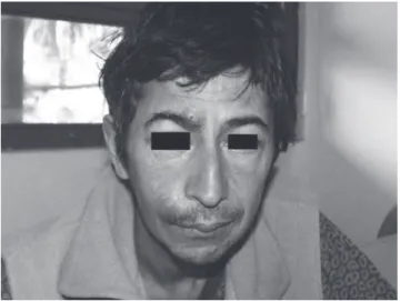
age was 36 years. All patients presented with B symp-tomsandnodularand/ortumourslesionswithdifferent cutaneouslocations(Figure1,2and3).Intwopatients, lymphomawasthefirstAIDS-definingillness.Extracu-taneous involvement was confirmed by biopsy in three patients(60%).Theothertwowereclassifiedasprimary cutaneouslymphomas(patients2and4).Mediancount of CD4+ T-cell at the time of neoplasm diagnosis was 56cells/μL.Alllymphomaswereextranodalandofhigh grade and B phenotype. Three of them were immuno-blastic, one plasmaimmuno-blastic, and one Burkitt lymphoma
Figure 2. A large tumoral lesion on the right shoulder corre-sponding to the diagnosis of plasmablastic lymphoma..
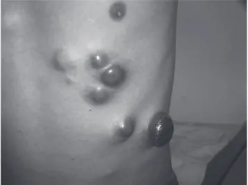
Figure 3. Multiple and reddish tumoral lesions on chest wall in a patient with plasmablastic lymphoma with cutaneous involvement..
Figure 4. Atypical lymphoid infiltrate with large and multinu-cleated cells with little or unapparent cytoplasm, the majority containing several and small nucleoli. Between the atypical elements we can see reactive histiocites situated within clear spaces, with “starred sky” aspect. The mitotic index was high with numerous cellular rests of picnotic nuclei, indicating the great cellular spare of this tumour. The histopathological
di-agnosis was Burkitt´s lymphoma.. (Figure 4). The EBV genome in the atypical cells was
detectedin2patients(onewithimmunoblasticandthe other with plasmablastic lymphoma). The HHV-8 ge- nomewasalsodetectedinthepatientwithplasmablas-tic lymphoma. Two patients could not receive chemo-therapyduetorapidprogressionofillnessorpresence ofotheropportunisticdiseases.Onepatientwithperi- analcutaneouslymphomareceivedantiretroviraltreat-ment(ART)only.TheothertwoweretreatedwithART plus chemotherapy based on CHOP (cyclofosfamide, doxorubicin, vincristine, and prednisone) completion ofsixcycles.Thepatientwithplasmablasticlymphoma diedafter6monthsofbeingdiagnosedwithneoplasm. Theothertwoarestillalivewithacompleteremissionof neoplasmdisease,after18and24months,respectively. ThemainclinicalfeaturesaresummarisedinTable1.
DISCUSSION
Patients infected with HIV are at increased risk of de-veloping NHL, including primary central nervous sys-tem lymphoma (PCNSL). Their frequency and clinical aggressive presentation has led to consider them as a criterionforstageCofAIDS,accordingtotheCenters for Disease Control (CDC) and prevention classifica-tion. AIDS-related NHL (AIDS-NHLs) are mostly, but notall,ofhigh-gradeandBphenotype,asintheseries
wepresented.7Cutaneouslymphomasarecharacterized
Primarycutaneouslymphomasrepresent5%to10% oftotalextranodalNHLandarethesecondinfrequency
afterlymphomasarisinggastrointestinaltract.9
Cutane-ous B-cell lymphomas are less frequent than primary cutaneous T-cell and include about 20% to 25% of all
primarycutaneouslymphomas.5Theyincludesubtypes
with an indolent course and evolution; and others, as the primary diffuse large B-cell lymphoma (DLBCL), moreaggressive.DLBCLaredividedintwosubcatego-ries,includingthecentroblasticandtheimmunoblastic lymphomas of the previous Kiel classification.9 In the
serieofBeylot-Barryet al. thatincluded21casesofcu-taneous lymphomas, the 10 B-cell cuthatincluded21casesofcu-taneous lympho-mas were immunoblastic or centroblastic lymphomas. Four of them expressed the EBV genome.10 In our
se-ries,DLBCLsubtypeimmunoblasticincludethreeofthe fivecases(60%)withoneplasmablasticandoneBurkitt lymphoma.
Clinical presentation of cutaneous NHL includes singleormultiplesubcutaneousnodes,dermispapules, andulcerativeandinfiltrativelesions.Specificcutaneous lesionsaremorefrequentsinNHLincomparisonwith the Hodgkin disease (HD). Patients with NHL showed cutaneous involvement in 15% to 20% of cases and in 5%to10%ofthem,skinlesionsarethefirstmanifesta- tionofthedisease.Incontrast,only5%to10%ofpa-tientswithHDpresentcutaneouslesions.5,9,10
Cutaneous compromise in NHL is an expression of advancedneoplasmdisease.Thefivepatientsofourse-ries had diagnosis of advanced HIV/AIDS disease with severeimmunosuppressionandlowCD4T-cellcounts
atthetimeofneoplasmdiagnosis.10
EBV appears to play a major role in certain AIDS-NHL, such as AIDS-related DLBCL, including PCNSL and AIDS anaplastic NHL. EBV is strongly associated withthepathogenesisofthesesubtypesoflymphomas. TherecentlyidentifiedHHV-8isrelatedwiththepatho-genesis of AIDS-primary effusion lymphoma and oral
plasmablasticlymphoma.11,12EBVcouldbeidentifiedby
ISHandIHQin2patientsofthisseries;inoneofthem alsowithHHV-8(plasmablasticlymphoma).
The number of lesions, single versus multiple, is a prognosticfactorinprimarycutaneousB-celllympho-mas.13 In this series, the patients with single perianal
skinlesionshadasignificantbettersurvivalastherewas noevidenceofdisseminateddisease(inspiteoftheco-existenceofanotherneoplasm,Kaposi’ssarcoma,inone ofthem)after18and24monthsofthediagnosis.The therapy was based on six cycles of chemotherapy fol-lowedbyHAARTinoneofthemandHAARTalonein theother,respectively.
Inconclusion,lymphomaswithcutaneouscompro-miseareuncommoninHIV-infectedpatients.Themost importantdiagnosiselucidationistodeterminewheth-er the cutaneous tumour is a primary cutaneous lym-phomaorwhenitrepresentscutaneousmetastasesofan
aggressivesystemicAIDS-relatedNHL.14
Inourexperience,earlydiagnosisfollowedbyspecif-ictherapybasedonhighlyactiveantiretroviraltherapy (HAART) plus chemotherapy improves the prognosis and the survival of patients with AIDS and cutaneous compromisebyNHL.
REFERENCES
1. Weisenburger DD. Epidemiology of non-Hodgkin’s lym-phoma. Recent findings regarding an emerging epidemic. AnnOncol1994;S19-S24.
2. CortiM,VillafañeMF,SoutoLet al.Burkitt’slymphomaof theduodenuminapatientwithAIDS.RevSocBrasMed Trop.2007;40:338-40.
3. GandhiMK,KhannaR.Virusesandlymphoma.Pathology 2005;37:420-3.
4. CortiM,SolariR,CangelosiDet al.Oralcavitylymphoma as a secondary AIDS-defining neoplasm in a patient on HAART with immune reconstitution. Rev Soc Bras Med Trop2007;40:582-4.
5. Willemze R, Meijer CJ. EORTC classification for primary cutaneous lymphomas: the best guide to good clinical management. European Organization for Research and TreatmentofCancer.AmJDermopathol1999;21:265-73. 6. GoteriG,RanaldiR,SimonettiOet al.Clinicopathological
featuresofprimarycutaneousB-celllymphomasfroman academicregionalhospitalincentralItaly:Noevidenceof Borrelia burgdorferi association. Leukemia & Lymphoma 2007;48:2184-88.
7. DiamondC,TaylorTH,IMT,Anton-CulverH.Presenta-tionandoutcomesofsystemicnon-Hodgkin’slymphomas: A comparisson between patients with acquired immu-nodeficiency syndrome (AIDS) treated with highly active antiretroviral therapy and patients without AIDS. Leuke-mia&Lymphoma2006;47:1822-9.
8. DummerR,AsagoeK,CozzioAet al.Recentadvancesin
cutaneouslymphomas.JDermatolSci2007;48:157-67.
9. WillemzeR,JaffeES,BurgGet al
.WHO-EORTCclassifica-tionforcutaneouslymphomas.Blood2005;105:3768-85. 10. Beylot-Barry M, Vergier B, Masquelier Bet al. The
spec-trumofcutaneouslymphomasinHIVinfection:Astudy of21cases.AmJSurgPathol1999;23:1208-16.
12. AllenC,KalmarJ,SusterS,BaiocchiR,NuovoG.Oralplas-mablasticlymphomasinAIDSpatientsareassociatedwith HumanHerpesvirus8.AmJSurgPathol2004;28:41-4.
13. GrangeF,BekkenkMW,WeschslerJet al
.Prognosticfac- torsinprimarycutaneouslargeB-celllymphomas:AEuro-peanmulticenterstudy.JClinOncol2001;19:3602-10. 14. LevineAM.Acquiredimmunodeficiencysyndrome-related