Bio m e chanical and histo lo gical
e valuatio n o f hydro ge l im plants
in articular cartilage
1Laboratório de Engenharia Biomecânica, Centro de Tecnologia,
2Departamento de Engenharia de Materiais, Faculdade de Engenharia Mecânica,
and 3Departamento de O rtopedia e Traumatologia, Faculdade de Medicina,
Universidade Estadual de Campinas, Campinas, SP, Brasil S.M. Malmonge1,
C.A.C. Zavaglia2 and
W.D. Belangero3
Abstract
We evaluated the mechanical behavior of the repaired surfaces of defective articular cartilage in the intercondylar region of the rat femur after a hydrogel graft implant. The results were compared to those for the adjacent normal articular cartilage and for control surfaces where the defects remained empty. Hydrogel synthesized by blending poly(2-hydroxyethyl methacrylate) and poly(methyl methacrylate-co-acrylic acid) was implanted in male Wistar rats. The animals were divided into five groups with postoperative follow-up periods of 3, 5, 8, 12 and 16 weeks. Indentation tests were performed on the neoformed surfaces in the knee joint (with or without a hydrogel implant) and on adjacent articular cartilage in order to assess the mechanical properties of the newly formed surface. Kruskal-Wallis analysis indicated that the mechanical behavior of the neoformed surfaces was significantly different from that of normal cartilage. Histological analysis of the repaired defects showed that the hydrogel implant filled the defect with no signs of inflammation as it was well anchored to the surround-ing tissues, resultsurround-ing in a newly formed articular surface. In the case of empty control defects, osseous tissue grew inside the defects and fibrous tissue formed on the articular surface of the defects. The repaired surface of the hydrogel implant was more compliant than normal articular cartilage throughout the 16 weeks following the operation, whereas the fibrous tissue that formed postoperatively over the empty defect was stiffer than normal articular cartilage after 5 weeks. This stiffness started to decrease 16 weeks after the operation, probably due to tissue degeneration. Thus, from the biomechanical and histological point of view, the hydrogel implant improved the articular surface repair.
Co rre spo nde nce S.M. Malmonge Laboratório de Engenharia Biomecânica, CT, UNICAMP Caixa Postal 6131 13083-970 Campinas, SP Brasil
Fax: + 55-19-788-7640 E-mail: malmonge@ ct.unicamp.br
Research supported by CNPq and FAPESP (No. 1996/10201-6).
Received December 30, 1998 Accepted January 3, 2000
Ke y wo rds
·Biomaterial
·Cartilage repair
·Hydrogel
·Biomechanical evaluation
Intro ductio n
Different synthetic and biological mate-rials have been studied for use in the repair of osteochondral defects (1-6), but to date no method has been reliable in restoring a func-tional articular surface. Among the different synthetic materials that have been studied,
physically similar to soft tissues, especially articular cartilage, these gels have appropri-ate mechanical properties and may be used as a graft for the repair of articular cartilage defects. Their surface and functional proper-ties should provide the necessary mechani-cal support for the joint (10).
The mechanical properties of articular cartilage as well as its functional perfor-mance are related to the tissue structure, which consists of a fluid phase (water and electrolytes) and a solid phase. The solid phase is a complex network of collagen with proteoglycan aggregates, forming an incom-pressible matrix permeable to synovial fluid (11). The indentation test is the most com-monly used method for evaluating the me-chanical behavior of articular cartilage (12-15).
In this study we evaluated the mechani-cal performance of the tissue formed in de-fects produced on the articular surface of the intercondylar region of rat femurs in the absence and presence of a hydrogel implant. We considered the intercondylar region the most appropriate for this study because of the large area available to perform the de-fects. This region is not a weight-bearing area, but is submitted to friction and wear due to the sliding of the patella during the movements.
Mate rial and Me tho ds
Hydrogel was obtained by thermal poly-merization (85oC/4 h) of a mixture of 96%
(w/w) 2-hydroxyethyl methacrylate (Aldrich Chemicals Co., Milwaukee, WI, USA), 5.0% (w/w) poly(methyl methacrylate-co-acrylic acid) (Metacril, Camaçarí, BA, Brazil), 1% (w/w) trimethyleneglycol trimethacrylate (Retilox, São Paulo, SP, Brazil) and 0.5% (w/w) benzoyl peroxide (Laport Chemicals, Santo André, SP, Brazil). Glucose crystals were added to this solution in order to obtain a porous structure. The resultant gel con-sisted of two layers: a porous layer 1.5 mm
thick (with a diameter of less than 45 µm) and a dense layer 0.5 mm thick. After syn-thesis the hydrogel was washed in distilled/ deionized water for glucose removal and immersed in saline solution at pH 7.0 for pH equilibrium. Before implantation it was ster-ilized in an autoclave for 30 min at 120o
C (16,17).
Animal mo de l
Male Wistar rats (~200 g) were anesthe-tized by ether inhalation. The knee joint was accessed by a medial parapatellar incision and a cylindrical hole 2 mm deep x 2.5 mm in diameter was made in the intercondylar re-gion of the femur. The orifice was washed with saline solution, and a sample of hydro-gel of the same size as the orifice was im-planted in the left knee while the control orifice in the right knee remained empty. The animals were divided into five groups of five animals per group and were observed 3, 5, 8, 12 and 16 weeks after the operation.
Me chanical analysis
The indentation test was performed on the repaired and normal articular surfaces of rat femurs. The advantage of this approach is that the mechanical behavior of the natural cartilage can be evaluated without removing it from the bone. The load was applied to the cartilage using a spherical stainless steel (310 L) tip (diameter 1.0 mm). A universal mate-rial test system (MTS model 810) was used for the test. The specimen was immersed in 0.15 M NaCl solution at 37o
the load versus time curves were recorded throughout the test.
Indentation tests were performed in the control and implanted defect areas. The nor-mal articular cartilage was evaluated in at least three places around the area of the defect. Five animals were evaluated per group and the average values from the five curves were used to analyze the results throughout the 16 weeks following the operation.
The values of the load measured to ob-tain a 0.16-mm indentation depth were used to calculate the elastic modulus of the in-dented areas. A small deformation of the surface was chosen for use in the calcula-tions of the elastic modulus.
Histo lo gical analysis
After the mechanical test, the joints were submitted to histological analysis. The speci-mens were fixed in 10% paraformaldehyde solution for 48 h, decalcified with 5.0% (v/v) nitric acid, washed and dehydrated with a gradual series of ethanol and xylene solu-tions. After this, they were embedded in wax blocks and sagittal sections, including the adjacent repair tissue, were cut using a rotary microtome (LEICA RM 2155) and stained with Massons trichrome. The stained sec-tions were observed and photomicrographed using an Olympus BX60 light microscope equipped with an automatic exposure photo-micrograph system PM 10AK3.
Statistical analysis
The Kruskal-Wallis test (a = 0.01) was used to compare the results of the indenta-tion tests for different situaindenta-tions (implant,
control and normal cartilage). Linear regres-sion was used to evaluate the influence of postoperative time on the stiffness of differ-ent repair tissues.
Re sults
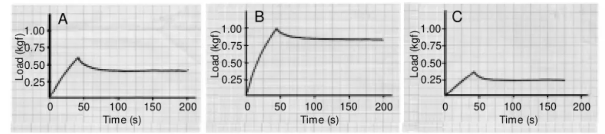
Figure 1 shows typical curves for the load pattern during the indentation test (load x time) for normal (A), control (B) and im-planted (C) articular surfaces. During the first 36 s, the indentation phase, the load was increased until a depth of 0.3 mm had been reached. The load necessary to reach this deformation was dependent on surface stiff-ness. In the second phase of the test, the load was decreased slightly as a consequence of tissue stress relaxation.
The pattern of the indentation curves ob-tained for normal cartilage remained un-changed during the 16 weeks of observation. Although the loads needed for deformation of the tissue formed over hydrogel increased significantly during the postoperative pe-riod, they did not reach the level of those for normal cartilage even within 16 weeks. In contrast, the loads needed for the tissue formed on the control defects were signifi-cantly higher than those for normal cartilage, but decreased over the 16-week period.
Statistical analysis by the Kruskal-Wallis test (a = 0.01) showed that the average curves resulting from the indentation tests on the normal, control and implanted surfaces were significantly different for all postoperative periods.
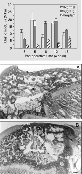
The results of the mechanical evaluation of normal and repaired articular surfaces can be observed in Figure 2. The figure shows the elastic modulus values obtained with the
Figure 1 - Typical load versus time curves obtained as a result of the indentation tests. A, Nor-mal articular cartilage surfaces; B, control repaired surfaces, and C, surfaces repaired w ith the hy-drogel implant.
L
o
a
d
(
k
g
f)1.00
0.75
0.50
0.25
0 50 100 150 200
Time (s)
L
o
a
d
(
k
g
f) 1.00
0.75
0.50 0.25
0 50 100 150 200
Time (s)
L
o
a
d
(
k
g
f) 1.00
0.75
0.50 0.25
0 50 100 150 200
Time (s)
indentation test of normal articular surfaces, control repaired surfaces and surfaces re-paired with the hydrogel for different post-operative periods. Variations in the elastic modulus of normal articular surfaces were observed for the different groups, probably due to experimental errors caused by the difficulty in finding a plane surface in the small knee joint of the rat. In the case of repaired surfaces there was a flat surface in the implanted area of the articular surface. For the surfaces repaired with the hydrogel implant the elastic modulus values were lower than those for the normal surfaces in all groups. Initially the elastic modulus of the surface was similar to that of the hydrogel
before implantation but increased with time following the operation. The elastic modu-lus of control repaired surfaces increased to values similar to those for articular cartilage after 5 weeks, showed higher values after 5 weeks but showed a decrease after 16 weeks. Linear regression of the elastic modulus values obtained for the different postopera-tive periods yielded a correlation coefficient of 0.77 for repair of the implanted surface and confirmed the tendency towards an in-crease in the stiffness of the neoformed sur-face during the period after implantation. For control repaired surfaces, the coefficient was 0.429, since the elastic modulus value decreased for 16 weeks after the operation. For normal cartilage, the correlation coeffi-cient value was -0.19.
The histological aspects of the repaired defects in the control and implanted groups 8 and 16 weeks after operation are shown in Figures 3 and 4, respectively. In the control repair situation, the defect was filled with osseous tissue with a very thin layer of fi-brous tissue on its surface (Figure 3). This fibrous tissue was not well integrated with the articular cartilage around the defect. The control defects were repaired by tissue grow-ing from the margins towards the center of the defect, resulting in irregular surfaces even after 16 weeks (Figure 3B).
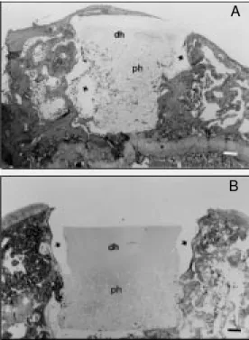
Figure 4 shows that in implanted surfaces the hydrogel graft filled the defect with no signs of significant inflammation, and was well anchored to the implant site due to the growth of surrounding tissue into the hydro-gel pores. The hydrohydro-gel implant repaired the defects, resulting in regular artificial articu-lar surfaces (Figure 4A and B). A detailed evaluation of the biological performance of this hydrogel in the repair of articular carti-lage defects was described in a previous report (18).
D iscussio n
Many studies involving indentation tests
E
la
s
ti
c
m
o
d
u
lu
s
(
M
P
a
)
30
25
20
15
123 123 123 123 123 123 123 12 12 12 12 123 123 123 123 123 123 123 123 123 123 12 12 12 10
5
0
3 5 8 12 16
Postoperative time (w eeks) Figure 2 - Average values of the
elastic modulus obtained from experimental measurements of load needed to make a 0.16-mm indent at ion dept h. Dat a are show n for normal articular carti-lage surfaces, control repaired surfaces and surfaces repaired w ith the hydrogel implant for dif-ferent postoperative periods.
A
B
Figure 3 - Section of a repaired control defect stained by M as-son’s trichrome (A) 8 w eeks af-ter operation and (B) 16 w eeks after operation. Note the new ly formed osseous tissue (ot) filling the defect and the fibrous tissue (fb) on the superficial zone. M ag-nification bar = 200 µm.
123 123
Normal Control Implant
fb
ot
ot
have been recently conducted to evaluate the mechanical behavior of articular cartilage (12-15,19). In the present study we chose to use the model used by Kempson et al. (12) and the results obtained showed that the mechanical performance of the repaired sur-faces of control and hydrogel graft implants differed from that of hyaline cartilage.
The neoformed tissue in the control group showed an increase in stiffness during 12 weeks following the operation and a de-crease for the 16th week. It is difficult to compare the stiffness of the control and im-plant repaired surfaces with the stiffness of normal articular cartilage surface since the latter showed wide variation in the elastic modulus values during the experiment (7.5-20.0 MPa). Nevertheless, if we consider an average value for the elastic modulus of normal cartilage of about 13.0 MPa with time, we can see that after the 5th week following the operation, the stiffness of the repaired surfaces was greater in the control group compared to normal articular carti-lage. The increased stiffness of the neoformed tissue in the control group can be justified by the occurrence of bone neoformation during the filling up of the defect and the formation of fibrous tissue on the surface (Figure 3). This tissue does not have the same compli-ance as normal cartilage and seems to start to degenerate after 16 weeks. According to some investigators (10,20), in the case of articular resurfacing by drilling the osteo-chondral surface, repair tissue is formed as a result of differentiation of mesenchymal cells into osteoblasts, chondroblasts or fibroblasts and does not show good integration with neighboring cartilage. This lack of integra-tion between tissues causes micromovements that contribute to degeneration of the neoformed surface.
Although the surface repaired by the hy-drogel implant was not as stiff as normal cartilage, it seemed to show a more appropri-ate mechanical and physiological perfor-mance. The lower degree of stiffness of the
surface will not cause damage to the oppo-site articular surface. The results of linear regression suggest that if the postoperative period were longer, the neoformed surface might behave in the same manner as articu-lar cartilage.
The hydrogel is able to work as a damp-ing material. This fact has to be considered when interpreting these data, since this ef-fect is probably important in determining the durability of a restored surface by protecting it from the wear and tear that normally oc-curs between two articular surfaces.
The growth of tissue into the hydrogel pores may have contributed to stabilizing the implant inside the defect, resulting in the improvement in surface stiffness throughout the postoperative period. In this study we did not observe tissue growth on the top of the hydrogel surface. It seems that if tissue was growing on the top surface during the first weeks after the operation, it did not adhere well to the hydrogel surface and consequently was removed due to the sliding of the patella against the repair site during the joint move-ment. In spite of this, the hydrogel remained inside the implant site working as an
artifi-A
B
cial articular surface.
No similar studies with hydrogel implants have been reported in the literature. In 1990, Corkhill et al. (7) published a study on the potential of synthetic hydrogels for the re-pair of articular cartilage, but to date no results of in vivo tests have been published. Reissis et al. (8) and Downes et al. (21) have studied the use of a poly(ethyl methacry-late)/tetrahydrofurfuryl methacrylate hydro-gel for the regeneration of articular cartilage,
but they have not evaluated the mechanical behavior of the repaired surfaces.
Although the results obtained here indi-cate that hydrogel is beneficial to the repair of osteochondral defects, they are not yet conclusive. Further studies on large animal species and with longer follow-up periods are currently underway in order to improve our understanding of the action of this poly-mer in articular cartilage repair.
Re fe re nce s
1. Bentley G (1992). Articular tissue grafts. Annals of the Rheumatic Diseases, 51: 292-296.
2. Solchaga L, Forriol F & Canadell J (1996). Réparation du cartilage articulaire par materiaux biologiques. Etude expérimen-tale sur le mouton. Revue de Chirurgie Orthopédique, 82: 101-107.
3. M oran M E, Kim HKW & Salter RB (1992). Biological resurfacing of full-thickness de-fects in patellar articular cartilage of the rabbit. Journal of Bone and Joint Surgery, 74-B: 659-667.
4. Kreder HJ, M oran M , Keeley FW & Salter RB (1994). Biologic resurfacing of a major joint defect w ith cryopreserved allogeneic periosteum under the influence of con-tinuous passive motion in a rabbit model. Clinical Orthopaedics and Related Re-search, 300: 288-296.
5. Vacanti C, Langer R, Shloo B & Vacanti JP (1991). Synthetic biodegradable polymers seeded w ith chondrocytes provide a tem-plate for new cartilage formation in vivo. Plastic and Reconstructive Surgery, 88: 753-759.
6. Freed LE, M arquis JC, Nohrie A, Emma-nueal J, M ikos AG & Langer R (1993). Neocartilage formation in vitro and in vivo using cells cultured on biodegradable polymers. Journal of Biomedical M ateri-als Research, 27: 11-23.
7. Corkhill PH, Trevett AS & Tighe BJ (1990). The potential of hydrogels as synthetic articular cartilage. Proceedings of the In-stitution of M echanical Engineers. Part H, Journal of Engineering in M edicine, 204:
147-155.
8. Reissis N, Kayser M , Bentley G & Dow nes S (1995). A hydrophylic polymer system enhanced articular cartilage regeneration in vivo. Journal of M aterials Science: M a-terials in M edicine, 6: 768-772.
9. Kudela V (1990). Hydrogels. In: Jacqueline IK (Editor), Encyclopedia of Polymer Sci-ence and Engineering. Wiley IntersciSci-ence, New York.
10. M ow VC, Ractliffe A, Rosenw asser M P & Buckw alter JA (1991). Experimental stud-ies on repair of large osteochondral de-fects at a high w eight bearing area of the knee joint: A tissue engineering study. Transactions of the American Society of M echanical Engineers. Journal of Biome-chanical Engineering, 113: 193-207. 11. M ow VC, Ractliffe A & Poole AR (1992).
Cartilage and diarthrodial joint as para-digms for hierarchical materials and struc-tures. Biomaterials, 13: 67-97.
12. Kempson GE, Freeman M AR & Sw anson SAV (1971). The determination of a creep modulus for articular cartilage from inden-tation tests on the human femoral head. Journal of Biomechanics, 4: 239-250. 13. Sw ann AC & Seedhom BB (1989).
Im-proved techniques for measuring the in-dentation and thickness of articular carti-lage. Proceedings of the Institution of M e-chanical Engineers. Part H, Journal of En-gineering in M edicine, 203: 143-150. 14. M ak AF, Lai WM & M ow VC (1987).
Biphasic indentation of articular cartilage. I. Theoretical solution. Journal of Biome-chanics, 20: 703-714.
15. M ow VC, Gibbs M C, Lai M W, Zhu WB & Athanasiou KA (1989). Biphasic indenta-tion of articular cartilage. II. A numerical algorithm and experimental study. Jour-nal of Biomechanics, 22: 853-861. 16. Ratner BD (1981). Biomedical applications
of hydrogels: Review and critical appraisal. In: Williams DF (Editor), Biocompatibility of Clinical Implant M aterials. CRC, Florida. 17. Chirila TV, Vijayasekaran S, Horne R, Chen Y, Dalton P, Constable IJ & Craw ford G (1945). Interpenetrating polymer netw ork (IPN) as a permanent joint betw een the elements of a new type of artificial cor-nea. Journal of Biomedical M aterials Re-search, 28: 745-753.
18. M almonge SM , Zavaglia CAC & Belangero W D (1997). Biocom pat ibilit y of poly-HEM A-poly(M M A-co-AA) sIPN blend for the repair of articular cartilage defects. Annals of the 13th European Conference on Biom at erials, 4-7 Sept em ber, Goteborg, Sw eden.
19. Haider M A & Holmes M H (1997). A math-ematical approximation for the solution of a static indentation test. Journal of Bio-mechanics, 30: 747-751.
20. Shapiro F, Koide S & Glimcher M (1993). Cell origin and differentiation in the repair of full-thickness defects of articular carti-lage. Journal of Bone and Joint Surgery, 75-A: 532-553.