Isolation of a ß-galactoside-binding
lectin from cat liver
1Cátedra de Bioquímica, Facultad de Química, Montevideo, Uruguay
2Center for Surface Biotechnology, BMC, Uppsala University, Uppsala, Sweden
L. Franco-Fraguas1,
F. Batista-Viera1
and J. Carlsson2
Abstract
A lectin from cat liver has been identified and purified by affinity chromatography on asialofetuin-Sepharose. One hundred micrograms of lectin was obtained from one cat liver with a purification factor of 1561. The lectin agglutinates trypsin-treated rabbit and cow erythro-cytes. Hemagglutination was inhibited only by saccharides containing ß-galactosyl residues, of which the 1-amine-1-deoxy-ß-D-galactose was the most potent one by inhibiting hemagglutination at a concen-tration of 12.5 mM, followed by melibiose, trehalose and galactose. The lectin has a subunit molecular mass of 14.4 kDa determined by SDS-PAGE under reducing conditions and a pI of 4.85. Compared with the composition of lectins from calf heart and porcine heart, cat liver lectin contains approximately the same amount of cysteine, half the amount of glycine, twice as much arginine and threonine, and three times the amounts of tyrosine and methionine. Cat liver lectin contains four cysteine residues per subunit, all of them in the reduced form. Their lack of reactivity towards thiol-reactive supports suggests they are not exposed on the lectin surface. The protein apparently has a blocked N-terminus. The purified lectin was stable for up to 20 months stored at +4ºC in buffer supplemented with 4 mM ß-mercaptoethanol. Results indicated that this lectin belongs to the family of soluble ß-galactoside-binding lectins, also known as galectins, which are ex-pressed in a wide range of vertebrate tissues.
Correspondence
L. Franco-Fraguas Cátedra de Bioquímica Facultad de Química CC 1157, Montevideo Uruguay
Fax: +598-2-924-1906 E-mail: [email protected]
Research supported by the International Program in the Chemical Sciences (IPICS), Uppsala University, Sweden.
Received March 28, 2002 Accepted November 18, 2002
Key words
·ß-Galactoside-binding lectin ·Galectins
·Lectin ·Cat liver lectin
·Affinity chromatography
Introduction
Lectins are proteins that bind to specific carbohydrate structures and thus can recog-nize particular glycoconjugates in different tissues or on the surface of cells (1). Within the animal lectins there is a family of closely related ß-galactoside-binding lectins, which have been called S-type (2) and S-lac lectins and also, more recently, galectins (3). Galec-tins possess a conserved carbohydrate-rec-ognition domain directed against
phyloge-netically distant animals like nematodes and sponges, the literature predominantly de-scribes galectins from mammalian sources (4) but a galectin has not been reported before in any cat organ. Mammalian galec-tins have been classified into ten families based on the amino acid sequence, but a more fundamental structural classification of subunits, which incorporates non-mam-malian galectins, is the division into proto-type, chimera-type and tandem-repeat-type galectins. Proto-type subunits consist of a single domain (Mr about 14 kDa) and usu-ally form homodimers with hemagglutinat-ing activity (4).
Galectins are involved in cell adhesion and the related function of regulation of cell proliferation and differentiation. They also participate in a number of other biological processes and thus it has been suggested that the function of a given galectin can vary from site to site depending on the nature of available ligands. Further research into the biological roles and a more complete charac-terization of these lectins will depend on the availability of reasonable amounts of highly purified material. To contribute to increas-ing the knowledge of the diversity, structure and functions of the galectins we report here the purification and partial characterization of a new ß-galactoside-binding lectin iso-lated from cat liver extracts.
Material and Methods
1,4-Dithiothreitol (DTT) and iodoacet-amide were obtained from Fluka Chemie AG, Buchs, Switzerland. Aldrithiol-4 (4-pyridyl-disulfide, 4-PDS) was from Aldrich Chemical Company Inc., Milwaukee, WI, USA. Fetuin (from fetal calf serum, type III), bovine serum albumin (BSA), transferrin, ß-mercaptoethanol, trypsin and all carbohy-drates were purchased from Sigma, St. Louis, MO, USA (except melibiose and galactose, which were from Merck, Darmstadt, Ger-many). Homogeneous 12.5 and 8-25
gradi-ent gels, the pI broad calibration kit for isoelectrofocusing (IEF) 3-9, buffer strips for SDS-PAGE and native PAGE, low-mo-lecular weight calibration kit, silver nitrate staining kit, CNBr-activated Sepharose 4B, thiopropyl-Sepharose 6B, epoxy-activated Sepharose 6B, and PD-10 columns (Sepha-dex G-25) were from Pharmacia-LKB, Upp-sala, Sweden. Microtiter U-shape plates were from Sigma. PM10 membranes were from Amicon, Danvers, MA, USA. Fresh livers were obtained from European shorthaired cats, 8 months old, at the Swedish Depart-ment of Animal Services, National Veteri-nary Institute, Uppsala, Sweden, and stored frozen at -80ºC.
Preparation of asialofetuin-Sepharose
0.1 M Tris buffer, pH 8.0, containing 0.5 M NaCl.
The adsorbent thus prepared contained 123.85 mg asialofetuin per g of dry gel, as determined by total amino acid analysis.
Coupling of 1-amine-1-deoxy-ß-D-galactose to epoxy-activated Sepharose 6B
This procedure was performed according to manufacturer instructions (Pharmacia): 1 g of freeze-dried epoxy-activated Sepharose 6B was suspended in distilled water and washed on a sintered glass filter. Five hun-dred milligrams of 1-amine-1-deoxy-ß-D-galactose was dissolved in 0.1 M Na2CO3, pH 9.5, and mixed with the gel using a shaker for 16 h at 25ºC. The excess of ligand was washed away and the remaining excess groups were blocked with 1 M ethanolamine overnight. The gel was washed with the cou-pling solution, water and 0.1 M acetate buf-fer, pH 4.0, and stored at 4ºC until use.
Preparation of soluble protein extract from cat liver
The material was treated as described by Beyer et al. (6). Each cat liver was cleaned and rinsed with buffer consisting of 75 mM NaCl, 75 mM Na2HPO4/KH2PO4, pH 7.2, containing 2 mM EDTA, 4 mM ß-mercapto-ethanol and 0.3 M lactose (Buffer A). The organ was cut into small pieces, mixed with 5 volumes of extraction buffer and homog-enized with an UltraTurrax Ika-Werk mixer at 4ºC for 1 min/load. The homogenate was centrifuged for 30 min at 15,000 g at 4ºC and the supernatant was collected and centri-fuged twice for 60 min at 30,000 g. The clear red supernatant fluid was stored at 4ºC.
Lactose-free extract
To eliminate the lactose from the crude extract, PD-10 columns for gel filtration were equilibrated with buffer consisting of 75
mM NaCl, 75 mM Na2HPO4/KH2PO4, pH 7.2, plus 2 mM EDTA and 4 mM ß-mercaptoethanol (Buffer B). The liver su-pernatant was submitted to gel filtration through the PD-10 columns by applying 1 ml and collecting 2 ml of the void volume. Alternatively, the lactose was removed by overnight dialysis against Buffer B. The lac-tose-free extract was ready to apply to asialofetuin-Sepharose or to be used for other purposes.
Affinity chromatography on asialofetuin-Sepharose
The lactose-free extract of cat liver (usu-ally 30 ml) was applied to a column (9 ml packed gel) of asialofetuin-Sepharose at a flow rate of 15 ml/h and 2-ml fractions were collected. The column was washed with Buffer B until the absorbance at 280 nm of the effluent was negligible. Desorption of the bound lectin was performed by elution with 300 mM lactose in Buffer B. The frac-tions were collected, filtered through PD-10 columns as described above and assayed for hemagglutinating activity. The fractions con-taining hemagglutinating activity were pooled and concentrated with an Amicon-10 ultra-filter. The concentrate, which was kept at 4ºC, was used for SDS-PAGE IEF, inhibi-tion studies and amino acid analysis, among others.
Affinity chromatography on 1-amine-1-deoxy-ß-D-galactosyl-Sepharose 6B
Hemagglutinating activity
Rabbit erythrocytes for the hemagglutina-tion test were prepared from fresh blood, col-lected in Alsevers medium, and washed four times with 50 mM sodium phosphate buffer, pH 7.4, containing 0.15 M NaCl (PBS buffer). The packed cells were diluted in PBS to give 4% red cell suspensions. Trypsin treatment was carried out essentially as described by Nowak et al. (7): a 4% erythrocyte suspension in PBS containing 1 mg/ml trypsin was incu-bated for 1 h at 37ºC. The trypsin-treated cells were washed four times with PBS buffer.
Agglutination assays were carried out on microtiter U-shaped plates by adding 25 µl 0.15 M NaCl, 25 µl 1% BSA in 0.15 M NaCl, 25 µl sample, and 25 µl 4% red cell suspension in PBS. The well contents were mixed by gentle shaking and covered with plastic wrap. The plate was incubated for 30 min at room temperature and examined for agglutination against a white or illuminated background to determine the resulting titer. The hemaggluti-nating activity of all samples was quantified by the two-fold serial dilution assay. Titer per ml was defined as the reciprocal of the highest dilution giving visible agglutination of the trypsin-treated rabbit erythrocytes after 30 min of incubation. Specific hemagglutinating ac-tivity was defined as the ratio of the titer/ml and protein concentration (mg/ml).
Red cells of different origins were used to test hemagglutination: fetal calf, rabbit, cow and porcine erythrocytes were tested with and without trypsin treatment.
Inhibition of lectin binding to erythrocytes by carbohydrates
Saccharide specificity of lectin binding to erythrocytes was determined by sugar in-hibition tests using 2-fold serial dilutions of the sugar solutions. To assess these inhibi-tion tests, standard soluinhibi-tions of the saccha-rides were prepared at the appropriate con-centration in 0.15 M NaCl. The lectin
dilu-tion used for these studies was the one able to cause 50% hemagglutination (the dilution before the last one able to cause clear hem-agglutination). The sugars tested were: 50 mM o-nitrophenyl-ß-D-galactopyranoside and p-nitro-phenyl-ß-D-glucopyranoside; 100 mM D-glucosamine, 6-desoxy-L-galac-tose, D-ribose, a-L-rhamnose, 1-amine-1-deoxy-ß-D-galactose, D-fructose, o- nitro-phenyl-ß-D-galactopyranoside-6-phosphate, N-acetyl-D-glucosamine, a
-methyl-D-man-noside, D-saccharose, D-glucose, thioglu-cose, a-D-fucose; 200 mM D-trehalose and
a-D-melibiose, and 300 mM D-galactose
and a-D-lactose. Heparin, asialofetuin, fetuin
and transferrin were also tested (all of them at 8 mg/ml concentration).
To determine the minimum concentra-tions required for inhibition of hemaggluti-nation by these different carbohydrates, a volume of 25 µl 0.15 M NaCl was added to each well of the corresponding lane on the microtiter plate. A volume of 25 µl of the sugar solution was added to the first well and mixed, 25 µl was withdrawn and added to the second well, its content mixed, and 25 µl was withdrawn and added to the third well, etc. The extra 25 µl from the well containing the highest sugar dilution was discarded. To each well containing the sugar dilution, 25 µl of the corresponding lectin solution, 25 µl 1% BSA in 0.15 M NaCl and 25 µl 4% red cell suspension in PBS were added. The well contents were mixed by gentle shaking, cov-ered with plastic wrap and analyzed.
Polyacrylamide gel electrophoresis
ß-mercaptoethanol, and boiled for 5 min. a
-Lactalbumin (14.4 kDa), trypsin inhibitor (20.1 kDa), carbonic anhydrase (30.4 kDa), ovalbumin (43 kDa), serum albumin (67 kDa) and phosphorylase b (94 kDa) were used as molecular mass calibration proteins. PAGE of the lectin was run using the 8-25 gradient Phast gels. Bromophenol blue was added to lectin samples as a migration marker. Pro-teins were silver stained, according to manu-facturer instructions.
IEF was determined using a Pharmacia Phast System apparatus (Pharmacia). Iso-electric point was determined using the broad pI calibration kit run on Phast gel IEF 3-9: lentil lectin (basic, 8.65), lentil lectin (middle, 8.45), lentil lectin (acidic, 8.15), horse myo-globin (basic, 7.35), horse myomyo-globin (acidic, 6.85), human carbonic anhydrase (6.55), bo-vine carbonic anhydrase (5.85), ß-lactoglo-bulin (5.20), soybean trypsin inhibitor (4.55), and amyloglucosidase (3.50). The kit pro-teins were reconstituted in 35 µl of distilled water. The gel was run according to manu-facturer instructions.
Protein determination
Protein concentration was determined by the method of Lowry et al. (8) using BSA as a standard, and by absorbance measurements at 280 nm.
Determination of amino acid composition
Amino acid analysis was performed at the University of Uppsala, Sweden. The lec-tin solution obtained by affinity chromatog-raphy on asialofetuin-Sepharose as described above was submitted to gel filtration through a PD-10 column with 10 mM ammonium acetate, pH 6.6, and hydrolyzed in 6 N HCl for 22 h at 110ºC in vacuum-sealed tubes.
Carboxymethylation of the cat liver lectin
Cat liver lectin (1.6 nmol) was incubated
with 20-fold excess iodoacetamide in PBS buffer for 1 h at 24ºC. Excess iodoacetamide was removed by gel filtration through a PD-10 column with PBS buffer.
Titration of free sulfhydryl groups
This procedure was carried out by the method of Grassetti and Murray (9). The lectin was treated with 4-PDS and the re-lease of 4-thiopyridone was monitored at 324 nm. The content of free thiol groups was determined using an extinction coefficient for 4-thiopyridone of 20.2 x 10³ M-1 cm-1. Results and Discussion
Extraction of cat liver lectin
agglutinate the untreated rabbit red cells. The extent of binding of the lectin to erythro-cytes from other species varied, depending on the source of the cells and the treatment (Table 1). Fresh cow erythrocytes were not agglutinated by the extract but were aggluti-nated after trypsin treatment. Porcine and calf red cells were not agglutinated by the extract even after trypsin treatment.
Purification of the lectin
Lectins have been purified by conven-tional procedures including ethanol precipi-tation, ion-exchange chromatography and gel filtration, or by affinity chromatography. The former methods rely on the physicochemical properties of proteins for separation while the affinity chromatography depends on the specific interaction between the lectin and a carbohydrate attached to a solid support. The lectin from cat liver was purified follow-ing general previously described affinity pro-cedures to purify S-type lectins from other sources (5,10,11). The lactose-free crude ex-tract was applied to either of two different affinity gels, i.e., the asialofetuin-Sepharose and 1-amine-1-deoxy-ß-D-galactosyl-Sepha-rose prepared as described in the Methods section. As the lectin was efficiently adsorbed onto the asialofetuin-Sepharose, the purifi-cation was performed using this affinity gel. Desorption was achieved using two differ-ent eludiffer-ents, 300 mM 1-amine-1-deoxy-ß-D-galactose (the best inhibitor in solution) and 300 mM lactose. The best results were ob-tained with 300 mM lactose. The fractions containing the protein eluted with lactose from the column were pooled, submitted to gel filtration through a PD-10 column with a buffer containing 4 mM ß-mercaptoethanol (Buffer B, see Material and Methods) and concentrated using the Amicon ultrafilters. Data showing the recovery of the lectin after the different purification steps are presented in Table 2. Approximately 0.1 mg of lectin containing 51,200 units of hemagglutinating
Table 1. Binding of various types of erythrocytes to cat liver galectin.
Source of Treatment Agglutination erythrocytes
Calf None
-Trypsin
-Cow None
-Trypsin +
Rabbit None
-Trypsin +
Porcine None -Trypsin
-Clear evidence of hemagglutination is indicated by a “+”. No detectable reaction is indicated by a “ -“.
Table 2. Purification of cat liver galectin by affinity chromatography on asialofetuin-Sepharose.
Step Volume Protein Activity Specific Total Purification Recovery (ml) (mg/ml) (IU/ml) activity activity -fold (%)
Crude extract 30 7.8 2,560 328 76,800 1 100 Affinity 1 0.1 51,200 512,000 51,200 1561 66.7 chromatography
14.4 20.1
30
43
67
94
8.66
8.45 8.15 7.35
5.85
5.2 6.85 6.55
4.55 3.5 pH kDa
A B
a b
a b c
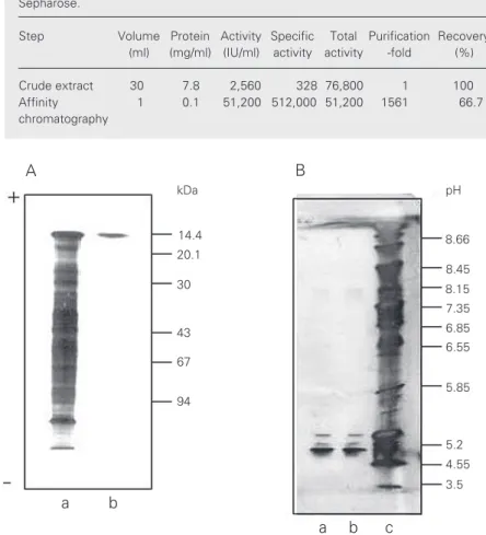
activity towards trypsin-treated rabbit eryth-rocytes was obtained from one cat liver (60 g). The procedure resulted into a purification factor of 1561. That this one-step affinity purification method using asialofetuin-Sepharose yielded a highly purified lectin preparation was demonstrated by the fact that only one band was obtained when the preparation was analyzed by SDS-PAGE under reducing conditions and with silver staining (Figure 1A). The protein migrated with a mobility corresponding to the protein standard of 14.4 kDa. We also examined the behavior of the cat liver lectin upon SDS-PAGE analysis under nonreducing condi-tions. The electrophoretic mobility and ap-parent molecular mass were identical to that observed under reducing conditions, thus indicating that the dimer is not held together by disulfide bonds. When electrophoresis was performed under native conditions the lectin migrated as a single band close to the 30-kDa standard, indicating that the native form is dimeric (data not shown). Calf heart lectin and porcine heart lectin have been reported to exhibit some tendency to form multimeric complexes or aggregates (12). We did not find any high molecular mass band other than the one of about 30 kDa when the purified cat liver lectin was run under native conditions using the 8-25 gradi-ent Phast gels.
Analysis of the purified lectin by IEF showed some heterodispersion in spite of the apparent subunit size homogeneity ob-served by PAGE and SDS-PAGE. The pro-file obtained comprised three bands: a major band at pI 4.85, and two faint bands located close together at pI 5.03 and 5.13 (Figure 1B). The former was by far the prevalent one, and the latter two more basic ones seemed to be present in equal but very low proportions.
Stability and storage
The crude extract showed no detectable
loss of hemagglutinating activity when kept for up to three weeks in the buffer containing 4 mM ß-mercaptoethanol and 300 mM lac-tose at +4ºC, and when kept in PBS for one week. The purified lectin was stable for up to 20 months when stored at +4ºC in the buffer containing 4 mM ß-mercaptoethanol.
Carbohydrate specificity of the lectin
To investigate the carbohydrate specific-ity of the lectin, we screened a panel of monosaccharides, disaccharides, monosac-charide derivatives and glycoproteins for their ability to inhibit the hemagglutination of trypsin-treated rabbit erythrocytes. In the hemagglutination inhibition assay, the sug-ars, by specific interactions, bind to the lec-tin before the leclec-tin can bind to the red blood cells. The result is no agglutination. The negative control shows positive agglutina-tion because no sugars with specificity for the lectin are present to prevent the lectin from agglutinating the red cells. The results, reported as the minimum concentrations re-quired for inhibition of the hemagglutination of cat liver lectin caused by different carbo-hydrates, are presented in Table 3. Among the saccharides tested, 1-amine-1-deoxy-ß-D-galactose was the most effective inhibitor, followed by melibiose, galactose and treha-lose. Within the concentration range studied (up to 300 mM), most of the monosaccha-rides were ineffective as inhibitors, includ-ing a-D-fucose, D-ribose, a-L-rhamnose, D-fructose, N-acetyl-D-glucosamine, a
-meth-yl-D-mannoside, D-glucose, o -nitro-phenyl-ß-D-glucopyranoside, and D-glucosamine.
inves-tigated heparin, fetuin, asialofetuin, trans-ferrin and asialotranstrans-ferrin as inhibitors. Asialofetuin inhibited at 1 mg/ml. None of the others inhibited hemagglutination at con-centrations lower than 8 mg/ml.
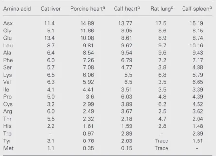
Amino acid analysis of the galectin
The amino acid composition of the galec-tin was determined and compared with those reported for other S-type lectins such as porcine heart lectin, calf heart and spleen lectins and rat lung lectin (Table 4). Com-pared with the composition of galectins from calf heart (14) and porcine heart (12), cat liver galectin contains half the amount of glycine, approximately twice as much argi-nine and threoargi-nine, and three times the amounts of tyrosine and methionine. It has 3 cysteines per 100 residues and therefore the subunit consisting of about 134 amino acids probably contains 4 cysteine residues. The cysteine residues do not seem to be involved in the dimerization of the lectin via disulfide bonds, as indicated by SDS-PAGE analysis under reducing and nonreducing conditions (see above) and by titration of the free thiol groups with 4-PDS (see below).
An N-terminal blockage of the galectin is suspected to be responsible for the failure to determine its NH2-terminal sequence.
Effect of sulfhydryl reagents on the galectin
A characteristic feature reported for the 14-kDa lectins from animal species that have been studied in detail is their dependence on a reducing medium to prevent oxidation and to maintain carbohydrate-binding activity after purification (12,15,16). Because of these repeated observations and after we estab-lished the need for ß-mercaptoethanol in the extraction buffer, we examined the require-ments for reducing conditions for maintain-ing the hemagglutinatmaintain-ing activity of the cat liver extract. ß-Mercaptoethanol was re-moved from the extract by gel filtration
Table 3. Effect of saccharides on the agglutinating activity of cat liver galectin.
Concentration that inhibits hemagglutination
by 50% (mM)*
1-amine-1-deoxy-ß-D-galactose 12.5
o
-nitro-phenyl-ß-D-galactopyranoside 25.0
a-D-melibiose 25.0
D-trehalose 50.0 D-galactose 50.0
a-D-lactose 75.0 a-methyl-D-mannoside >100.0
D-glucosamine >100.0
a-D-fucose >100.0
D-ribose >100.0
a-L-rhamnose >100.0
D-fructose >100.0
p
-nitro-phenyl-ß-D-glucopyranoside >100.0 N-acetyl-D-glucosamine >100.0 D-saccharose >100.0 D-glucose >100.0
*Corresponds to the dilution before the last one which caused agglutination of red cells in the two-fold serial dilution experiment.
Table 4. Comparison of the amino acid composition of cat liver lectin with other S-type mammalian galectins.
Amino acid Cat liver Porcine hearta Calf heartb Rat lungc Calf spleenb
Asx 11.4 14.89 13.77 17.5 15.19 Gly 5.1 11.86 8.95 8.6 8.15 Glu 13.4 10.08 8.61 8.9 8.74 Leu 8.7 9.81 9.62 9.7 10.16
Ala 6.4 8.54 9.54 9.6 9.43
Phe 6.0 7.26 6.79 7.2 7.17
Ser 5.7 7.08 4.77 3.8 4.88
Lys 6.5 6.06 5.5 6.8 5.79
Val 6.3 5.92 6.5 3.5 6.65
Ile 4.1 4.41 3.51 3.5 3.39
Pro 5.0 3.6 6.03 4.8 4.39
Cys 3.2 2.99 3.89 6.2 4.52
Arg 6.0 2.49 3.67 2.5 3.62
Thr 5.5 2.32 2.18 4.7 2.04
His 2.2 1.61 1.59 2.8 1.48
Trp - 0.97 2.89 - 2.89
Tyr 3.1 0.76 2.03 Trace 1.51
Met 1.1 0.35 0.15 Trace
-Data are reported as the number of residues/100 residues (mol%).
through PD-10 columns equilibrated with PBS buffer. In order to ensure complete reduction and to prevent formation of mixed disulfides of the lectin and of mercaptoetha-nol in the crude extract after gel filtration, incubation with 4 mM DTT was performed, and then excess reducing agent was removed by gel filtration. The eluate was immediately assayed by the hemagglutination test and the rest of the eluate was kept at +4ºC and examined 8 days later. The lectin in the extract kept its carbohydrate-binding ability in the absence of reducing agents even when stored for 8 days; after this period the activ-ity dropped to half the initial level.
We also tried to determine the require-ment of the purified lectin for reducing con-ditions during storage. A solution of the lectin in PBS was incubated for 3 h with 4 mM ß-mercaptoethanol. The reducing agent was then removed by gel filtration of the sample on a PD-10 column with PBS and the void material was kept at +4ºC for 24 h. A parallel control was run with the constant presence of the reducing agent. Hemaggluti-nation analysis at regular intervals showed that within the first 4 h the lectin lost part of its activity and 5-6 h after the removal of the reducing agent the lectin was completely inactive, while the activity of the control with the reducing agent remained active. We again added ß-mercaptoethanol at a concen-tration of 40 mM to the inactive sample and incubated the preparation for 24 and 48 h to determine whether the inactivation was re-versible or not. The sample was still inactive after these time periods in the presence of the reducing agent, so the inactivation of the purified lectin appears to be irreversible.
The importance of thiol groups in main-taining the activity of these lectins (14,17,18) has been extensively discussed. Whitney et al. (14) have proposed that oxygen inacti-vates rat lung lectin by oxidation of cysteine residues, resulting in intrachain disulfide bond formation rather than disulfide bonds between the subunits of the dimeric lectin. In
addition, it has been pointed out that no cysteine residues are found in the highly conserved region of these lectins and that the thiol groups therefore are not directly in-volved in the carbohydrate-binding function of the lectins (19).
Different mechanisms have been pro-posed to account for the oxidative inactiva-tion of the soluble galactose-binding lectins of the rat (14) and the electric eel (21). In both cases oxidation of the lectin solutions was accelerated by flushing them with oxy-gen or bubbling oxyoxy-gen through them for 30 min. In the case of the rat 14-kDa lectin, it was proposed that intramolecular disulfide bonds are formed leading to a change in the secondary structure. This conclusion was based on the observations that, upon oxida-tion, the decrease in hemagglutinating activ-ity was paralleled by a change in circular dichroism and a decrease in the number of free thiol groups, but without evidence of intermolecular cross-linking as assessed by SDS-PAGE in the absence of ß-mercapto-ethanol. In the 14-kDa lectin of the electric eel, where cysteine residues have not been detected, it was suggested that inactivation could be due to oxidation of a tryptophan residue to form an oxindol. Abbott and Feizi (22) reported that oxidation of the bovine lectin was achieved by removal of the reduc-ing agent durreduc-ing a period of 72 h by dialysis and their results were consistent with oxida-tive inactivation resulting from disulfide bond formation between a number of cysteine resi-dues. Tracey et al. (15) demonstrated that intramolecular disulfide bond formation in the soluble ß-galactoside-binding lectin from bovine heart muscle is associated with oxi-dative inactivation. The effect of amino acid substitution by site-directed mutagenesis in human 14-kDa ß-galactoside-binding lectin has been studied (23), suggesting that oxida-tion of one cysteine could be the key to the inactivation of this lectin. It has been re-ported for other mammalian 14-kDa lectins of the galectin-2 type that one of these cys-teine residues is located close to the sugar-binding site but does not participate in the actual sugar-lectin interaction. Nevertheless, the presence of reducing agents such as ß-mercaptoethanol is needed for these lectins to remain in a reduced state. It has been
found, however, that the activity is preserved for a reasonable time even in the absence of reducing agents after alkylation of the lectin thiol groups with iodoacetamide (14,16). Consistent with this, we found that, after carboxymethylation, the cat liver galectin kept its activity for at least one week regard-less of whether ß-mercaptoethanol was pres-ent or not. The carboxymethyl group is obvi-ously too small to interfere sterically with the sugar binding but, on the other hand, it seems to protect the lectin from inactivation. A possible explanation for these results might be that the loss of activity is due to a confor-mational change of the lectin caused by oxi-dation of the thiol groups to disulfides.
This hypothesis is supported by the fact that reaction of the cat liver galectin with the thiol titration reagent 4,4'-dipyridyl disulfide led to instantaneous loss of hemagglutinat-ing activity. This reagent is well known to react with free thiols with formation of mixed reactive disulfides, which in turn can react with other thiols to form disulfide bonds. Another possibility for the observed loss of activity might of course also be that the introduced 4-pyridyl-disulfide structure is large enough to block the sugar-binding site of the lectins.
These findings may contribute to the un-derstanding of the biological role of this type of lectins since there is evidence that they function in an extracellular environment (4), which is generally considered to be less re-ducing than inside the cell. Cooper et al. (24) suggested that oxidative inactivation of the L-14 lectin carbohydrate-binding activity might be a way to limit the duration and physical range of its activity, which opens up interesting research possibilities in vivo.
Acknowledgments
References
1. Barondes SH (1984). Soluble lectins: A new class of extracellular proteins. Science, 223: 1259-1264.
2. Drickamer K (1988). Two distinct classes of carbohydrate-recogni-tion domains in animal lectins. Journal of Biological Chemistry,263:
9557-9560.
3. Barondes SH, Castronovo V, Cooper DN, Cummings RD, Drickamer K, Feizi T, Gitt MA, Hirabayashi J, Hughes RC, Kasai K, Leffler H, Liu F-T, Lotan R, Mercurio AM, Monsigny M, Pillai S, Poirier F, Raz A, Rigby P, Rini JM & Wang JL (1994). Galectins: a family of animal ß-galactoside binding lectins. Cell,76: 597-598.
4. Kilpatrick DC (2000). Vertebrate lectins. In: Kilpatrick DC (Editor),
Handbook of Animal Lectins: Properties and Biomedical Applica-tions. John Wiley & Sons, Chichester, Sussex, UK, 31-49.
5. de Waard A, Hickman S & Kornfeld S (1976). Isolation and properties of ß-galactoside-binding lectins of calf heart and lung. Journal of Biological Chemistry,251: 7581-7587.
6. Beyer EC, Zweig SC & Barondes SH (1980). Two lactose binding lectins from chicken tissues. Journal of Biological Chemistry,255: 4236-4239.
7. Nowak TP, Haywood PL & Barondes SH (1976). Developmentally regulated lectin in embryonic chick muscle and a myogenic cell line.
Biochemical and Biophysical Research Communications,68:
650-657.
8. Lowry OH, Rosebrough NJ, Farr AL & Randall RJ (1951). Protein measurement with the Folin phenol reagent. Journal of Biological Chemistry,193: 265-275.
9. Grassetti DR & Murray JR (1967). Sulfhydryl groups with 2,2' or 4,4’dithiodipyridine. Archives of Biochemistry and Biophysics,119:
41-49.
10. Cerra RF, Gitt MA & Barondes SH (1985). Three soluble rat ß-galactoside-binding lectins. Journal of Biological Chemistry, 260: 10474-10477.
11. Briles EB, Gregory W, Fletcher P & Kornfeld S (1979). Comparison of properties of ß-galactoside-binding lectins from tissues of calf and chicken. Journal of Cell Biology,81: 528-537.
12. Merkle RK, Zhou Q, Schultz TK, Harder WB & Cummings RD (1989). Characterization of an S-type lectin purified from porcine heart.
Archives of Biochemistry and Biophysics,274: 404-416.
13. Lobsanov Y, Gitt MA, Leffler H, Barondes SH & Rini J (1993). X-ray crystal structure of the human dimeric S-lac lectin, L-14-II, in
com-plex with lactose at 2.9 Å-resolution. Journal of Biological Chemis-try,268: 27034-27038.
14. Whitney PL, Powell JT & Sanford GL (1986). Oxidation and chemical modification of lung ß-galactoside-specific lectin. Biochemical Jour-nal,238: 683-689.
15. Tracey BM, Feizi T, Abbott MW, Carruthers RA, Green BN & Lawson AM (1992). Subunit molecular mass assignment of 14,654 Da to the soluble ß-galactoside-binding lectin from bovine heart muscle and demonstration of intramolecular disulfide bonding associated with oxidative inactivation. Journal of Biological Chemistry,267: 10342-10347.
16. Den H & Malinzak D (1977). Isolation and properties of ß-galacto-side-specific lectin from chick embryo thigh muscle. Journal of Biological Chemistry,252: 5444-5448.
17. Clerch LB, Whitney P, Hass M, Brew K, Miller T, Werner R & Massaro D (1988).Sequence of a full-length cDNA for rat lung ß-galactoside-binding protein: primary and secondary structure of the lectin. Biochemistry,27: 692-699.
18. Liao DL, Kapadia G, Ahmed H, Vasta GR & Herzberg O (1994). Structure of S-lectin, a developmentally regulated vertebrate ß-ga-lactoside-binding protein. Proceedings of the National Academy of Sciences, USA,91: 1428-1432.
19. Hirabayashi J, Kawasaki H, Suzuki K & Kasai K (1987). Further characterization and structural studies on human placenta lectin.
Journal of Biochemistry,101: 987-995.
20. Drickamer K & Taylor ME (1993). Biology of animal lectins. Annual Review of Cell Biology,9: 237-264.
21. Levi G & Teichberg VI (1981). Isolation and characterization of chicken thymic electrolectin. Journal of Biological Chemistry,256: 5735-5740.
22. Abbott W & Feizi T (1991). Soluble 14-kDa ß-galactoside-specific bovine lectin. Journal of Biological Chemistry,266: 5552-5557. 23. Hirabayashi J & Kasai K (1991). Effect of amino acid substitution by
site-directed mutagenesis on the carbohydrate recognition and sta-bility of human 14-kDa ß-galactoside-binding lectin. Journal of Bio-logical Chemistry,266: 23648-23653.