Research paper
Contribution of the carbohydrate-binding ability of
Vatairea guianensis
lectin to induce edematogenic activity
Gabriela F.O. Marques
a,1, Vinicius J.S. Osterne
b,1, Livia M. Almeida
a,
Messias V. Oliveira
b,2, Luiz A.C. Brizeno
a, Vanir R. Pinto-Junior
b, Mayara Q. Santiago
b,
Antonio H.B. Neco
b, Mario R.L. Mota
c, Luiz A.G. Souza
d, Kyria S. Nascimento
b,
Alana F. Pires
a, Benildo S. Cavada
b,*, Ana M.S. Assreuy
a,** aInstitute of Biomedical Sciences, State University of Ceara, Av. Paranjana, 1700, 60740-000, Fortaleza, CE, BrazilbDepartment of Biochemistry and Molecular Biology, Federal University of Ceara, Mr. Hull s/n Building 907, 60445-970, Fortaleza, CE, Brazil cDepartment of Dental Clinic, Division of Oral Pathology, Federal University of Ceara, R. Alexandre Baraúna, 949, 60430-160, Fortaleza, CE, Brazil dInstituto Nacional de Pesquisas da Amaz^onia - INPA, Manaus, Amazonas, Brazil
a r t i c l e
i n f o
Article history:
Received 23 March 2017 Accepted 9 June 2017 Available online 16 June 2017
Keywords: Vatairea guianensis
Lectin
Molecular modeling Molecular docking Inflammation
a b s t r a c t
Vatairea guianensislectin (VGL), Dalbergiae tribe, is a N-acetyl-galactosamine (GalNAc)/Galactose (Gal) lectin previously purified and characterized. In this work, we report its structural features, obtained from bioinformatics tools, and its inflammatory effect, obtained from a rat paw edema model. The VGL model was obtained by homology with the lectin ofVatairea macrocarpa(VML) as template, and we used it to demonstrate the common characteristics of legume lectins, such as the jellyroll motif and presence of a metal-binding site in the vicinity of the carbohydrate-recognition domain (CRD). Protein-ligand docking revealed favorable interactions withN-acetyl-D-galactosamine,D-galactose and related sugars as well as several biologically relevantN-andO-glycans.In vivotesting of paw edema revealed that VGL induces edematogenic effect involving prostaglandins, interleukins and VGL CRD. Taken together, these data corroborate with previous reports showing that VGL interacts withN-and/orO-glycans of molecular targets, particularly in those presenting galactosides in their structure, contributing to the lectin in-flammatory effect.
©2017 Elsevier B.V. and Société Française de Biochimie et Biologie Moléculaire (SFBBM). All rights reserved.
1. Introduction
Widely distributed among living organisms and viruses, lectins are proteins or glycoproteins capable of forming complexes with molecules and biological structures containing saccharides [1]. Since lectins can reversibly bind to carbohydrates, these molecules play major roles in cell communication, such as that occurring in the inflammatory process via glycocode decoding in the structure of soluble and integral cell membrane glycoconjugates[2].
Among lectins, those purified from leguminous plants are
widely studied. This group comprises a large family of closely related lectins with similarity in physicochemical and structural properties, but significant differences in their biological activities
[3]. Some lectins of the Dalbergieae tribe (Fabaceae, Papilionoideae) have now been purified and characterized. Moreover, the structures ofPterocarpus angolensis[4],Centrolobium tomentosum [5], Platy-podium elegans[6],Arachis hypogaea[7]andVatairea macrocarpa
[8,9] lectins have already been solved. Those lectins possessing binding affinity for N-acetyl-glucosamine present anti-inflammatory property, such as the lectin ofLonchocarpus sericeus
[10e12]andLonchocarpus araripensis[13]. However, those lectins with binding affinity for galactose, such as Vatairea macrocarpa lectin, present inflammatory property [14e16]. In addition, the
inflammatory effect ofV. macrocarpalectin occurs via activation of macrophages with release of cytokines[16].
The N-acetyl-D-galactosamine/D-galactose-specific lectin of Vatairea guianensis(VGL) is a homotetrameric glycoprotein with *Corresponding author.
**Corresponding author.
E-mail addresses: [email protected] (M.V. Oliveira),[email protected]
(B.S. Cavada),[email protected](A.M.S. Assreuy). 1 Equal contribution.
2 Participated in the new docking experiments and in paper writing.
Contents lists available atScienceDirect
Biochimie
j o u r n a l h o m e p a g e :w w w . e l s e v i e r . c o m / l o c a t e / b i o c h i
http://dx.doi.org/10.1016/j.biochi.2017.06.008
twoN-glycosylations at Asn111 and Asn183. It was purified by
af-finity chromatography and possesses a molecular mass of 120 kDa. VGL presents in vitro vasodilator effect, inducing relaxation in endothelialized aorta via nitric oxide[17]. We aimed to gain a better understanding of VGL-saccharide binding (CRD) in the context of VGL biological effects. To accomplish this, we modeled the three-dimensional structure of VGL and focused on itsin vivo vasodi-lator effects on a rat paw edema model.
2. Materials and methods
2.1. Lectin isolation
VGL was isolated fromVatairea guianensisseeds by ion exchange chromatography (DEAE-Sephacel column), followed by affinity chromatography (guar gum)[17]. The pure protein was diluted in sterile saline (0.9% NaCl) before biological assays.
2.2. Rat paw edema model
VGL (0.01, 0.1 and 1 mg/kg) was administered by subcutaneous (s.c.) route in Wistar rats (150e200 g) as inflammatory stimulus. Controls received sterile saline (0.1 mL/100 g body mass). The experimental protocols were approved by our Institutional Ethical Committee (UECE No. 10130208-8/40).
Paw edema was measured by hydroplethysmometry immedi-ately before VGL injection (zero time), and from 0.5 to 72 h there-after and was expressed as the variation in paw volume (mL) or area under curve (arbitrary units)[18]compared to zero time.
The participation of inflammatory mediators in the lectin effect was evaluated by treating the animals with the following phar-macological inhibitors: nitric oxide synthase (N-Nitro-L-arginine
methyl ester/L-NAME; 25 mg/kg; intravenous), cyclooxygenase
(indomethacin; 5 mg/kg; subcutaneous) andinterleukin-1
b
(thalidomide; 45 mg/kg; intraperitoneal)[19]30 min before VGL administration (1 mg/Kg; s.c.).The participation of the lectin carbohydrate-recognition domain (CRD) was evaluated by the injection of the most active dose of VGL (1 mg/kg) after incubation (30 min/37C) with its binding sugar
galactose (0.1 M). Galactose was individually incubated at the same conditions as control.
2.3. Histological analysis
Paw tissues were removed 6 h after VGL (1 mg/kg) adminis-tration,fixed with 10% buffered formalin for 24 h, embedded in paraffin, cut into 5-
m
m thick slices, stained with hematoxylin &eosin (HE) and analyzed by light microscopy coupled to image acquisition systems (ScopePhoto; Image Manager 50). The intensity of tissue inflammation was graded according to the following scores: 0. normal tissue (no distinguishable change, 0%), absence of inflammatory infiltrate; 1. discrete tissue changes (initiation of changes, up to 30%), slight inflammatory infiltrate; 2. moderate tissue changes (patent changes, 31e60%), moderate inflammatory infiltrate; and 3. severe tissue changes (widespread changes, 61e100%), severe inflammatory infiltrate.
2.4. Immunohistochemistry
Fragments of paw tissue were sectioned to a thickness of 3
m
m, placed on silanized slides and processed as described in the following protocol. Samples were deparaffinized, subjected to rehydration and antigen-recovery using citrate buffer (pH 6.0), incubated (10 min; r.t.) with 6% H2O2in methanol (1:1) and washedwith TRIS pH 7.6 (TRIS) in order to inactivate endogenous
peroxidases. Samples were re-incubated for 1 h (r.t.) with the pri-mary antibody (Ab) against IL-1
b
(monoclonal; Abcam“AB9787”;1:100), washed and further incubated (30 min; r. t.) with bio-tinylated immunoglobulin (Ig; DAKO E0468) and streptavidin (DAKO P0397). Diaminobenzidine chromogen (DAKO K3469) was applied for 10 min, and Mayer's hematoxylin was used for coun-terstaining. Samples were dehydrated (ethanol and xylene) and cover-slipped with permanent Mounting medium. Parallel sections were treated with control IgG instead of the primary Ab.
For semi quantitative immunohistochemical evaluation, sec-tions were randomly selected in 5fields (400x magnification) in areas of greater concentration of immunostained cells located in connective or epithelial tissue. The percentage of cells with cyto-plasmic or nuclear expression was scored as follows: (0) no positive cells; (1 - mild) 1e33% positive cells; (2 - moderate) 34e66% pos-itive cells; (3 - intense) 67e100% positive cells (adapted from Minal Chaudhary et al. 2012)[20].
2.5. Statistical analysis
Thein vivodata were presented as mean±SEM, and the sta-tistical analysis was performed by ANOVA, followed by Bonferroni's test. P values<0.05 were considered significant. Histopathological and immunohistochemical data were expressed as median (maximum and minimum) and analyzed by Mann-Whitney test.
2.6. Template determination and secondary structure prediction
VGL sequence was downloaded from the Universal Protein Resource (Uniprot) (ID: P86893). The template for homology modeling was obtained from BLASTp search on the Protein Data Bank (PDB) with default parameters. Proteins most similar to VGL were chosen and ranked based on their resolution and geometric parameters.
Secondary structure prediction was carried out using PsiPro server, an automated system for secondary structure prediction
[21]. The secondary structure was applied as one of the validation factors for homology model selection.
2.7. Homology modeling and validation
The 3D structure of VML in complex with Tn antigen (PDB ID: 4U36) was downloaded from PDB as the template structure. The homology model of VGL was built with MODELLER v.9.16[22]. VGL and VML structures were aligned using salign module, followed by manual optimization. Initially, a hundred models were generated and ranked based on the Modeller objective score function (molpdf) and Discrete Optimized Protein Energy (DOPE) scores. Several models with lower molpdf and DOPE were selected and submitted to validation of stereochemical properties like Ram-achandran plot, steric overlaps, C
b
deviation parameters, rotamers, and bond angle deviations using PROCHECK [23]. Side-chain acceptability was obtained by the Verify3D server [24]. QMEAN and Z-scores were also assessed by Protein Structure and Assess-ment tools[25e27]. The model having the best values in all vali-dations was selected and applied in the subsequent analyses. Molecular drawings were prepared with PyMol (Shrodinger, LLC).2.8. Molecular docking
a standard precision mode to determine the favorable binding poses and detect several flexible ligand conformations, while holding the protein as a rigid structure. The location of VGL CRD was obtained by superposition with VML using. As docking pa-rameters, the binding radius set to 10 Å3around VGL CRD and the
number of iterations set to 5000. The PLANTSPLP algorithm was applied to calculate docking score[29], where more negative values indicate stronger interaction, and the best poses for each ligand were selected based on hydrogen bonds and hydrophobic in-teractions. LIGPLOTþ[30]and PyMol were applied to generate the
2D interaction plots and molecular representations, respectively. The result for GalNAc pose was compared to that in the VML crystal structure (PDB ID: 4U2A).
2.9. N- and O-glycans docking
VGL structure was also submitted to molecular docking with severalN- andO-glycans commonly found in glycoproteins. Gly-cans structures were obtained from several scientific works
[31e33]and built with the carbohydrate builder tool of Glycam-Web[34]All glycans were submitted to energy minimizations us-ing AMBER 12[35]with GLYCAM_06j-1 forcefield[36]via Glycam-Web built in modules. Missing hydrogen atoms and bond type corrections were performed by ligand preparation module of Her-mes v. 1.8.2. Glycans structures representations are shown inFig. S1. Docking simulations were performed with GOLD v. 5.5 (Genetic Optimization for Ligand Docking e CCDC, Cambridge, England). GOLD implements a generic algorithm to dock ligands into protein binding sites exploring a great range of ligands conformations with partial proteinflexibility[37]. VGL was prepared for docking by removal of solvent and ligand molecules. Binding site was defined in center of carbohydrate-recognition domain and all atoms comprising 12 Å of radius. Docking parameters were: population size of 100, selection pressure of 1.1, number of operations of 10,000, number of islands of 5, niche size of 2 and crossover fre-quency of 95. For all ligands, 20 poses were generated andfiltered by docking score, oligosaccharide geometry, hydrogen bonds and hydrophobic interactions coherence. Further validation was per-formed by comparing interactions with those from VGL-Galactose complex and removing poses with serious geometric strains. PLANTSPLP was chosen as score function[29]and VGL-Galactose score was used as comparison. All other options were program
default. VGL-glycans complexesfigures were generated in PyMol.
3. Results and discussion
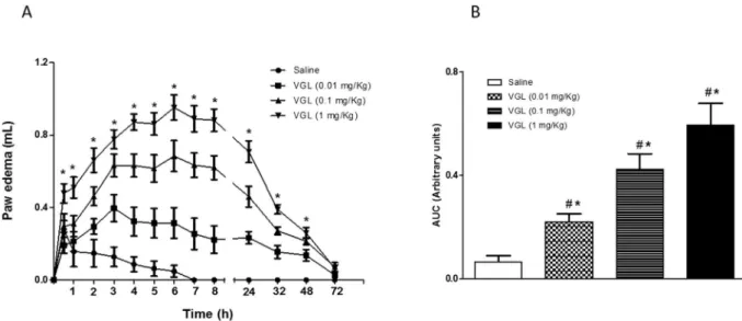
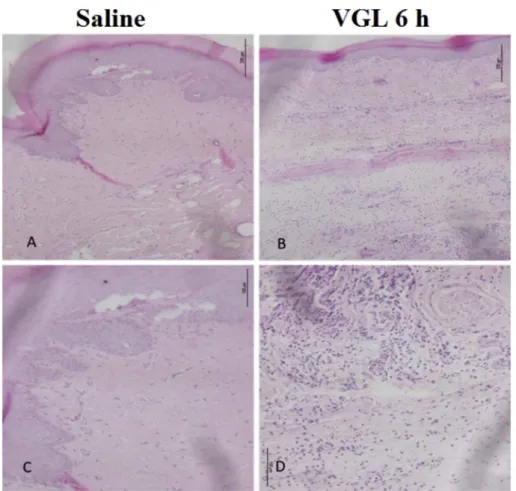
VGL induced a time- and dose-dependent paw edema that las-ted 48 h (Fig. 1a) at 0.01 mg/Kg (0.21 ± 0.03 AUC); 0.1 mg/Kg (0.42±0.06 AUC) and 1 mg/Kg (0.59 ± 0.08 AUC) compared to saline (0.06±0.02 AUC) (Fig. 1b). At 1 mg/Kg, VGL showed maximal efficacy (4e8 h), initiating edematogenic effect 30 min after administration (Fig. 1a). VGL at 1 mg/Kg also induced poly-morphonuclear infiltrate along the edema time-course, which was accentuated at 6 h [VGL: median 3 (3.3)*; p¼0.0022vs. saline:
median 0 (0.1)] (Fig. 2). These results corroborate the typical acute inflammatory process demonstrated by the macroscopic evaluation shown inFig. 1and are in accordance with previous studies per-formed with the homologous lectin VML that elicited acute ede-matogenic activity accompanied with leukocyte infiltration[15].
Pharmacological modulation, as performed by treating animals with inhibitors of inflammatory mediators before VGL injection, implied the participation of prostaglandins in thein vivo vasodi-lator effect (edema) of VGL since indomethacin (inhibitor of the enzyme cyclooxygenase that catalyzes the synthesis of prosta-glandins caused moderate (37%) inhibitory effect. In contrast,L
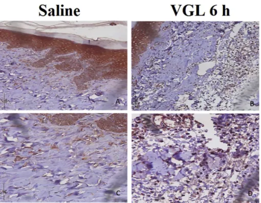
-NAME (inhibitor of the enzyme NOS that catalyzes the synthesis of NO) did not modify the lectin edematogenic effect, despite the important vasodilator effect of NO. These data seem to contradict the vasodilator effect of VGL as previously shown in vitro [17]. However, this could be explained by the different NOS isoforms expressed in the noninflamed vessels (endothelial NOS) and the vessels in inflamed paw tissues (inducible NOS)[38]. In addition to the implication of prostaglandins, also a vasodilator mediator, we demonstrated an important participation of interleukins since thalidomide inhibited the edema induced by VGL by 62% (Fig. 3).Corroborating the results obtained by pharmacological modulation, IL-1
b
immunostaining was revealed at 6 h in the epithelial and connective tissues (fibroblasts and inflammatory cells): epithelium [saline: 2 (1.2)vs.VGL: 3 (2.3)*]; connective tis-sue [saline: 0 (0.1)vs. VGL: 3 (2.3)*] (Fig. 4). These data are in line with the role of prostaglandins and IL-1b
, both mediators of cellular origin, in acute inflammation inducing vasodilatation and tissue damage[39], as well as the demonstrated effect of VML on acute models of inflammation with the participation of prostaglandinsFig. 1.VGL induces dose- and time-dependent paw edema. VGL (0.01, 0.1, 1 mg/kg; s.c.) or saline was injected intraplantar, and edema was measured before (zero time) and from 0.5 to 72 h after VGL. (a) Time course and (b) area under curve (AUC). Mean±S.E.M. (n¼5e6). *p<0.05vs. saline; #p<0.05vs. VGL at all doses.
and interleukins[15].
Importantly, classical literature has implicated the participation of the carbohydrate-recognition domain (CRD) in the inflammatory effects of leguminous lectins isolated from the tribes Phaseoleae
[32,33]and Dalbergieae[10,13,15], demonstrated by the partial or total inhibition of the lectin activity by its binding sugar[40].
The protein BLAST analysis of VGL sequence demonstrated 93% identity with VML sequence (4U36) and because of this, VML was chosen as the template for homology modeling of VGL using the MODELLER 9.16 suite. Secondary structure prediction resulted in a
prevalence of
b
-sheet structures and loops with absence ofa
-helix (Fig. S2).This result agrees with previously reported data for other lectins. VGL monomer is shown inFig. 5.Reliability of the VGL model obtained by homology modeling was assessed by various validation parameters. For the chosen model, PROCHECK analysis showed that local and global stereo-chemical parameters had favorable values. Ramachandran plot indicated that 100% of the residues are in favorable and allowed regions of the graph. The QMEAN global and QMEAN z-score, as obtained by the protein assessment tools, were 0.755 and 0.156, both within the range of high-quality models. Compatibility of the amino acid sequence and three-dimensional structure was ob-tained by the Verify3D program, in which 92.89% of the residues were compatible, suggesting that the side-chain environment is acceptable. Also, the superposition between the best model and the template resulted in a root mean square deviation (RMSD) of 0.143, indicating reliable prediction. Altogether, these analyses demon-strated that the VGL model was comparable to those of experi-mental structures, indicating that the modeled structure was adequate for the subsequent tests. VGL monomer was shown to be typical of legume lectin fold, consisting of a
b
-sandwich presenting the jellyroll motif with an antiparallelb
-sheet of six strands partially extended and another curved antiparallelb
-sheet of seven strands interconnected by loops of variable length. The monomer presents a single CRD stabilized by two divalent cations: calcium and manganese, both present in the metal bind site (MBS) in the vicinity of CRD.Tetrameric biological assembly of VGL was experimentally determined by Silva and colleagues[17]. Tetramer is composed of Fig. 2.VGL induces paw edema with polymorphonuclear leukocyte infiltrate. VGL (1 mg/kg; s.c.) or saline was injected intraplantar, and histological analysis was performed at 6 h. (a,c) Saline, (b,d) VGL.
Fig. 3.The paw edema induced by VGL involves prostaglandins and interleukins. VGL (1 mg/kg; s.c.) was injected in the paw of naïve or treated animals 30 min before indomethacin (5 mg/kg, s.c.),L-NAME (30 mg/kg; i.v.) or thalidomide (45 mg/kg; i.p.). Mean±S.E.M. (n¼5e6). *p<0.05vs. saline; #p<0.05vs. VGL.
two dimers oppositely arranged to form large central cavity which is, in turn, formed by interactions between the outermost loops of the six-stranded
b
-sheets of each monomer, generating a type 2 canonical interface. Other lectins, such as VLLB4, PHA and SBA, also present this kind of oligomerization[41].Similar to VML, CRD and metal binding sites (MBS)are conserved in VGL. Carbohydrate-recognition domain is an exposed region in protein surface formed by four loops (see more details in molecular docking section), and for MBS, calcium ion is coordinated by Phe127, Asp125, Asp132, while the manganese ion is coordinated by Glu123, Asp125, Asp132 and His137 (Fig. S3).
The results demonstrated that VGL presents favorable in-teractions with GalNAc and D-galactose, corroborating previous
inhibition assays[17]. Like other lectins, the binding with sugars is mediated by Van der Waals, hydrophobic and hydrogen in-teractions[4,5,8].
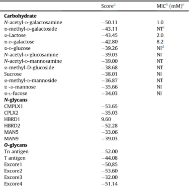
The results demonstrated that VGL presents favorable in-teractions with GalNAc (Score: 50.11), Gal (Score: 42.80),
a
-methyl-D-galactoside ( 43.11) anda
-Lactose ( 43.45) corrobo-rating with previous sugar inhibition assays performed by Silva and colleagues [17]. Comparison of scores and experimental data is shown in Table 1, and indicates the lectin specificity for galactosides.The set of interactions established between the VGL model and galactose is shown inFig. 6a. The galactose residue was stabilized by a network of H-bonds and hydrophobic interactions. The amino acid residues Asn87, Asn129, Leu213 and Ser214 interact by H-bonds with oxygen atoms O2, O3, O4 and O6 from carbohydrate structure. Gly104, Phe127, Gly212 and His217 residues are responsible for hydrophobic interactions which stabilize the galactose residue in the CRD. TheN-acetyl-D-galactosamine residue
complexed in the CRD was stabilized by a network of H-bonds connecting Asp87, Gly105, Asn129, Leu213, Ser214 and His217 residues to oxygen atoms O3, O4, O5 and O6 present in the mole-cule. Hydrophobic interactions involving the amino acid residues Gly104, Phe106, Phe127, Trp131 and Gly212 also contribute to the binding of lectin with this carbohydrate (Fig. 6b). The larger num-ber of interactions with GalNAc in relation to Gal was suggested in previous study [17] and was confirmed here. Previous results demonstrated the strong binding of Vatairea macrocarpa lectin (VML) with galactosides[8,9]. As shown inFig. S4, superposition of VGL with the structure of VML complexed with GalNAc demon-strated remarkable similarity of ligand binding validating the docking experiments.
These results demonstrated the efficiency of homology modeling and molecular docking for VGL. In fact, the animal Fig. 4.IL-1bplays an important role in VGL-induced paw edema. VGL (1 mg/kg; s.c.) or saline was injected intraplantar, and immunohistochemistry for IL-1bin epithelial and connective paw tissues fragment was performed at 6 h. (a,c) Saline, (b,d) VGL.
Fig. 5.Overall structure of VGL monomer. VGL chain is shown in cartoon represen-tation colored in blue. Spheres represent calcium ion (in gray) and manganese ion (in purple).
experiments indicated partial inhibition (40%) of VGL edemato-genic effect (0.35±0.05 AUC) in response to the association of VGL and galactose (0.22± 0.03 AUC) (Fig. 6c). This partial inhibition could be explained by the involvement of other binding sites on the molecule, such as metal or hydrophobic cavity [42,43]. Alterna-tively, the lectin would have high affinity for N- and O-glicans, present in the cell membrane, despite of the high galactose con-centration used in the reversion assay. Attempting to confirm VGL capacity of interactions with glycoproteins a total of 13 glycans, 6 N-and 7O-glycans, were chosen for docking based primarily on its relevance and relative high presence in glycoproteins. Best docking poses and scores are shown in Fig. S5 and Table 1 respectively. Among N-glycans, complex type presenting galactosyl terminal moieties demonstrated favorable interactions with the lectin (CPLX1 score: 53.65) (Fig. 7A) but the addition of a sialic acid moiety reduced the score drastically (CPLX2 Score: 35.03) due to galactosyl capping. Similarly, hybridN-glycans presenting galactose in terminal region interact strongly with VGL (HBRD2 Score: 52.28) differently to that occurs with its sialylated coun-terpart (HBRD1 Score: 9.60). Unsurprisingly high-mannose type did not show important interactions in CRD.
Sugar sequences found in glycoproteins normally share a com-mon core, in case of N-glycans Man
a
1e6 (Mana
1e3)Man-b
1e4GlcNAcb
1e4GlcNAcb
1-Asn-X-Ser/Thr[44] and are classified in three types: high-mannose, in which only mannose residues are attached to the core, like man5 and man9 used in the present study. Man5 is precursor of several high-mannose glycans, while man9 is found in a number of glycoproteins including insulin receptor and HIV gp120[31,45,46], other potential therapeutical targets.Complex type glycans, in which branches initiated by N-acetylglucosamine (GlcNAc) are attached to the core, being not uncommon the subsequent addition of galactose residues in
b
1e4 Table 1Docking score results and minimum inhibitory concentration of carbohydrates on VGL hemagglutination activity.
Scorea MICb(mM)e
Carbohydrate
N-acetyl-D-galactosamine 50.11 1.0
a-methyl-D-galactoside 43.11 NTc
a-Lactose 43.45 2.0
a-D-galactose 42.80 8.2
a-D-glucose 39.26 NId
N-acetyl-D-glucosamine 39.03 NI
N-acetyl-D-mannosamine 39.00 NT
a-methyl-D-glucoside 38.68 NT
Sucrose 38.01 NI
a-methyl-D-mannoside 36.87 NT
a-D-mannose 35.66 NI
a-L-fucose 34.03 NI
N-glycans
CMPLX1 53.65
CPLX2 35.03
HBRD1 9.60
HBRD2 52.28
MAN5 33.06
MAN9 39.03
O-glycans
Tn antigen 52.00
T antigen 44.08
Excore1 50,85
Excore2 53.60
Excore3 32.00
Excore4 51.14
aMoldock score (MDS)¼E
interþEintra, whereEinteris the ligand protein inter-action energy: .
b MIC: minimum inhibitory concentration. c NT: not tested.
d NI: not inhibitory on tested concentrations. e Data from Silva and colleagues[17].
Fig. 6.CRD mediates VGL edematogenic effect. Representation of carbohydrate-recognition domain of VGL in complex with (a)D-galactose and (b)N-acetyl-D-galactosamine. Blue dashes represent polar contacts. (c) Inhibition of VGL-induced edema (1 mg/Kg; s.c.) by galactose (0.1 M). Mean±S.E.M. (n¼5e6). *p<0.05vs. saline #p<0.05vs. VGL.
bond with GlcNAc, named N-acetyllactosamine (LacNAc). Poly-LacNAc chains are found in glycans in various cell types and may serve as scaffold for insertion of specific glycosyl moieties[31]. Alternatively, formation of GalNAc
b
1e4GlcNAc branches are also present in several structures[47]. Complex glycan CPLX1 presents LacNAc and fucosylated GalNAcb
1e4GlcNAc branches, while CPLX2 has similar structure to CPLX1 with LacNAc galactosyl ter-minal capped by a sialic acid.HybridN-glycans have branches with unsubstituted mannose residues and others with GlcNAc linkage[48]. HBRD1 glycan pre-sents the mannose residues branch and a sialic acid capped LacNAc branch, while HBRD2 have similar structure with uncapped galac-tosyl moiety.
AmongO-glycans, docking revealed that, similar to VML[8,9], VGL possibly interacts with tumour-associated antigens T and Tn (Scores: 52.00 and 44.08), best pose of Tn antigen is shown in
Fig. 7B, indicating a possible application in cancer research. Results also revealed very favorable interactions withO-glycans extended cores with exception of extended core 3.
O-glycans are important post-translational modification of mammalian proteins. Usually these glycans are linked via N-ace-tylgalactosamine (GalNAc) moiety to a serine or threonine residue
[32]. Among the tested glycans, Tn antigen (GalNAc
a
Ser/Thr) and T antigen (Galb
1-3GalNAca
Ser/Thr) were chosen based on its importance in cancer studies and prevalence as cores of O-glycans[32,49,50]. Other chosen sugars include Excore1 that contains sialic acid capped LacNAc branch and are found in many glycoproteins and mucins, excore2 that contains a branching GlcNAc attached to core 1 and are found in both glycoproteins in specific cells and tissues, excore3 and excore4 that contains some LacNAc branches and are found in mucins of certain mucin-secreting tissues[51e53].
Most of biological activities triggered by lectins occur due to interaction between proteins and molecular targets via glycosyl residues[54e57] and together, these results suggest that VGL is
capable of binding toO-glycans due to favorable docking scores and interactions suggestive of high specificity for this glycan type, while for N-glycans, the lectin binds to preferentially those possessing galactose as terminal residue. It is likely that the molecular target of VGL in order to elicit the edematogenic effect is a glycosylated protein presenting galactosyl moieties on its terminal regions or common types ofO-glycans.
4. Conclusion
The theoretical three-dimensional structure of Vatairea
guianensis lectin (VGL) presents high similarity with Vatairea macrocarpa lectin (VML). VGL elicited edematogenic activity, involving prostaglandins, IL-1
b
and CRD. In silico tests demon-strated the binding capacity of VGL with galactosides and impor-tantN-andO-glycans, corroborating with the hypothesis that VGL interaction with glycosylated molecular targets are one of the main factors responsible for itsin vivoeffects.Conflict of interest
The authors declare that they have no conflict of interest.
Ethical approval
All procedures performed in animals were in accordance with the ethical standards of the institution at which the studies were conducted e UECE Institutional Ethical Committee (UECE No. 10130208-8/40).
This article does not contain any studies with human partici-pants performed by any of the authors.
Acknowledgments
This study was supported by grants from the Conselho Nacional de Desenvolvimento Científico e Tecnologico (CNPq), Coordenaç ~ao de Aperfeiçoamento de Pessoal de Nível Superior (CAPES) and Fundaç~ao Cearense de Apoio ao Desenvolvimento Científico e Tecnologico (FUNCAP). B.S.C., K.S.N., and A.M.S.A. are senior in- vestigators of CNPq. David Martin helped with the English editing of the manuscript.
Appendix A. Supplementary data
Supplementary data related to this article can be found athttp:// dx.doi.org/10.1016/j.biochi.2017.06.008.
References
[1] H.S. Gabius, S. Gabius, Glycoscience: Status and Perspectives, WILEY-VCH Verlag, 1997.
[2] A.G. Rothfuchs, E. Roffe, A. Gibson, et al., Mannose-binding lectin regulates host resistance and pathology during experimental infection with Trypano-soma cruzi, Plos One 7 (11) (2012) e47835.
[3] B.S. Cavada, T. Barbosa, S. Arruda, et al., Revisiting proteus: do minor changes in lectin structure matter in biological activity? Lessons from and potential biotechnological uses of the Diocleinae subtribe lectins, Curr. Protein Pept. Sci. 2 (2) (2001) 123e135.
Fig. 7.Best docking poses of VGL complexed with A) CPLX1 glycan and B) Tn antigen.
[4] A. Loris, A. Imberty, S. Beeckmans, et al., Crystal structure ofPterocarpus angolensislectin in complex with glucose, sucrose, and turanose, J. Biol. Chem. 278 (18) (2003) 16297e16303.
[5] A.C. Almeida, V.J.S. Osterne, M.Q. Santiago, et al., Structural analysis of Cen-trolobium tomentosumseed lectin with inflammatory activity, Arch. Biochem. 15 (596) (2016) 73e83.
[6] R.G. Benevides, G. Ganne, R.C. Sim~oes, et al., A lectin fromPlatypodium elegans
with unusual specificity and affinity for asymmetric complex N-glycans, J. Biol. Chem. 287 (31) (2012) 26352e26364.
[7] R. Banerjee, S.V. Mande, V. Ganesh, et al., Crystal structure of peanut lectin, a protein with an unusual quaternary structure, Proc. Natl. Acad. Sci. 91 (1994) 227e231.
[8] B.L. Sousa, J.C. Silva-Filho, P. Kumar, et al., High-resolution structure of a new Tn antigen-binding lectin fromVatairea macrocarpaand a comparative anal-ysis of Tn-binding legume lectins, Int. J. Biochem. Cell Biol. 59 (2015) 103e110.
[9] B.L. Sousa, J.C. Silva-Filho, P. Kumar, et al., Structural characterization of a
Vatairea macrocarpalectin in complex with a tumor-associated antigen: a new tool for cancer research, Int. J. Biochem. Cell Biol. 72 (2016) 27e39. [10] N.M.N. Alencar, E.H. Teixeira, A.M.S. Assreuy, et al., Leguminous lectins as tools
for studying the role of sugar residues in leukocyte recruitment, Mediat. Inflamm. 8 (2) (1999) 107e113.
[11] N.M.N. Alencar, C.F. Cavalcante, M.P. Vasconcelos, et al., Antiinflammatory and antimicrobial effect of lectin fromLonchocarpus sericeusseeds in an experi-mental model of infectious peritonitis, J. Pharm. Pharmacol. 57 (7) (2005) 919e922.
[12] M.H. Napimoga, B.S. Cavada, N.M. Alencar, et al.,Lonchocarpus sericeuslectin decreases leukocyte migration and mechanical hypernociception by inhibiting cytokine and chemokines production, Int. Immunopharmacol. 7 (6) (2007) 824e835.
[13] A.F. Pires, N.V. Rodrigues, P.M. Soares, et al., A novel N-acetyl-glucosamine lectin ofLonchocarpus araripensisattenuates acute cellular inflammation in mice, Inflamm. Res. 65 (1) (2016) 43e52.
[14] N.M.N. Alencar, A.M.S. Assreuy, V.B. Alencar, et al., The galactose-binding lectin fromVatairea macrocarpaseeds inducesin vivoneutrophil migration by indirect mechanism, Int. J. Biochem. Cell Biol. 35 (12) (2003) 1674e1681. [15] N.M.N. Alencar, A.M.S. Assreuy, D.N. Criddle, et al.,Vatairea macrocarpalectin induces paw edema with leukocyte infiltration, Protein Pept. Lett. 11 (2) (2004) 195e200.
[16] N.M.N. Alencar, A.M.S. Assreuy, A. Havt, et al.,Vatairea macrocarpa (Legumi-nosae) lectin activates cultured macrophages to release chemotactic media-tors, Naunyn Schmiedeb. Arch. Pharmacol. 374 (4) (2007) 275e282. [17] H.C. Silva, C.S. Nagano, L.A.G. Souza, et al., Purification and primary structure
determination of a galactose-specific lectin fromVatairea guianensisAublet seeds that exhibits vasorelaxant effect, Process Biochem. 47 (12) (2012) 2347e2355.
[18] E.C. Landucci, E. Antunes, J.L. Donato, et al., Inhibition of carrageenan -induced rat paw o edema by crotapotin, a polypeptide complexed with phospholipase A2, Br. J. Pharmacol. 114 (3) (1995) 578e583.
[19] R.F.G. Feitosa, G.B. Melcíades, A.M.S. Assreuy, et al., The pharmacological profile of ovalbumin-induced paw oedema in rats, Mediat. Inflamm. 11 (3) (2002) 155e163.
[20] M. Chaudhary, A.R. Gadbail, G. Vidhale, M.P. MankarGadbail, et al., Compari-son of myofibroblasts expression in oral squamous cell carcinoma, verrucous carcinoma, high risk epithelial dysplasia, low risk epithelial dysplasia and normal oral mucosa, Head Neck Pathol. 6 (3) (2012) 305e313.
[21] D.W.A. Buchan, F. Minneci, T.C.O. Nugent, et al., Scalable web services for the PSIPRED protein analysis Workbench, Nucleic Acids Res. 41 (2013) 340e348. [22] B. Webb, A. Sali, Comparative protein structure modeling using MODELLER,
Curr. Protoc Protein Sci. 86 (2016), 2.9.1-2.9.37.
[23] R.A. Laskowski, M.W. MacArthur, D.S. Moss, et al., PROCHECK: a program to check the stereochemical quality of protein structures, J. Appl. Cryst. 26 (1993) 283e291.
[24] R. Lüthy, J.U. Bowie, D. Eisenberg, Assessment of protein models with three-dimensional profiles, Nature 356 (1992) 83e85.
[25] P. Benkert, M. Biasini, T. Schwede, Toward the estimation of the absolute quality of individual protein structure models, Bioinformatics 27 (2011) 343e350.
[26] P. Benkert, M. Kunzli, T. Schwede, QMEAN server for protein model quality estimation, Nucleic Acids Res. 37 (2009) 510e514.
[27] P. Benkert, S.C.E. Tosatto, D. Schomburg, QMEAN: a comprehensive scoring function for model quality assessment, Proteins 71 (2008) 261e277. [28] The Pubchem Projecthttps://pubchem.ncbi.nlm.nih.gov/, 2017 (Accessed 18
May 2017).
[29] O. Korb, T. Stützle, T.E. Exner, Empirical scoring functions for advanced protein-ligand docking with PLANTS, J. Chem. Inf. Model. 49 (2009) 84e96. [30] A.C. Wallace, R.A. Laskowski, J.M. Thornton, LIGPLOT: a program to generate
schematic diagram of protein-ligand interactions, Protein Eng. 8 (1995) 127e134.
[31] P. Stanley, H. Schachter, N. Taniguchi, Chapter 8: N-Glycans, in: A. Varki, R.D. Cummings, J.D. Esko, et al. (Eds.), Essentials of Glycobiology, second ed., Cold Spring Harbor, New York, 2009.
[32] I. Brockhausen, H. Schachter, P. Stanley, Chapter 9: O-GalNAc glycans, in: A. Varki, R.D. Cummings, J.D. Esko, et al. (Eds.), Essentials of Glycobiology, second ed., Cold Spring Harbor, New York, 2009.
[33] B.L. Parker, M. Thaysen-Andersen, N. Solis, et al., Site-specific glycan-peptide analysis for determination of N-glycoproteome heterogeneity, J. Proteome Res. 12 (12) (2013) 5791e5800.
[34] Glycan-Web, carbohydrate builder. http://glycam.org/tools/molecular-dynamics/oligosaccharide-builder/build-glycan?id¼1, 2017 (Accessed 18 May 2017).
[35] D.A. Case, T.A. Darden, T.E. Cheatham, et al., AMBER 12, University of Cali-fornia, San Francisco, 2012.
[36] K.N. Kirschner, A.B. Yongye, S.M. Tschampel, et al., GLYCAM06: a generalizable biomolecular forcefield. Carbohydrates, J. Comput. 29 (4) (2008) 622e655. [37] G. Jones, P. Willett, R.C. Glen, et al., Development and validation of a genetic
algorithm forflexible docking, J. Mol. Biol. 267 (1997) 727e748.
[38] D. Salvemini, Z.Q. Wang, P.S. Wyatt, et al., Nitric oxide: a key mediator in the early and late phase of carrageenan-induced rat paw inflammation, Br. J. Pharmacol. 4 (1996) 829e838.
[39] S. Nikolaus, J. Bauditz, P. Gionchetti, et al., Increased secretion of pro-inflammatory cytokines by circulating polymorphonuclear neutrophils and regulation by interleukin 10 during intestinal inflammation, Gut 42 (1998) 470e476.
[40] A.M.S. Assreuy, M.D. Shibuya, G.J. Martins, et al., Anti-inflammatory effect of glucose-mannose binding lectins isolated from Brazilian beans, Mediat. Inflamm. 6 (1997) 201e210.
[41] K.V. Brinda, A. Surolia, S. Vishveshwara, Insights into the quaternary associ-ation of proteins through structure graphs: a case study of lectins, Biochem. J. 39 (2005) 1e15.
[42] T.K. Dam, B.S. Cavada, T.B. Grangeiro, et al., Diocleinae lectins are a group of proteins with conserved binding sites for the core trimannoside of asparagine-linked oligosaccharides and differential specificities for complex carbohydrates, J. Biol. Chem. 273 (1998) 12082e12088.
[43] R. Loris, F. Casset, J. Bouckaert, et al., The monosaccharide binding site of lentil lectin: an X-ray and molecular modelling study, Glycoconj. J. 11 (6) (1994) 507e517.
[44] S.S. Pinho, C.A. Reis, Glycosylation in cancer: mechanisms and clinical impli-cations, Nat. Rev. Cancer 15 (9) (2015) 540e555.
[45] A.P. Cornfield, Structure/function of O-glycans. In: encyclopedia of genetics, genomics, Proteomics Bioinformatics 3 (3) (2005), 5:65.
[46] J. Mitoma, B. Petryniak, N. Hiraoka, et al., Extended core 1 and core 2 branched O-glycans differentially modulate sialyl Lewis X-type L-selectin ligand activ-ity, J. Biol. Chem. 278 (11) (2003) 9953e9961.
[47] A. Guzman-Aranquez, P. Arqueso, Structure and biological roles of mucin-type O-glycans at the ocular surface, Ocul. Surf. 8 (1) (2010) 8e17.
[48] V.R. Pinto-Junior, M.Q. Osterne VJS Santiago, et al., Structural studies of a vasorelaxant lectin fromDioclea reflexahook seeds: crystal structure, mo-lecular docking and dynamics, Int. J. Biol. Macromol. 98 (2017) 12e23. [49] H. Yao, X. Xie, Y. Li, Legume lectin FRIL preserves neural progenitor cells in
suspension culture in vitro, Clin. Dev. Immunol. 2008 (2008) 531317. [50] D.O. Croci, J.P. Cerliani, T. Dalotto-Moreno, et al., Glycosylation-dependent
lectin-receptor interactions preserve angiogenesis in anti-VEGF refractory tumors, Cell 156 (4) (2014) 744e758.
[51] G.A. Bezerra, R. Viertlmayr, T.R. Moura, et al., Structural studies of an anti-inflammatory lectin fromCanavalia bolivianaseeds in complex with diman-nosides, PLoS One 9 (5) (2014) e97015.
[52] E. Bieberich, Synthesis, processing, and function of N-glycans in N-glycopro-teins, Adv. Neurobiol. 9 (2015) 47e70.
[53] L.G. Sparrow, M.C. Lawrence, J.J. Gorman, et al., N-linked glycans of the human insulin receptor and their distribution over the crystal structure, Proteins 71 (1) (2008) 426e439.
[54] S. Sirois, M. Touaibia, K.C. Chou, et al., Glycosylation of HIV-1 gp120 V3 loop: towards the rational design of a synthetic carbohydrate vaccine, Curr. Med. Chem. 14 (30) (2007) 3332e3342.
[55] D.H. Van den Eijnden, A.P. Neeleman, H. Bakker, et al., Novel pathways in complex-type oligosaccharide synthesis. New vistas opened by studies in invertebrates, Adv. Exp. Med. Biol. 435 (1998) 3e7.
[56] B. Schiller, A. Hykollari, S. Yan, Complicated N-linked glycans in simple or-ganisms, Biol. Chem. 393 (8) (2012) 661e673.
[57] K. Marino, J. Bones, J.J. Kattla, et al., A systematic approach to protein glyco-~
sylation analysis: a path through the maze, Nat. Chem. Biol. 6 (10) (2010) 713e723.