Offi cial Publication of the Brazilian Society of Anesthesiology www.sba.com.br
REVISTA
BRASILEIRA DE
ANESTESIOLOGIA
Abstract
Background and Objectives: In our study we aimed to investigate the effect of bupivacaine and levobupivacaine on QT, corrected QT (QTc), and P wave dispersion durations during spinal anesthesia in cesarean section.
Methods: Sixty parturients scheduled for elective cesarean section in ASA I-II risk groups were included in the study. Baseline electrocardiographic (ECG) records of the patients were obtained in the operation room. Heart rate (HR), non-invasive blood pressure (NIBP), peripheral oxygen saturation (SpO2) and respiration rates (RR) were recorded. Venous cannulation was performed
with 18G cannula and fl uid preload made with 10 mL.kg-1. Lactated Ringer solution. After fl uid
preload, second ECG recordings were taken and the patients were randomly separated into two groups. Group B (n = 30) received 10 mg of bupivacaine and Group L (n = 30) received 10 mg of levobupivacaine for spinal anesthesia. ECG recordings were repeated at 1, 5 and 10 minutes after spinal block. HR, NIBP, SpO2 , RR and sensory block levels were also recorded at the same time intervals. At predetermined time intervals of spinal anesthesia, P wave dispersion (Pwd), QT dispersion (QTd), and QTc dispersion (QTcd) durations were measured from ECG records. QT and QTc durations are calculated with Bazzett formula.
The Effect of Levobupivacaine and Bupivacaine on QT,
Corrected QT (Qtc), and P Wave Dispersions in Cesarean Section
Yeliz Deniz
1, Dilek Okyay
2, Volkan Hancı*
3, Serhan Yurtlu
2, Hilal Ayoğlu
4,
Işıl Özkoçak Turan
51. Staff Doctor, MD, Karaelmas University, School of Medicine, Department of Anaesthesiology and Reanimation, Zonguldak, Turkey
2. Associate Professor, MD, Dokuz Eylül University, School of Medicine, Department of Anaesthesiology and Reanimation, İzmir, Turkey (Formerly Karaelmas University, Zonguldak, Turkey)
3. Associate Professor, MD, Dokuz Eylül University, School of Medicine, Department of Anaesthesiology and Reanimation, İzmir, Turkey (Formerly Karaelmas University, School of Medicine, Department of Anaesthesiology and Reanimation, Zonguldak, Turkey)
4. Associate Professor, MD, Karaelmas University, School of Medicine, Department of Anaesthesiology and Reanimation, Zonguldak, Turkey
5. Professor, MD, Karaelmas University, School of Medicine, Department of Anaesthesiology and Reanimation, Zonguldak, Turkey Received from Zonguldak Karaelmas University, School of Medicine, Department of Anaesthesiology
and Reanimation, Zonguldak, Turkey. A part of this study was presented as an abstract poster at the Turkish Anaesthesia and Reanimation Congress 27-30 October 2010.
Submitted on December 21, 2011. Approved on April 12, 2012.
Keywords:
Anesthesia, Spinal; Cesarean Section; Bupivacaine/ levobupivacaine; Electrocardiography.
SCIENTIFIC ARTICLE
*Corresponding author: Dokuz Eylül University, School of Medicine, Department of Anaesthesiology and Reanimation, İzmir, Turkey. Phone: +90.530.643.32.40. Reprint: Serhan Yurtlu, MD (+90.372.261.23.83.).
E-mail: [email protected]
Results: There was no difference between two groups according to block levels, hemodynamic parameters, Pwd, QTd, QTc and QTcd durations.
Conclusion: Bupivacaine and levobupivacaine may be preferred in spinal anesthesia in pregnant patients who have extended Pwd and QTcd preoperatively.
© 2013 Sociedade Brasileira de Anestesiologia. Published by Elsevier Editora Ltda. All rights reserved.
Introduction
Anesthetic agents may display proarrhythmic and antiarrhyth-mic activity by inducing electrical activity with various
mechanisms 1. Other than the anesthetic agents used,
exist-ing heart disease and other concomitant systemic diseases, surgical manipulation, procedures performed on the patient and medication may also cause arrhythmias in the
intraop-erative stage 2. Many hormonal and hemodynamic changes
that take place during pregnancy may also result in proar-rhythmic effects. Pregnancy may trigger the development of new arrhythmia or exacerbate already existing ones. Left axis deviation may be present in ECG as a result of the shift in the position of the heart due to the enlargement of the uterus during pregnancy. Premature atrial and ventricular
beats are common 3.
Regional anesthesia in cesarean surgeries has the advan-tages of allowing the mother to be awake during delivery, not neeeding airway manipulation, keeping mother’s airway refl exes, decreasing blood loss, reducing the risk of drug-induced fetal depression, and carrying the need for analgesia over to the postoperative stage. Regional anesthesia is the most common method of anesthesia used in cesarean
surger-ies in developed countrsurger-ies 4-6. One of the most commonly used
local anesthetics in obstetrics is bupivacaine. A relatively new agent, levobupivacaine is also increasingly being used
in obstetric patients 7. Bupivacaine and levobupivacaine may
increase the PR interval and QRS duration and prolong cardiac
conduction 8. Bupivacaine in spinal anesthesia had been
re-ported to induce ECG changes 9,10. However, levobupivacaine
has been shown to be less cardiotoxic 11. On the other hand,
high sympathetic blockage and hemodynamic changes that occur due to regional anesthesia and the inotropic agents
used may cause proarrhythmic effects 10. However, this has
not been investigated adequately through research. In our study, we aimed to examine the effects of bupi-vacaine and levobupibupi-vacaine on QT, QTc and P wave disper-sion in pregnant women.
Material and Method
This prospective randomized study was conducted in 2009-2010 at Zonguldak Karaelmas University’s School of Medicine Research and Practice Hospital, Department of Anesthesiology and Reanimation, after obtaining the approval of the Hospital Ethics Board (06. 12. 2007, Meeting decision No.: 2007 /09 /04) and patient consents.
Sixty pregnant women aged between 16 and 50, height ≥ 1.60 cm, weight between 60 and 100 kg, placed in the ASA risk group I-II in their preanesthetic evaluation and
scheduled for elective cesarean surgery were included in the study. They were randomly allocated into two groups: bupivacaine (Group B) and levobupivacaine (Group L) by using a randomized numbers table.
Exclusion criteria were refusal to participate in the study, the existence of brain tumors, scalded skin syndrome (SSS infection), spinal cord and peripheral nervous system diseases (poliomyelitis, multiple sclerosis, demyelinating diseases), hemorrhagic and hypovolemic shock, severe anemia, increased intracranial pressure, aortic and valvular heart disease, cardiac decompensation, systemic infection (generalized sepsis and bacteremia), local infection (dermal infections in puncture site of spinal needle, etc.), congenital spinal anomalies, scoliosis, post-traumatic vertebral injuries, vertebral colon metastatic lesions, increased abdominal pressure, chronic severe headache, anticoagulant drug use and anatomic diffi culties, electrolyte disturbances diabetes mellitus, hypothyroidism, hyperthyroidism, cardiomyopathy, atrial and/or ventricular hypertrophy on ECG, cardiomegaly, valvular disease, cardiac failure or chronic disease, patients with excessive smoking and alcohol consumption and used medication causing QT interval prolongation.
Premedication was not administered to our subjects. Following their admission into the operation room, ECG monitorization was performed and their control ECG (T0) records were taken. Heart rate, noninvasive blood pressure, peripheral oxygen saturation values and respiration rates were recorded.
Vascular access was obtained by using 18G catheter.
Preloading was performed with a 10 mL.kg-1 Lactated Ringer’s
solution. Following the preloading, second ECG (T1) records were taken and the patients were placed in lateral decubitus position. Dural puncture was performed from the L2-L3 or L3-L4 interval by using a 27G quincke spinal needle. After the fl ow of cerebrospinal fl uid we administered 10 mg bupi-vacaine in Group B (n = 30) and 10 mg levobupibupi-vacaine in
Group L (n = 30) in two minutes (at a speed of 1.5 mL.min-1)
intrathecal. We brought patients to a supine position after the injection.
Other ECG records were taken 1 (T2), 5 (T3), and 10 (T4) minutes after the block. Bromage scale (BS) scores, heart rate, blood pressure, peripheral oxygen saturation, respira-tion rate values and sensory block levels with pinprick test were recorded at minutes 1, 5 and 10 of spinal anesthesia and every 5 minutes thereafter.
Additional fl uid loading and stabilization of hemodynam-ics with 5 mg ephedrine was planned for cases where blood pressure values fell 20% below control values; 0.5 mg IV atropine was planned for cases where heart rate fell below
patients with nausea and vomiting. Standard 2 L.min-1 oxygen
was delivered to all patients via nasal cannula. Sensory and motor block levels were identifi ed and noted until 10 min and surgery started later.
Electrocardiography
Standard 12 derivation ECG recordings obtained with a
pa-per speed of 25 mm.sec-1 and a defl ection of 10 mm.mV-1 of
patients participating in the study was analyzed (Hewlett Packard®, Pagewriter 300pı). We calculated heart rate using mean RR time.
Analysis of QT dispersion
The QT interval was defi ned as between the beginning of QRS complex and the point where T waves descend onto the TP isoelectric line. When a U wave interrupted the T wave before returning to baseline, the QT interval was measured
to the nadir of the curve between the T and U waves 1. The
corrected QT interval (QTc) was calculated using the Bazett formula; QTc (ms) = QT measured/√RR (where RR is the RR interval). The QTd value was determined as the difference between the longest and shortest QT intervals in the 12 ECG leads. The QTc dispersion (QTcd) duration according to heart rate was identifi ed with the Bazett formula; QTcd (ms) = QTd
measured/√RR 1.
Analysis of P-wave dispersion
The beginning of P-wave was defi ned as positive defl ection from the isoelectric line, and the end point when the
posi-tive defl ection returned to the isoelectric line 1. Derivations
where the beginning and end of P-waves were not obvious were excluded from the study. Pwd was the difference
be-tween the longest and shortest P-wave durations 1.
Subjects who had less than 9 derivations assessed on the ECG were excluded from the study. All ECG measurements were evaluated three times by two experts who were not
aware of which group the subject belonged to 1.
Statistical Analysis
Statistical analyses were performed by using the Statistical Package for the Social Sciences (SPSS) 13.0 (SPSS Inc., Chicago, IL, USA). Descriptive statistics included arithmetic
mean ± standard deviation for numerical data, and num-bers and percentages for categorical data. We used the Kolmogorov-Smirnov test to examine compatibility between measured variables and normal distribution. We used a signifi -cance test when parametric test assumptions were met for intergroup differences between the measured variables, and the Mann-Whitney U test when they were not. We analyzed the differences between groups for categorical variables by using Chi-Square analysis. For measured variables, we analyzed differences between groups and time-dependent changes by two-way analysis of variance in repeated measurements. When we found a difference as a result of two-way analysis of variance in repeated measurements, comparisons between pairs were made with the Bonferroni test. For ordinary variables, differences between groups and time-dependent changes were analyzed by using analysis of variance in repeated measurements. The results were evalu-ated at 95% confi dence interval and p < 0.05 was accepted as statistically signifi cant difference.
Results
Our subjects were allocated randomly into two groups. Groups were similar in terms of age, body mass, height and
American Society of Anesthesiologists
risk class (Table 1). When we compared the groups with respect to sensory block levels, we found no statistically signifi cant difference between groups at any time (p > 0.05) (Table 2).Compared with respect to systolic and diastolic arterial blood pressure values at all times, Groups B and L did not display a statistically signifi cant difference (p > 0.05). When we compared T3 and T4 times against control values, we saw a signifi cant decrease in systolic and diastolic arterial blood pressure values in both Groups B and L (p < 0.05) (Table 3). When the mean heart rates in the two groups were compared, a statistically signifi cant difference did not exist between or within groups at any time (p > 0.05) (Table 3).
When the groups were compared with regard to maxi-mum P-wave values, no statistically signifi cant difference was found between the values measured in Groups B and L at any time (p = 0.146). When T4 time was compared with the control value, a signifi cant decrease was detected in the maximum P-wave value of Group L (p = 0.015) (Table 3).
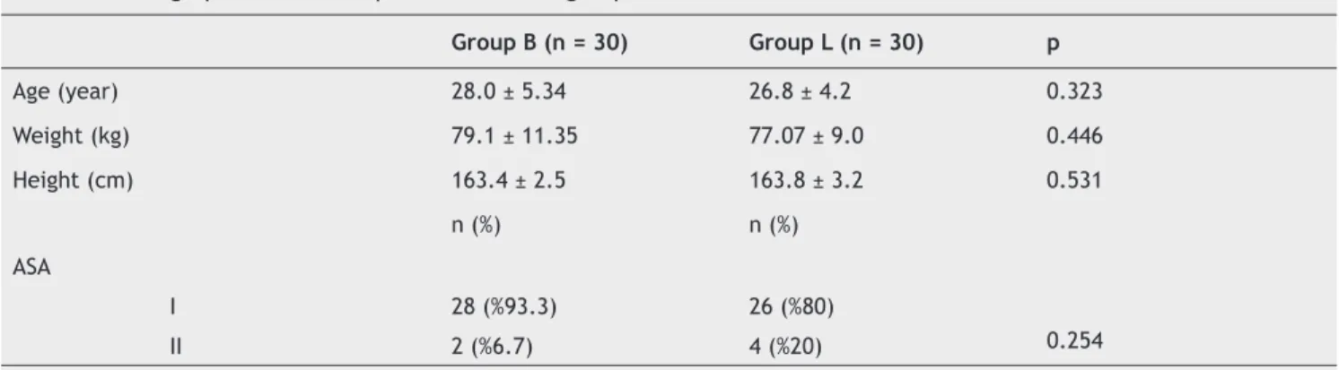
Table 1 Demographic and anthropometric data of groups.
Group B (n = 30) Group L (n = 30) p
Age (year) 28.0 ± 5.34 26.8 ± 4.2 0.323
Weight (kg) 79.1 ± 11.35 77.07 ± 9.0 0.446
Height (cm) 163.4 ± 2.5 163.8 ± 3.2 0.531
n (%) n (%)
ASA
I 28 (%93.3) 26 (%80)
0.254
II 2 (%6.7) 4 (%20)
Table 2 Sensory block levels of groups.
Time Group B (n = 30) Group L (n = 30) p
T0 – – –
T1 – – –
T2 L1 (Th10-5) L1 (Th11-L5) 0.150
T3 Th8 (Th4- T12) Th8 (Th6-Th11) 0.106
T4 Th4 (Th2-Th6) Th4 (Th3-Th5) 0.327
T0: Control, T1: After the preloading, T2: 1 minute after spinal anesthesia, T3: 5 minutes after spinal anesthesia, T4: 10 minutes after spinal anesthesia.
Table 3 Hemodynamic and Electrocardiographic data of groups.
T0 T1 T2 T3 T4
HR (beats.min-1)
Group B (n = 30) 90.6 ± 13.7 91.5 ± 15.9 95.6 ± 15.7 91.9 ± 21.6 96.0 ± 23.7 Group L (n = 30) 90.6 ± 12.9 90.1 ± 13.3 92.2 ± 20.0 88.2 ± 17.8 87.6 ± 18.5 SAP (mm Hg)
Group B (n = 30) 122.6 ± 13.5 128.7 ± 14.4 123.2 ± 15.2 104.6 ± 21.4* 110.4 ± 21.7* Group L (n = 30) 127.8 ± 9.1 131.5 ± 10.9 121.1 ± 15.3 111.4 ± 18.6† 113.1 ± 20.8† DAP (mm Hg)
Group B (n = 30) 75.1 ± 11.2 79.0 ± 9.2 72.9 ± 14.2 58.2 ± 16.1* 64.3 ± 15.1* Group L (n = 30) 79.6 ± 8.2 80.3 ± 7.5 72.9 ± 13.0 58.2 ± 16.2† 64.4 ± 15.7† Max. P-wave duration (ms)
Group B (n = 30) 99.0 ± 16.5 96.0 ± 18.0 96.3 ± 15.2 96.3 ± 13.3 93.3 ± 13.5 Group L (n = 30) 99.7 ± 14.5 94.6 ± 15.0 92.7 ± 13.1 92.0 ± 14.7 90.3 ± 11.6† Min P-wave duration (ms)
Group B (n = 30) 42.9 ± 16.0 42.3 ± 13.8 42.7 ± 13.9 41.3 ± 12.8 42.7 ± 11.4 Group L (n = 30) 38.3 ± 13.2 35.6 ± 12.2 33.3 ± 9.2 35.7 ± 10.4 34.3 ± 11.0 P-wave dispersion (ms)
Group B (n = 30) 56.3 ± 19.2 53.7 ± 20.4 53.7 ± 17.7 55.7 ± 16.1 52.7 ± 17.2 Group L (n = 30) 62.0 ± 16.7 59.0 ± 19.0 59.3 ± 11.1 56.3 ± 13.0 54.3 ± 14.5 QT interval (ms)
Group B (n = 30) 352.7 ± 21.4 348.0 ± 20.2 339.3 ± 22.4 340.0 ± 32.1* 346.0 ± 30.1* Group L (n = 30) 341.0 ± 29.0 344.1±28.5 339.3±23.4 340.1±26.3 350.7±36.8 QTc interval (ms)a
Group B (n = 30) 428.0 ± 26.0 419.0±19.1 422.7±23.5 422.1±30.6 422.8±29.3 Group L (n = 30) 422.1 ± 19.1 421.7±25.1 418.1±23.0 422.4±24.2 420.6±32.8 QTd interval (ms)
Group B (n = 30) 55.0 ± 10.7 49.3±13.8 49.0±11.2 53.3±15.8 55.0±11.9 Group L (n = 30) 55.3 ± 19.7 55.0±19.6 54.6±16.7 57.0±15.5 56.0±19.5 QTcd interval (ms)b
Group B (n = 30) 67.2 ± 16.1 64.7±17.3 71.2±36.1 65.2±35.0 69.5±17.5
Group L (n = 30) 66.7 ± 26.7 66.0±27.8 67.7±21.0 68.0±18.0 70.3±23.0
T0: Control, T1: After the preloading, T2: 1 minute after spinal anesthesia, T3: 5 minutes after spinal anesthesia, T4: 10 minutes after spinal anesthesia; Max: maximum; Min: minimum; HR: Heart Rate; SAP: Systolic Arterial Pressure; DAP: Diastolic Arterial Pressure
Values are mean ± SD.
When the two groups were compared with respect to minimum P-wave values, no statistically signifi cant difference was seen between or within groups at any time (p > 0.05) (Table 3).
Similarly, when we studied the groups’ P-wave disper-sion values, no statistically signifi cant difference was found between or within groups at any time (p > 0.05) (Table 3).
When we compared the groups with respect to QT values, we did not detect a statistically signifi cant difference be-tween Groups B and L at different measurements (p > 0.05). When we compared T3 and T4 times to the control value within Group B, we found a decrease in the QT value. This decrease was statistically signifi cant (p < 0.05). When we compared the values obtained at all times in Group L to control values, no statistically signifi cant difference existed in the QT value (p > 0.05) (Table 3).
When we compared the groups with respect to QTd, QTc and QTcd values, we found a statistically signifi cant differ-ence both between and within groups at all times (p > 0.05) (Table 3).
Ephedrine consumption was 13.3 ± 14.7 mg in Group B and 6.5 ± 6.2 mg in Group L. No statistically signifi cant dif-ference existed between the total ephedrine, atropine and metoclopramide consumption in the two groups (p > 0.05).
Discussion
In our study we examined the effects of using bupivacaine and levobupivacaine in spinal anesthesia for cesarean surgeries on P-wave in ECG, QT, and QTc dispersion times, and we found no signifi cant difference between the p wave, QT and QTc dispersions of the levobupivacaine and bupivacaine groups.
In pregnancy, a dramatic hormonal and hemodynamic change can be observed in the organism, which may lead to a proarrhythmic effect. Pregnancy may trigger the de-velopment of new arrhythmia or exacerbate existing ones. Hemodynamic changes, increased cardiac output and cir-culating blood volume cause an arrhythmogenic effect by increasing end diastolic volume and myocardial regression. In addition, elevated catecholamine levels also cause the development of arrhythmia. Arrhythmia and tachycardia are very common occurrences among pregnant women. Heart rate may increase by 20% during pregnancy and left axis deviation shows up in ECG due to the enlargement of the uterus. Premature atrial and ventricular beats are also
very common 3.
Even though spinal block may be a safe anesthesia technique, severe tachycardia, cardiac arrest and other arrhythmia are reported during spinal anesthesia practices. In an ASA study of closed claims project, sudden cardiac arrest was reported during spinal anesthesia performed on 14 hemodynamically stable, young and healthy patients. In another study of 952 patients who received spinal anesthesia, risk factors for bradycardia and hypotension during spinal anesthesia were defi ned as being female, a control heart rate below 60, use of beta-blockers, and a sensory block
above T5 12.
In a multiple center study conducted on over 17,000
patients, Youngs PJ et al. 13 studied the effects of spinal
anesthesia that cause arrhythmia. Of their subjects, 70.2% had tachycardia, bradycardia or arrhythmia. Most of these
were spontaneously recovering minor arrhythmia. Sinusal arrhythmia was found in 30.3% of the patients, premature beats in 27.2%, and bradycardia in 13.8%.
The incidence of arrhythmia and hypotension among pregnant women who receive spinal anesthesia in cesarean surgeries is greater than expected. Most of these incidents are spontaneous and temporary. They may occur suddenly and require urgent treatment. In a study of 254 healthy pregnant
women who underwent cesarean surgery, Shen CL et al. 14
administered 10 mg bupivacaine + 0.2 mg morphine for spinal
anesthesia, and observed 1st degree atrioventricular block in
9 patients (3.5%), 2nd degree atrioventricular block in 9 (3.5%),
severe bradycardia in 17 (heart rate < 50 beat.min-1) (6.7%)
and multiple ventricular premature complexes in 3. During cesarean surgeries, one should take care about arrhythmia and handle monitoring attentively. Among our subjects, we observed premature ventricular beats in one pregnant woman in the bupivacaine group and in two women in the levobupi-vacaine group.
Many clinical studies have shown both bupivacaine and levobupivacaine to have equal effectiveness in spinal
an-esthesia over doses of 10 mg 15. Alley EA et al. 15 used 4 mg,
8 mg and 12 mg hyperbaric bupivacaine and hyperbaric levobupivacaine for spinal anesthesia in 18 healthy volun-teers and observed similar sensory and motor block levels in the two groups. Similarly, in our study we observed no signifi cant difference between the sensory block levels, systolic blood pressure, diastolic blood pressure, heart rate changes, and amounts of atropine used in the two groups until minute 10.
In spinal anesthesia, cardiovascular effects are related to the sympathetic blockage that develops with spinal an-esthesia rather than the systemic absorption of local
anesthetics 9.
However, there are a limited number of studies that ex-plore the effects of spinal anesthesia on QT and QTc intervals,
the results of which seem to vary 10,16,17. In 20 adult males who
did not receive premedication, Owczuk et al. 10 performed
spinal anesthesia with 3 or 4 mL 5% hyperbaric bupivacaine and calculated the QTc interval from the ECG records taken at minutes one, three, fi ve and 15, and observed a signifi cantly prolonged QTc interval starting from minute 1 after spinal anesthesia induction and in later measurements. With mean values, they detected no signifi cant difference at the onset of spinal block between QTc intervals and heart rate. However, starting from min three, a signifi cant decrease was observed in systolic, diastolic and mean blood pressure as compared to control values. The QTc interval exceeded 440 msec in a total of eight patients, and QTc interval > 440 msec occurred once in two patients, twice in fi ve patients, and three times in two patients. Prolonged QTc interval was seen at min one following spinal anesthesia in fi ve patients, after mins three and fi ve in three patients, and after min 15 in two patients. QTc interval > 500 msec occurred only in one patient, but severe arrhythmia or conduction did not. Ventricular ectopic beats were observed in one patient who had normal QTc interval.
At the same time, many case studies report that spinal anesthesia can be used safely in pregnant women with
during combined spinal-epidural anesthesia with 9 mg bupi-vacaine and 100 mg lidocaine in a pregnant woman with asymptomatic idiopathic prolonged QT interval syndrome
who underwent elective cesarean surgery. Kameyam et al. 19
also emphasized that spinal anesthesia with bupivacaine is a safe option for patients with asymptomatic idiopathic prolonged QT syndrome.
Şen et al. 9 studied the effects of spinal anesthesia on
the QTc interval of preeclamptic patients. They reported that even though preeclamptic patients have higher QTc intervals than the control group prior to spinal anesthesia, their interval shortens following spinal anesthesia while no change occurs in that of patients without preeclampsia. They also wrote that the sympathetic blockage effect of spinal anesthesia might normalize patients with prolonged QTc interval.
In our study, we found that both levobupivacaine and bupivacaine for spinal anesthesia shortens the QT intervals of patients, but no statistically signifi cant difference existed between the groups. Even though a signifi cant difference did not exist among the subjects in the levobupivacaine group, the QT interval of patients who received bupivacaine for spinal anesthesia were signifi cantly shorter than control values at minutes fi ve and 10 after spinal anesthesia. We are of the opinion that this shortening in the QTc interval may be related to the sympathetic suppression caused by spinal anesthesia. Supporting fi ndings were observed in a previous
study of Owczuk et al. 20, where they had compared lumbar
and thoracic epidural block by using isobaric bupivacaine and found out that QTcd was signifi cantly shorter in patients who received thoracic epidural block, suggesting the role of higher symphathetic block level.
Pregnancy may also affect QTd and QTcd times.
Lechmanova et al. 21 compared QTds of 37 healthy pregnant
women in their late pregnancy and postnatal days, and found out that parturients had signifi cantly longer QTd in late pregnancy. In our study, we found that the QTd and QTcd interval of patients who received spinal anesthesia with levobupivacaine and bupivacaine were prolonged after spinal anesthesia, but a statistical difference did not exist within groups or in comparison with control values.
Anesthetic substances may affect P wave dispersion (Pwd). The general anesthetic sevofl urane has been reported to prolong Pwd, desfl urane to have no effect on it, and
propofol to shorten it 22-24. At the same time, our literature
survey found no study that investigated the effects of local or spinal anesthesia on Pwd. However, in one study that evalu-ated Pwd changes in pregnant women, Pwd was reported to
prolong due to the shortening of minimum P wave time 25.
In our study, we found no signifi cant difference between the Pwd, Pmax and Pmin times of the groups following spinal anesthesia with either local anesthetic.
Our study has several limitations. First, P wave, QT, QTc dispersions were manually calculated from the ECG record. Even though there are many studies stating that these pa-rameters can be measured manually with a minimal room for
error 26,27, others mention about the reliability of this type of
measurement 26,28. The second limitation of our study is that
it has lasted until 10 minutes after spinal anesthesia applica-tion and ECG records were taken with fi ve-minute intervals. However, there are studies that show in ECG that changes
can occur in P-wave, QT times after minute 10 10,13. We are
of the opinion that using continuous Holter monitorization in addition to intermittent ECG records in future studies may help longer-term and more detailed identifi cation of perioperative arrhythmia and ECG changes. Third, effects of fl uid therapy and vasopressors on ECG records cannot be excluded in this study, because one has to administer these therapies due to ethical reasons.
In conclusion, our double blind randomized prospective study which compared the electrocardiographic effects of spinal anesthesia by using 10 mg of bupivacaine and 10 mg of levobupivacaine in cesarean surgeries showed that P wave, QT and corrected QT dispersion values were not affected. For this reason, levobupivacaine and bupivacaine may be preferred for spinal anesthesia of the pregnant women with prolonged P wave and QT dispersion determined in the preoperative stage.
References
1. Hancı V, Ayoğlu H, Yurtlu S et al. - An evaluation of P-wave dispersion, QT, corrected QT and corrected QT dispersion intervals on the electrocardiograms of malnourished adults. Anaesth Intensive Care. 2010;38:122-127.
2. Akçay M, Albayrak D, Akçay FK et al. - Sevofl oran ile yapılan VİMA ve bupivakainle yapılan spinal anestezi yöntemlerinin QT dispersiyonuna olan etkilerinin karşılaştırılması. Türkiye Klinikleri J Anest Reanim. 2004;2:137-143.
3. Emmanuel M, Kanoupakis, Panos EV - Arrhythmias and Pregnancy. Cardiology Department, Heraklion University Hospital, Crete, Greece. Hell J Cardiol. 2005;46:317-319.
4. Wee MY - Brown H, Reynolds F - The National Institute of Clinical Excellence (NICE) guidelines for Caesarean sections: implications for the anesthetist. Int J Obstet Anesth 2005;14:147-158. 5. Gori F, Pasqualucci A, Corradetti F, Milli M, Peduto VA - Maternal
and neonatal outcome after Cesarean section: the impact of anesthesia. J Matern Fetal Neonatal Med. 2007;20:53-57. 6. Paech MJ - Anesthesia for Cesarean Section. In: Palmer CM,
D’angelo R, Paech MJ, editors. Handbook of Obstetric Anesthesia 1 st ed. Oxford: BIOS, 2002;82-113.
7. Zi-gang Li, Liang Zhou, and Hui-fang Tang - Effects of levobupivacaine and bupivacaine on rat myometrium. J Zhejiang Univ Sci B. 2006;7:757–62.
8. Leone S, Di Cianni S, Casati A, Fanelli G - Pharmacology, toxicology, and clinical use of new long acting local anesthetics, ropivacaine and levobupivacaine. Acta Biomed. 2008;79:92-105.
9. Sen S, Ozmert G, Turan H, Caliskan E, Onbasili A, Kaya D - The effects of spinal anesthesia on QT interval in preeclamptic patients. Anesth Analg. 2006;103:1250-5.
10. Owczuk R, Sawicka W, Wujtewicz MA, Kawecka A, Lasek J, Wujtewicz M - Infl uence of spinal anesthesia on corrected QT interval. Reg Anesth Pain Med. 2005;30:548-52.
11. Udelsmann A, Lorena SE, Grioli SU, Silva WA, Moraes AC, Andreollo NA - Hemodynamic effects of local anesthetics intoxication: experimental study in swine with levobupivacaine and bupivacaine. Acta Cir Bras. 2008;23:55-64.
12. Benumof JL, Saidman LJ - Anesthesia and perioperative complications. Second edition. 1999;50-63.
13. Youngs PJ, Littleford J - Arrhythmias during spinal anesthesia. Can J Anaesth. 2000;47:385-390.
15. Alley EA, Kopacz DJ, McDonald SB, Liu SS - Hyperbaric spinal levobupivacaine: a comparison to racemic bupivacaine in volunteers. Anesth Analg. 2002;94:188-193.
16. Pedroviejo Saez V, Lasa Unzue C - Intradural anesthesia for emergency cesarean section in a woman with congenital long QT syndrome. Rev Esp Anestesiol Reanim. 2011;58:189-191. 17. Palkar NV, Crawford MW - Spinal anaesthesia in prolonged Q-T
interval syndrome. Br J Anaesth. 1986;58:575-576.
18. Al-Refai A, Gunka V, Douglas J - Spinal anesthesia for Cesarean section in a parturient with long QT syndrome. Can J Anaesth. 2004;51:993-996.
19. Kameyama E, Ito Y, Ito J et al - Anesthetic management of caesarean section in a patient with asymptomatic idiopathic prolonged QT interval syndrome. Eur J Anaesthesiol. 2004;21:566-570.
20. Owczuk R, Steffek M, Wujtewicz MA - Infl uence of reversible adrenergic blockade of the heart obtained through thoracic epidural anaesthesia on cardiac repolarisation effects on cardiac repolarisation of reversible adrenergic blockade through thoracic epidural anaesthesia. Clin Exp Pharmacol Physiol. 2009 [in press]. Available at: [http://www.biomedsearch.com/nih/Infl uence-reversible-adrenergic-blockade-heart/19298541.html] 21. Lechmanová M, Kittnar O, Mlcek M et al. - QT dispersion and
T-loop morphology in late pregnancy and after delivery. Physiol Res. 2002;51:121-129.
22. Kazanci D, Unver S, Karadeniz U et al. - A comparison of the effects of desfl urane, sevofl urane and propofol on QT, QTc, and P dispersion on ECG. Ann Card Anaesth. 2009;12:107-112. 23. Owczuk R, Wujtewicz MA, Sawicka W et al - Effect of anaesthetic
agents on p-wave dispersion on the electrocardiogram: comparison of propofol and desfl urane. Clin Exp Pharmacol Physiol. 2008;35:1071-1076.
24. Hanci V, Aydin M, Yurtlu BS et al. - Anesthesia induction with sevofl urane and propofol: evaluation of P-wave dispersion, QT and corrected QT intervals.
Kaohsiung J Med Sci
. 2010;26(9):470-477.25. Ozmen N, Cebeci BS, Yiginer O, Muhcu M, Kardesoglu E, Dincturk M - P-wave dispersion is increased in pregnancy due to shortening of minimum duration of P: does this have clinical signifi cance. J Int Med Res. 2006;34:468-474.
26. Dilaveris PE, Gialafos JE - P-wave dispersion: a novel predictor of paroxysmal atrial fi brillation. Ann Noninvasive Electrocardiol. 2001;6:159-165.
27. Ciaroni S, Cuenoud L, Bloch A - Clinical study to investigate the predictive parameters for the onset of atrial fi brillation in patients with essential hypertension. Am Heart J. 2000;139:814-819.