MINISTÉRIODAEDUCAÇÃO
UNIVERSIDADEFEDERALDORIOGRANDEDONORTE
CENTRODECIÊNCIASDASAÚDE
PROGRAMADEPÓS-GRADUAÇÃOEMCIÊNCIASDASAÚDE
AVALIAÇÃODAEFICÁCIAANALGÉSICANOPÓS-OPERATÓRIODE
REVASCULARIZAÇÃODOMIOCÁRDIO:ENSAIOCLÍNICORANDOMIZADO
CONTROLADO
VALDECYFERREIRADEOLIVEIRAPINHEIRO
NATAL–RN
VALDECYFERREIRADEOLIVEIRAPINHEIRO
AVALIAÇÃODAEFICÁCIAANALGÉSICANO
PÓS-OPERATÓRIODEREVASCULARIZAÇÃO
DOMIOCÁRDIO:ENSAIOCLÍNICO
RANDOMIZADOCONTROLADO
Tese apresentada ao Programa de Pós-Graduação do Centro de Ciências da Saúde da Universidade Federal do Rio Grande do Norte, como requisito para a obtenção do título de Doutor em Ciências da Saúde.
ORIENTADORA: Profª. Drª. Ivonete Batista de Araújo
NATAL –RN
MINISTÉRIODAEDUCAÇÃO
UNIVERSIDADEFEDERALDORIOGRANDEDONORTE
CENTRODECIÊNCIASDASAÚDE
PROGRAMADEPÓS-GRADUAÇÃOEMCIÊNCIASDASAÚDE
VALDECY FERREIRA DE OLIVEIRA PINHEIRO
AVALIAÇÃODAEFICÁCIAANALGÉSICANOPÓS-OPERATÓRIODE
REVASCULARIZAÇÃODOMIOCÁRDIO:ENSAIOCLÍNICORANDOMIZADO
CONTROLADO
Aprovada em 09/05/2014
Banca Examinadora:
Presidente da Banca: Profa Dr aIvonete Batista de Araújo (UFRN)
Membros da Banca:
Prof. Dr.Irami Araújo Filho (UFRN)
Profa Dr a Milva Marina Figueiredo de Martino (UFRN)
Profa Dr a Ana Cristina Mancussi e Faro (USP)
DEDICATÓRIA
Aos meus pais, Valfredo Xavier de Oliveira (in memoriam) e Rita Ferreira de Oliveira, exemplos de vida e motivadores para obtenção deste título.
AGRADECIMENTOS
A Deus. À Profa. Dra Ivonete Batista de Araújo pela adoção, oportunidade,
confiança, acima de tudo, pela sua humanidade e amizade. Assim como por todo o aprendizado transmitido.
À Profa. Dra Ana Cristina Pinheiro Fernandes de Araújo, referência acadêmica, competência, exemplo de determinação e de coragem. Obrigada pela ajuda em todos os momentos.
À Profa. Dra Ângela Maria Ferreira Fernandes, pelo apoio e incentivo que foram decisivos para a realização deste trabalho. Por compartilhar seu conhecimento e me ensinar a buscar sempre mais.
A meus familiares e amigos, fonte de vitalidade e fiel torcida nesta
jornada.
Às enfermeiras Neyse Patrícia do Nascimento Mendes, Keila Olga
Aquino de Queiros, Maria Gorete Lourenço da S. Araújo, pelo apoio e boa vontade.
Aos alunos de Iniciação Científica do Departamento de Enfermagem, Civa
Cynara dos Santos Lima e Lícia Regina Siqueira Garcia, e do Departamento
de Estatística, Rumenick Pereira da Silva.
Aos técnicos de enfermagem da Unidade de terapia intensiva do Hospital
Geral Promater, especialmente à Joseane Luciana dos Santos.
Ao anestesista José Madson Vidal da Costa, por ter sido o responsável
técnico do experimento perante o Comitê de Ética em Pesquisa da Universidade
Federal do Rio Grande do Norte e aos cirurgiões da equipe de cirurgia
cardíaca e aos cardiologistas e plantonistas da UTI do Hospital Promater e Incor Natal, pela solicitude com que compartilharam seus conhecimentos em todos os momentos.
A todos os pacientes que ainda não perceberam o direito que têm a uma
assistência sem dor, principalmente àqueles que consentiram participar desse experimento.
Às bibliotecárias Cecilia Isabel dos Santos, Departamento de
Odontologia, e à Eveline Knychala Jambo, pelo auxílio constante.
A todos os funcionários da Secretaria do PPGCSA - UFRN pelo apoio
em todos os momentos, especialmente à Kalieny Moreno.
A todos aqueles que contribuíram de alguma forma para a realização deste estudo.
“... A dor é inevitável. O sofrimento é opcional...”
RESUMO
A analgesia pós-operatória eficaz é especialmente importante após cirurgias torácicas, pois, além de aliviar a dor, facilita a retomada de atividades normais, incluindo a deambulação, a respiração e a tosse. Dessa forma, os objetivos deste estudo são: avaliar a eficácia analgésica da associação entre anestesia geral e raquianestesia com morfina e ropivacaína mais esquema multimodal em relação à anestesia geral e esquema multimodal em cirurgia de revascularização do miocárdio; analisar a eficácia analgésica da injeção subcutânea de lidocaína e analgesia multimodal na remoção de tubos torácicos em cirurgia de revascularização do miocárdio. A metodologia consiste em ensaio clínico randomizado, controlado, envolvendo 58 pacientes, de ambos os sexos, com
idade média de 59,8 8,9 anos, estado físico ASA II e III. Os participantes foram
alocados em dois grupos, sendo o GI composto por indivíduos submetidos à anestesia geral combinada à raquianestesia com morfina 400µg e 6 ml (30mg) a 8 ml (40mg) de ropivacaína a 0,5% e analgesia multimodal; já o GII foi composto por indivíduos submetidos à anestesia geral associada à analgesia multimodal. Foi avaliada a dor, ao despertar, nas primeiras 24 horas, e ao realizar exercício respiratório, ao retirar drenos de torácicos e o tempo para extubação. A análise estatística foi realizada pelos testes do Qui-quadrado e Teste t de Student e o
teste de Fisher. O resultado obtido foi o seguinte: o GI apresentou menor
intensidade de dor ao despertar (p= 0,001), nas primeiras 24 horas (p= 0,001) e durante a realização dos exercícios respiratórios (p= 0,004). Houve maior necessidade de analgesia complementar no grupo GII, com maior consumo de morfina (p= 0,05), e os efeitos colaterais leves, como náuseas (p= 0,001), vômito (p= 0,002), prurido (p= 0,030), predominaram no GI. Não houve diferença estatisticamente significante entre os grupos (P= 0,47), em relação à intensidade
de dor na remoção dos drenos.Após as observações feitas, o estudo sugere que
a anestesia geral combinada à raquianestesia com morfina associada à ropivacaína oferece melhor efeito analgésico no pós-operatório de cirurgia cardíaca. Adicionalmente, o estudo sugere que o efeito analgésico da injeção subcutânea de lidocaína 1% associado à analgesia multimodal não é eficaz.
LISTA DE ABREVIATURAS E SIGLAS
AINH- Anti-inflamatórios não hormonais
ASA- American Society of Anesthesiologist
ATIE- Artéria Torácica Interna Esquerda
CEC- Circulação Extracorpórea
EVA- Escala Visual Analógica
EVN- Escala Verbal Numérica
IASP- Associação Internacional para o Estudo da Dor
IDATE- Inventário de Ansiedade Traço-Estado
JCOCAS- Juntas de Creditação de Organizações de Cuidados na Área da Saúde
OMS- Organização Mundial da Saúde
OR- Odds Ratio
RM- Revascularização Miocárdica
SNC- Sistema Nervoso Central
TCLE - Termo Consentimento Livre e Esclarecido
UTI- Unidade de Terapia Intensiva
SUMÁRIO
RESUMO x
LISTA DE ABREVIATURA E SIGLAS xi
1. INTRODUÇÃO 12
2. JUSTIFICATIVA 14
3. OBJETIVOS 15
4. MÉTODOS 16
5. ARTIGOS PRODUZIDOS
ARTIGO 1 ARTIGO 2
23
24 46
6. COMENTÁRIOS, CRÍTICAS E SUGESTÕES 63
REFERÊNCIAS 65
APÊNDICE 67
1- INTRODUÇÃO
A dor é conceituada pelo comitê de taxonomia da Associação Internacional
para o Estudo da Dor (IASP) como "uma experiência sensorial e emocional
desagradável, que está associada a lesões teciduais reais ou potenciais ou descrita em termos de tais lesões. A dor é sempre subjetiva. Cada indivíduo
aprende a utilizar este termo através de suas experiências traumáticas prévias" 1.
A dor é a principal manifestação relatada por pacientes submetidos à
cirurgia cardíaca, apresentado características multifatoriais. Ela pode surgir devido à incisão cirúrgica, à retração e à dissecção tecidual durante o procedimento cirúrgico, a múltiplas canulações intravenosas, ao procedimento
cirúrgico, a tubos torácicos e a outros procedimentos dolorosos invasivos2.
A cirurgia cardíaca provoca alteração de diversos mecanismos fisiológicos, devido ao contato com medicamentos e com materiais que podem causar danos ao organismo, além de gerar grande estresse orgânico. Apesar da aplicação de terapêutica analgésica simples ou do uso da técnica considerada avançada, persistem os relatos de dor. Embora a dor seja frequente após essa cirurgia, entre
50% e 75% dos pacientes não recebem tratamento analgésico apropriado3.
A esternotomia ou toracotomia mediana longitudinal é a abordagem mais usada para as cirurgias cardíacas. Esse procedimento pode alterar significativamente a função pulmonar pela consequente instabilidade do tórax superior. A redução da função pulmonar é resultado da combinação de diversos
fatores, como anestesia geral, esternotomia, circulação extracorpórea (CEC)4 e
drenagem pleural, utilizando enxertos de Artéria Torácica Interna Esquerda (ATIE)
com pleurotomia5. Esta, associada à presença do dreno pleural, contribui para
maior desconforto do paciente, funcionando como um fator adicional de
deterioração da mecânica respiratória4,5.
O papel da anestesia regional em cirurgia cardíaca aumentou com o advento de programas de alta hospitalar precoce em cirurgia cardíaca, e o manuseio das cirurgias cardíacas sem CEC trouxe mudanças na técnica
anestésica, como o uso de opióides de curta duração em menor dose6.
O uso de Bypass cardiopulmonar requer o sistema anticoagulante com
incidência de hematomas espinhais após bloqueios regionais é baixa, porém aumenta com o uso de anticoagulação sistêmica. O crescimento de riscos relativos no paciente anticoagulado foi estimado para a raquianestesia-1:1, 528 e para a anestesia epidural de - 1:1, 360. O risco aumenta quando há necessidade
de múltiplas punções, sendo menor quando se utiliza raquianestesia7.
A analgesia pós-cirurgia de tórax tem sido feita com técnicas sistêmicas ou regionais, administrando-se antinflamatórios não hormonais, anestésicos locais,
opióides e outros8. Estudos clínicos6,9 já demonstraram a eficácia e a segurança
da ropivacaína aplicada a estruturas nervosas por via subaraquinóidea.
Um recente ensaio clínico randomizado10 comparou a anestesia peridural
torácica com ropivacaína a 2% mais fentanil e associação do bloqueio paravertebral torácico com ropivacaína a 5% à raquianestesia com sulfentanil e morfina, para o controle da dor pós-operatória em pacientes submetidos à toracatomia, e concluiu-se que a associação do bloqueio paravertebral torácico, ropivacaína a 5% associada à raquianestesia com morfina e fentanil, pode ser considerada uma alternativa satisfatória em relação à anestesia peridural torácica com ropivacaína a 2%.
Não existem investigações prévias sobre a eficácia da associação entre a anestesia geral com propofol, 2mg/kg, e o sulfentanil na dose de 1 µg/kg, seguido de 0,05 µg/kg e raquianestesia com morfina, na dose de 400 µg/kg, seguida da infusão de 06 ml (30mg) a 0,8 ml (40mg) de ropivacaína a 0,5%, mais analgesia multimodal em cirurgia de revascularização do miocárdio ou toracotomia. Adicionalmente, também não se tem conhecimento sobre o efeito analgésico da injeção subcutânea de 8,0 ml lidocaína a 1%, associado à analgesia multimodal ou esquema analgésico administrados por via intravenosa (IV) para remoção de tubos de tórax.
2- JUSTIFICATIVA
Foram estabelecidas normas pelas Juntas de Creditação de Organizações de Cuidados na Área da Saúde (JCOCAS) para a abordagem da dor, que classificam a dor como um dos sinais vitais, devendo ser avaliada e registrada com a mesma acurácia da frequência cardíaca, da frequência respiratória, da pressão arterial e da temperatura, embora, sendo um sintoma, a Sociedade Americana da Dor a tenha denominado como 5º sinal vital pela atenção que deve ser dada à sua avaliação e registro. Tais normas precisam ser integradas à prática clínica usando uma abordagem mais dinâmica, multidisciplinar e
multiprofissional11.
O efeito do opióide espinhal no controle da dor no pós-operatório de cirurgia cardíaca tem evidência clínica comprovada, permitindo a utilização de
baixas doses do fármaco para obtenção de analgesia prolongada6. A morfina
combinada com outros analgésicos vem sendo utilizada para controlar dores pós-operatórias, com resultados satisfatórios. Dessa forma, diversos estudos têm
evidenciado os benefícios dessa prática 6,7,9, 10.
3- OBJETIVOS
3.1 Objetivo geral
Avaliar a eficácia analgésica da associação entre anestesia geral, associada à sulfentanil e à raquianestesia com morfina, seguida da infusão de ropivacaína mais esquema multimodal em cirurgia de revascularização do miocárdio.
3.2 Objetivos específicos
- Caracterizar a resposta dolorosa quanto à intensidade da dor ao despertar, nas primeiras 24 horas, ao realizar exercício respiratório;
- qualificar e localizar a dor;
- avaliar a interferência do tempo de cirurgia, o tempo de extubação, o tempo de permanência no leito, na UTI e no hospital;
- verificar o consumo de analgésico e a frequência de efeitos colaterais da morfina (náuseas, vômitos e prurido, depressão respiratória);
- avaliar a ansiedade estado e o traço na manifestação dolorosa;
- verificar a intensidade da dor ao infiltrar lidocaína para retirar tubos de tórax;
4- METODOLOGIA
4.1 Desenho do estudo
Trata-se de um ensaio clínico, randomizado e controlado, conduzido em um único centro. O estudo foi aprovado pelo Comitê de Ética em Pesquisa da
Universidade Federal do Rio Grande do Norte no 186/05 CAAE 0109.0.051.000-06
(Anexo 1) e registrado no Registro Brasileiro de Ensaios Clínicos no RBR
8M444Q.
4.2 População e amostra
Em relação à escolha do tamanho da amostra, foi realizada uma pesquisa retrospectiva no âmbito do banco de dados da instituição promotora do estudo. Primeiramente, ao observar esses dados, verificou-se que, em média, o hospital recebia aproximadamente vinte cinco pacientes ao mês com indicações de cirurgias cardíacas do tipo revascularização do miocárdio, ou seja, população com aproximadamente trezentos indivíduos ao ano. Em seguida, avaliaram-se os dados segundo os critérios de inclusão, com o intuito de identificar o número de pacientes que apresentavam tais características, visualizando que essa amostra representava aproximadamente sessenta e quatro indivíduos ao ano.
Para o cálculo do tamanho da amostra, consideramos os seguintes
aspectos: tamanho populacional de 300 sujeitos, probabilidade do erro tipo 1 (α)
igual a 0,05, poder do teste (1-ß) igual a 0,80 e diferença entre os grupos de 20%. Segundo esses critérios, o tamanho da amostra foi estimado em 64 pacientes, sendo 32 para cada um dos dois grupos.
Devido aos critérios utilizados para inclusão, a amostra final do estudo constituiu-se de 58 pacientes. Em ambos os subgrupos, foram recrutados 207 pacientes, sendo que 147 não atenderam aos critérios de inclusão. Foram incluídos 60, com idade entre 35 e 75 anos, com avaliação do estado físico da
Sociedade Americana de Anestesiologia (ASA) II e III12,indicação de cirurgia de
revascularização do miocárdio, com capacidade de verbalização e compreensão adequadas para participar da entrevista e que não apresentava dor no pré-operatório.
4.3 Treinamentos de pacientes e avaliadores
Esta etapa envolveu o treinamento desenvolvido pela pesquisadora e por duas alunas de iniciação científica, sobre o controle da dor e o uso da ficha sistematizada para avaliar a dor e os efeitos colaterais, para 16 técnicos de enfermagem envolvidos com assistência ao paciente no pós-operatório de cirurgia cardíaca. Esse treinamento ocorreu em serviço, dada a dificuldade de retirar os funcionários da Unidade. As orientações foram individuais e/ou em duplas, com duração de uma hora, durante 05 dias consecutivos, somando 22 horas de treinamento.
Para a coleta dos dados, a equipe foi formada por 16 técnicos de enfermagem, 03 enfermeiras, 02 alunas de iniciação científica, 05 médicos e a pesquisadora, totalizando 28 pessoas.
4.4 Instrumentos para coleta de dados
Foi elaborado um instrumento para a coleta de dados, composto por 03 partes (Apêndice1):
- a 1a parte, que visava caracterizar os pesquisados nos aspectos relativos aos
dados de identificação (nome, idade, sexo, escolaridade, peso, altura). Posteriormente, foi aferida a ansiedade (Traço-Estado) por meio do teste de
ansiedade do referencial teórico de SPIELBEGER13.
Para avaliação dos resultados, os escores obtidos nos testes de ansiedade foram categorizados de acordo com os níveis a seguir:
20 – 40 pontos= baixo nível de ansiedade.
41 – 60 pontos = médio nível de ansiedade.
42 – 80 pontos = alto nível de ansiedade.
- A 2a parte destinou-se a obter informações acerca das variáveis, tempo de
avaliar a capacidade de compreensão e de verbalização foi utilizada a escala de
Ramsay que avaliou o nível de sedação adequada14.
- A 3ª parte destinou-se a caracterizar a dor quanto à intensidade, por meio
da Escala Analógica Visual (EVA)15 de 0 a 10 para avaliar a intensidade da dor;
Em 0 significa ausência de dor e em 10 dor insuportável. O local da queixa álgica foi avaliado por meio de diagrama corporal, a partir do qual o doente indicava com o dedo o local da dor.
Para apontar outras características da dor, além da intensidade e da localização, foi utilizado o inventário de dor McGill, na versão reduzida elaborada
por Pimenta16. Esse instrumento permitiu qualificar a dor por meio de descritores
verbais; para sua aplicação, leu-se em voz alta a instrução do questionário McGill e, após cada questão, o doente escolheu a palavra que melhor explicava como era a sua dor.
4.5 Procedimentos para coleta de dados
Somente após a seleção da amostra pelo anestesista responsável pelo doente e responsável técnico pelo experimento perante o comitê de Ética, de acordo com os critérios de inclusão, os pesquisadores realizaram visita pré-operatória ao doente, com objetivo de obter autorização para a sua participação na pesquisa, assinando-se o Termo de Consentimento Livre e Esclarecido (TCLE) (apêndice 2).
Todos os pacientes foram acompanhados até a retirada dos tubos de tórax, sendo o nível de sedação avaliado por meio da escala de Ramsay, que se baseia em critérios clínicos segundo escore de sedação de 6 pontos (1 = ansiedade e/ou
agitação; 2 = tranquilidade, cooperação, orientação, cochilando
intermitentemente; 3 = dormindo, atende ao comando verbal; 4 = sonolento, dificuldade de responder a comandos verbais; 5 = resposta débil ao estímulo auditivo ou doloroso; 6= irresponsividade). Indivíduos muito sedados, definidos pelo escore de sedação > 4 combinado à frequência respiratória < 8 bpm, foram tratados com naloxona.
características da dor, de acordo com o inventário de dor McGill, bem como sua intensidade, localização e qualidade foram registradas em ficha própria, mediante as respostas do paciente em diversos momentos do pós-operatório, como ao despertar da anestesia, a cada 02 horas, durante as primeiras 24 horas, ao realizar exercício respiratório e no momento da retirada dos tubos de tórax.
Foram também registradas as necessidades individuais de analgesia complementar, obedecendo à padronização de analgesia pós-operatória da UTI, que teve como base o conceito multimodal para melhorar o resultado analgésico no pós-operatório. Indivíduos que apresentaram dor na escala de intensidade menor que 3 recebiam dipirona endovenosa 30mg/kg a cada 6h, ou paracetamol 500mg VO 6/6 horas; já aqueles com dor de intensidade entre 4 e 7 recebiam tramadol endovenoso 50mg/kg a cada 6h ou nimesulida 100mg 12/12h (observando contra indicações). Quanto aos pacientes com dor de intensidade maior que 8, recebiam 1 a 2mg de morfina endovenosa.
As estratégias para controlar os possíveis efeitos da morfina foram a administração de cloridrato de ondasentrona 4mg, a cada 8 horas, ou cloridrato de metoclopramida 10mg de 6/6h, para controle da náusea ou vômito, e a utilização de cloridrato de difenidramina 50mg a cada 8h, em caso de prurido moderado a intenso.
Todos os pacientes conheciam os instrumentos de coleta de dados.
Após 24 horas, abriu-se o experimento 1. Antes da remoção dos tubos, os pacientes foram alocados aleatoriamente por um cirurgião, utilizando um programa de computador. Os pacientes foram inseridos no banco de dados obedecendo a distribuição dos elementos do subestudo 1 de 1 a 60, invertendo os grupos de estudo. Portanto, criou-se outro banco de dados no qual os pacientes que foram do grupo experimental no estudo 1 passavam a compor o grupo controle do estudo 2, respectivamente.
Após a realização do procedimento, na ausência do cirurgião, os pacientes foram avaliados por enfermeiras e por alunas de graduação treinadas que desconheciam a que grupo os pacientes pertenciam.
4.6 Análise estatística
A análise da normalidade dos dados foi realizada pelo método
Shapiro-wilk. Os resultados estão expressos em média desvio padrão e em frequências
absolutas (n) relativas (%). Para a comparação dos grupos foi usado o teste de Mann-Whitney ou teste T. Para as variáveis categóricas, empregaram-se os testes estatísticos de independência/homogeneidade; para a comparação de proporções das variáveis categóricas foi utilizado o teste Qui-Quadrado ou o teste exato de Fisher, quando pertinente.
O efeito da intervenção foi avaliado nos principais desfechos a partir do cálculo da Odds Ratio (OR).
Em todas as etapas da análise, considerou-se um intervalo de confiança
(IC) de 95% e p ≤ 0,05. Todas as análises foram feitas em SPSS, versão 17,0
5- ARTIGOS PRODUZIDOS
Artigo1. Pinheiro VFO, Costa JMV, Cascudo MM, Pinheiro EO, ARAÚJO, IB. Analgesic efficacy the analgesic effectiveness of the association between general anesthesia and rachianesthesia with morphine associated ropivacaine compared
with to general anesthesia. 2014.Pain. [Artigo Submetido à publicação].
Periódico classificado como A1, CAPES –Medicina I.
Artigo 2. Pinheiro VFO, Costa JMV, Cascudo MM, Pinheiro EO, ARAÚJO, IB. Analgesic efficacy of subcutaneous lidocaine and multimodal analgesia injection in
the removal of tubes thoracic.2014.Pain. [Artigo submetido à publicação].
Periódico classificado como A1, CAPES –Medicina I.
ARTIGO 1 TITLE
Analgesic efficacy of spinal anesthesia plus general anesthesia in cardiac surgery: randomized clinical trial
AUTHOR NAMES AND AFFILIATIONS
Valdecy Ferreira de Oliveira Pinheiroa, José Madson Vidal da Costab, Marcelo
Matos Cascudoc, Ênio de Oliveira Pinheirod, Ivonete Batista de Araujoe
a Ph.D. student at the Graduate Program, Center of Health Sciences, Federal
University of Rio Grande do Norte (Universidade Federal do Rio Grande do Norte - UFRN), Natal; Professor at the Department of Nursing, UFRN, email:
valdecyfopinheiro@gmail.com, Natal, Rio Grande do Norte (RN), Brazil.
b Anesthesiologist at the Promater General Hospital, Natal, RN, Brazil email:
madson@hotmail.com.br
c Cardiovascular Surgeon at the Promater General Hospital, Natal, RN, Brazil.
d Cardiologist at the Promater General Hospital, Natal, RN, Brazil. email:
pinheiro@cardiol.br, Natal, RN, Brazil.
e Ph.D. in Health Sciences from UFRN, Natal, RN, Brazil; Professor at the
Department of Pharmacy and the Graduate Program in Health Sciences, UFRN, Natal, RN, Brazil. email: ivoneteba@ufrnet.br.
CORRESPONDING AUTHOR
Valdecy Ferreira de Oliveira Pinheiro
Departamento de Enfermagem – Universidade Federal do Rio Grande do Norte –
Campus Universitário Lagoa Nova, CEP: 59072-970 Natal/RN – Brasil. Phone: 084-3215-3615, fax: 084-3215-3615, email: valdecyfopinheiro@gmail.br
NUMBER OF PAGES
ABSTRACT
The objective of this study was to assess the analgesic effectiveness of the combination of general anesthesia and spinal anesthesia with morphine and 0.5% ropivacaine, as compared to general anesthesia, during the postoperative period of a coronary artery bypass graft procedure. We report a randomized controlled
trial that included 58 participants of both genders, with a mean age of 59.88.9
years and ASA physical status II and III. Subjects were allocated to two groups, as follows: group GI, subjected to general anesthesia combined with spinal anesthesia and 400 µg morphine and 6 ml (30 mg) to 8 ml (40 mg) of 0.5% ropivacaine + multimodal analgesia; and group GII, composed of individuals subjected to general anesthesia + multimodal analgesia. Pain was evaluated upon awakening, in the first 24 hours, and when performing breathing exercises.
Statistical analysis was conducted using chi-square and Student’s t-tests. GI
experienced lower pain intensity upon awakening (p=0.001), in the first 24 hours (p=0.001), and during breathing exercises (p=0.004). There was a greater need for complementary analgesia in the GII group, with a higher morphine intake (p=0.05). Mild side effects, such as nausea (p=0.001), vomiting (p=0.002), and itching (p=0.030), were frequent in GI. The study suggests that general anesthesia in combination with spinal anesthesia, morphine, and ropivacaine provides better pain relief during the postoperative period of cardiac surgery.
Keywords: thoracic surgery, cardiac surgery, pain measurement, spinal anesthesia, morphine.
Summary:
INTRODUCTION
Adequate analgesia during the postoperative period may reduce morbidity, time to extubation, and the length and costs of hospitalization, therefore contributing to better patient recovery [2].
Spinal anesthesia has been the method of choice by anesthesiologists, with the aim of reducing the postoperative pain associated with the reduced risk of postoperative hematoma. This procedure has been used in the last 20 years without major neurological complications, unlike peridural anesthesia, which leads to increased risk of postoperative hematoma [11]. Spinal anesthesia with morphine produces an intense and prolonged analgesia by stimulating the opioid receptors, gelatinous substance, and dorsal root receptors of the spinal cord. The main benefits of spinal anesthesia include reduced pain, morphine request, and reduced use of morphine postoperatively [5].
Ropivacaine is a local anesthetic of the amide group, which has a prolonged action and less toxic effect on the central nervous system and cardiovascular system, leading to reduced motor block when compared with equivalent doses of bupivacaine [14]. However, its use in major surgical procedures has been little studied. McNamee et al. [15] were the first to confirm the safety and efficacy of two concentrations of intrathecal isobaric ropivacaine
(7.5 and 10 mg·ml-1) in patients undergoing primary total hip arthroplasty.
A recently published meta-analysis [13] involving 3,000 patients in 60 clinical trials reported that ropivacaine produces few neurological complications and low cardiotoxicity, even in situations involving the accidental intravenous injection of ropivacaine. Due to its low cardiotoxicity, ropivacaine has emerged as an important anesthetic agent and is potentially beneficial for patients with previous heart disease [13].
A randomized clinical trial [6] compared thoracic epidural anesthesia with 2% ropivacaine plus fentanyl (G1) and evaluated the effect of a thoracic paravertebral block with 5% ropivacaine combined with spinal anesthesia using sufentanil and morphine (G2) for the control of postoperative pain in patients undergoing thoracotomy. This study concluded that this combination (G2) could be a satisfactory alternative to thoracic epidural anesthesia with 2% ropivacaine.
However, the application of spinal anesthesia using the
little studied. Although thoracic epidural analgesia is considered the gold standard for the relief of post-thoracotomy pain, the subarachnoid administration of opioids has also been effective.
Therefore, the present study aimed to evaluate the analgesic efficacy of the combination of general anesthesia and spinal anesthesia with morphine at 400
µg/kg, followed by the infusion of 6–8 ml (30–40 mg) of 0.5% ropivacaine for a
coronary artery bypass graft procedure, in addition to determining the pain intensity upon awakening, in the first 24 hours after surgery, and during the performance of breathing exercises. Moreover, this study aimed to determine the time to extubation and length of surgery, in addition to the duration of stay in the ICU, bed, and hospital. Lastly, the present study aimed to assess the quality and location of pain, the postoperative analgesic consumption, and the frequency of side effects resulting from this combination of analgesics.
METHODO Study design
This study was a randomized and controlled clinical trial, and it was approved by the Research Ethics Committee of the Federal University of Rio Grande do Norte (Universidade Federal do Rio Grande do Norte - UFRN), according to protocol 186/05 CAAE 0109.0.051.000-06 and registered in the Brazilian Record of Clinical Trials under code RBR 8M444Q.
Study sample
To calculate the sample size, the following aspects were considered: a
population size of 354 subjects, probability of type 1 error (α) equal to 0.05, test
power (1-ß) of 0.80, and difference between groups of 20%. According to these criteria, the sample size was estimated to be 60 patients, i.e., 30 for each of the two groups. However, due to the inclusion criteria and loss of participants during the study period, the final sample comprised 58 patients.
treated with heparin and aspirin derivatives in the seven days prior to surgery. The patients that developed postoperative complications, including severe cardiac and/or respiratory failure, stroke, and the need for reoperation, were excluded from the study.
Procedures
After signing the informed consent form, the patients were evaluated by ICU nursing staff who were trained to assess anxiety using the STAI (State-Trait Anxiety Inventory) of Spielberger et al. [20], which is composed of two scales designed to quantitatively measure the following two stages of anxiety: State Anxiety and Trait Anxiety. The characteristics of state anxiety include transient anxiety, tension, nervousness, worry, and unpleasant and conscious feelings of tension and apprehension. Trait anxiety involves increased tendency toward anxiety, a reaction to situations perceived as threatening, threatening to
self-esteem, and situations involving interpersonal relations.
To assess the analgesic efficacy, the verbal rating scale (VRS) and the visual analogue scale (VAS) [9] were used, with levels of pain intensity of 0 (absent), 1 to 3 (mild pain), 4 to 7 (medium), 8 to 9 (severe), and 10 (maximum pain intensity). The location of pain was assessed with a body chart, and the quality of pain was investigated using the McGill Pain Inventory, which was translated and abridged by Pepper et al. [17].
Prior to surgery, all patients received an oral dose of 7.5 mg of midazolam as the pre-anesthetic medication. In the operating room, the patients were randomly assigned to one of the two groups by the drawing of lots using 64 ballots present in a metal box attached to a support table, which was performed by an anesthesiologist of the surgical team. The patients in the experimental group GI underwent a subarachnoid puncture in the sitting position, after a previous infiltration of the skin and subcutaneous tissue with 2% lidocaine without epinephrine.
After the puncture, spinal anesthesia was administered with 25G to 27G spinal needles in the midline at the L3 and L4 levels, with a first infusion of
morphine at a dose of 400 mg/kg, which was followed by the infusion of 6–8 ml
(30–40 mg) of 0.5% ropivacaine. Subsequently, the induction of general
sufentanil, for target-controlled infusion anesthesia with the aim of reaching a bispectral index (BIS) between 40 and 50. For muscle relaxation, 0.1 mg/kg pancuronium was used, followed by orotracheal intubation. Individuals in the control group (GII) underwent general anesthesia using the same procedure and anesthetic concentrations that were used for GI. After surgery, all patients were transferred to the ICU, and the multimodal analgesia protocol was attached to their respective beds (Appendix 1).
All patients were monitored until the removal of the catheters, and the level of sedation was assessed by applying the Ramsay scale [18], which is based on the clinical criteria for classifying the level of sedation according to a 6-point sedation score (1=anxiety and/or agitation; 2=tranquility, cooperation, orientation, and intermittent sleeping; 3=sleeping and responsive to verbal commands; 4=sleepiness and difficulty responding to verbal commands; 5=weak response to auditory or pain stimuli; and 6=unresponsiveness). Very sedated individuals, who were defined by a sedation score >4 combined with a heart rate <8 bpm, were treated with naloxone.
The assessment of postoperative pain was conducted by the ICU nursing staff using the VRS and VAS, and the staff was blinded to the group identity of the patients [9]. The pain characteristics, according to the McGill Pain Inventory [17], and pain intensity, location, and quality were recorded in individual forms by collecting the responses of patients during the postoperative period, i.e., upon awakening from anesthesia, at every 2 hours during the first 24 hours after surgery, and during breathing exercises.
Furthermore, the individual needs for complementary analgesia were recorded, following the standards of postoperative analgesia in the ICU, which was based on the multimodal concept to improve the analgesic outcome postoperatively [10]. Individuals with a pain intensity of <3 on the intensity scale received intravenous dipyrone at 30 mg/kg every 6 h or 500 mg of paracetamol orally every 6 h. Those with a pain intensity between 4 and 7 received intravenous tramadol at 50 mg/kg every 6 h or 100 mg of nimesulide every 12 h (taking into
consideration contraindications). Patients with a pain intensity of >8 received 1–2
mg of morphine intravenously.
metoclopramide hydrochloride every 6 h for the control of nausea and vomiting, or 50 mg of diphenhydramine hydrochloride every 8 h in cases of moderate or severe itching. The flowchart of the experiment is shown in Figure 1.
Statistical analysis
In the descriptive analysis, the categorical variables are expressed as the absolute and relative frequency, whereas the quantitative variables are expressed as the mean and standard deviation.
For the categorical variables, the chi-square test and Fisher's exact test
were used. For quantitative variable, Student’s t-test was used, and p values <0.05 were considered significant. The effect of the intervention was evaluated in medication-related outcomes from the calculation of the odds ratio (OR).
RESULTS
Characteristics of the study population
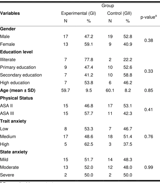
Table 1 shows the demographic characteristics of the study population, with no significant difference between the groups considered; therefore, the groups can be considered homogeneous. Male participants predominated in both groups, with 36 (62.1%) men and 22 (37.9%) women. The mean age was 59.8 ± 8.9 years.
Regarding the classification of surgical risk, as defined by the American Society of Anesthesiologists (ASA), all patients were rated as levels II or III, amounting to 55.2% (N=32) and 44.8% (N=26) of the population, respectively. There was no significant difference between these two groups (p=0.412).
Table 1 - The sample characteristics for the independent variables at baseline.
Variables
Group
Experimental (GI) Control (GII)
p-valuea
N % N %
Gender
Male 17 47.2 19 52.8
0.38
Female 13 59.1 9 40.9
Education level
Illiterate 7 77.8 2 22.2
0.33
Primary education 9 47.4 10 52.6
Secondary education 7 41.2 10 58.8
High education 7 53.8 6 46.2
Age (mean ± SD) 59.7 9.5 60.1 8.2 0.85
Physical Status
ASA II 15 46.8 17 53.1
0.41
ASA III 15 57.7 11 42.3
Trait anxiety
Low 8 53.3 7 46.7
0.76
Medium 17 48.6 18 51.4
High 5 62.5 3 37.5
State anxiety
Mild 15 51.7 14 48.3
0.99
Moderate 13 52.0 12 48.0
Severe 2 50.0 2 50.0
Assessment of postoperative pain
A significantly lower pain intensity was observed for GI, when compared
with GII, in all three conditions, as follows: upon awakening (0.30 1.21 and 3.43
3.29 for GI and GII, respectively, p=0.001), within the first 24 hours (1.93 2.84
and 5.11 3.61 for GI and GII, respectively, p=0.001), and during the breathing
exercises (43 1.94 and 3.07 2.21 for GI and GII, respectively, p=0.004), as
shown in Table 2.
Table 2 – The intensity of pain in the postoperative period of the coronary artery
bypass graft procedure for both study groups, according to the verbal rating scale.
Pain intensity
Groupa
p-valueb
Experimental (GI, N=30)
Control (GII, N=28)
Upon awakening 0.30 1.21 3.43 3.29 <0.001
Greater intensity in the first 24 hours 1.93 2.84 5.11 3.61 <0.001
During breathing exercises 1.43 1.94 3.07 2.21 0.004
a Values are expressed as the means ± standard deviation.
b Student's t-test for independent samples.
The most commonly reported pain descriptors were uncomfortable (25%) and nauseating (13.7%) pain in GI and intolerable (17.6%) and burning (17.6%) pain in GII.
The site of greater pain occurrence was at the chest incision, and it was similar in both groups (30.0% and 39.7% in GI and GII, respectively), which was followed by pain in the anterior chest (25.7% and 16.7% in GI and GII, respectively) and at the site of the sternotomy and the placement of catheters (21.7% and 14.1% in GI and GII, respectively).
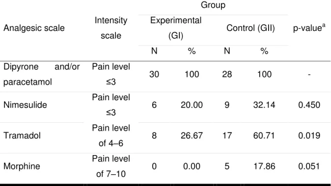
allergic to dipyrone. Concerning the other analgesics, significant differences were observed only for the consumption of tramadol, which was higher in GII.
Table 3 – The absolute and relative frequencies of analgesic use between the
groups.
Analgesic scale Intensity
scale
Group
p-valuea
Experimental
(GI) Control (GII)
N % N %
Dipyrone and/or
paracetamol
Pain level
≤3 30 100 28 100 -
Nimesulide Pain level
≤3 6 20.00 9 32.14 0.450
Tramadol Pain level
of 4–6 8 26.67 17 60.71 0.019
Morphine Pain level
of 7–10 0 0.00 5 17.86 0.051
a - Pearson’s chi-square test.
Table 4 – The relationship between the postoperative complications for each study group.
Variables
Group
p-valuea OR
Experimental (GI) Control (GII)
N % N %
Vomiting
Yes 10 90.9 1 8.1
0.006 13.50
No 20 42.6 27 57.4
Nausea
Yes 13 76.5 4 23.5
0.021 4.59
No 17 41.5 24 58.5
Itching
Yes 12 80.0 3 20.0
0.016 5.56
No 18 41.9 25 58.1
Respiratory depression
Yes 0 0 1 100
0.483 -
No 30 52.6 27 47.4
a Fisher's exact test.
The time to extubation exhibited a decreasing trend in individuals who used the combination of general anesthesia with spinal anesthesia (GI), and the mean
time periods (expressed in hours) were 5.3 1.1 and 6.4 2.9 (p=0.057) in GI and
Table 5 – The hospital stay for each study group. Time Groupa p-valueb Experimental (GI, N=30) Control (GII) (N=28)
Extubation (h) 5.27 1.11 6.43 2.92 0.057
Surgery (h) 4.13 0.82 4.39 0.88 0.248
ICU (h) 24.03 6.79 29.18 7.49 0.152
Length of stay in bed (h) 24.52 7.82 24.04 3.72 0.771
Hospitalization (days) 7.17 0.95 7.50 0.79 0.152
a Values are expressed as the means ± standard deviation.
b Student's t-test for two independent samples.
DISCUSSION
Post-thoracotomy pain is caused by the surgical incision, rib and intercostal nerve trauma, chest wall inflammation, incisions on the pleura and lung parenchyma, and the insertion of chest catheters [2]. This pain is most frequently
described as intense within the first 48 hours, and it affects the patient’s health and
quality of life [16]. These pain episodes, when untreated, appear to represent a risk factor for the development of chronic pain [21].
The present study indicated that the combination of general anesthesia and spinal anesthesia with ropivacaine and morphine promoted a significant reduction in the pain intensity at three distinct timepoints after surgery, as follows: upon awakening, during the first 24 hours after surgery, and during breathing exercises. This result is extremely important because during the postoperative period of thoracic surgery, 70% of patients experience a considerable amount of pain, and this condition may cause some complications, including weak cough, decreased tidal volume and atelectasis, hypoxemia, postoperative infection, and dyspnea [19].
be a protective factor against pain. Before assessing the pain intensity, the Ramsay sedation scale was used to reduce the bias of that variable.
However, the significantly higher pain scores in GII were not associated with worse postoperative outcomes (e.g., morbidity, duration of stay at ICU, and hospital), with this group exhibiting an increased consumption of analgesics and a decreased incidence of nausea and vomiting.
These results corroborate those published in a recent meta-analysis [22] on spinal anesthesia during cardiac surgery, wherein it was reported that spinal anesthesia did not affect clinically relevant outcomes, such as mortality and cardiovascular morbidity. Moreover, the study concluded that no clinical trials conducted to date could discourage the use of spinal anesthesia because no clinical trials were multicenter or adequately conducted to assess the risks and benefits of spinal anesthesia in cardiac surgery. These findings confirmed the results of a previous meta-analysis [12], which evaluated 17 clinical trials involving 668 patients undergoing coronary artery bypass graft procedure. The latter study concluded that spinal anesthesia had no significant effect on mortality, myocardial infarction, and arrhythmias.
Macias et al. [14] conducted a randomized double-blind study involving 80 patients undergoing an elective thoracotomy. The authors observed no significant differences between the groups treated with ropivacaine and those treated with epidural bupivacaine in combination with fentanyl, with all administered epidurally. In the present study, we used a combination of general anesthesia and spinal anesthesia with morphine and ropivacaine, and we evaluated whether the multimodal analgesia was effective for pain control.
Regarding the sites of pain, both groups reported a higher frequency of local pain at the site of the sternotomy and at the anterior chest, which was followed by the site of the catheter insertion, particularly pleural catheters, and at the site of the saphenectomy. These results are in agreement with previous studies [8,16], which demonstrate the occurrence of pain at the site of the sternotomy until the third postoperative day.
An important aspect of our study involved the lower morphine consumption in GI (p=0.051), which corroborates the findings of Alhashemi et al. [1], who used
a placebo group and spinal anesthesia with 250 μg or 500 μg of morphine. These
authors observed a significant difference between the groups (p=0.001) and
concluded that the spinal anesthesia with 250 μg of morphine was effective in
controlling the postoperative pain for a coronary artery bypass graft procedure, with a lower consumption of intravenous morphine.
Although the GI group had a higher incidence of nausea, vomiting, and itching, these side effects are considered mild and did not affect the patient outcome. These findings confirmed the results of a previous meta-analysis [12], wherein spinal anesthesia using morphine alone or in combination with other anesthetics did not significantly affect nausea and vomiting. However, in the clinical trials of that meta-analysis and in the present study, the risk factors for postoperative nausea and vomiting (PONV) were not evaluated [3], thereby limiting the scope of the present study. Strategies to reduce the baseline risk may involve understanding the physiological mechanisms of nausea and vomiting when associated with the multimodal approach [7]. These studies may contribute to the successful treatment of PONV [3,7,12] in future clinical trials.
A potential limitation of the present study is that the reduced number of
patients used in the initial sample size may have affected the study’s power
(approximately 77.0%). However, given the consistency of the results, we can infer that the findings should be similar to those that would be found if the intended 60 patients had participated in the study.
Conclusions
This study suggests that the combination of general anesthesia and spinal
anesthesia using 400 µg of morphine and 6-8 ml (30–40 mg) of 0.5% ropivacaine
REFERENCES
[1] Alhashemi JA, Sharpe MD, Harris CL, Sherman V, Boyd D. Effect of
subarachnoid morphine administration on extubation time after coronary artery bypass graft surgery. J Cardiothorac Vasc Anesth 2000;14:639-44.
[2] American Society of Anesthesiologists Task Force on Acute Pain
Management. Practice guidelines for acute pain management in the perioperative setting: an updated report by the American Society of Anesthesiologists Task Force on Acute Pain Management. Anesthesiology 2012;116:248-73.
[3] Apfel CC, Lããrã E, Koivuranta M, Greim CA, Roewer N. A simplified risk
score for predicting postoperative nausea and vomiting: conclusions from cross-validations between two centers. Anesthesiology 1999;91:693-700.
[4] Baumgarten MC, Garcia GK, Frantzeski MH, Giacomazzi CM, Lagni VB,
Dias AS, Monteiro MB. Pain and pulmonary function in patients submitted to heart surgery via sternotomy. Rev Bras Cir Cardiovasc 2009;24:497-505.
[5] Chaney MA, Nikolov MP, Blakeman BP, Bakhos M. Intrathecal morphine for
coronary artery bypass graft procedure and early extubation revisited. J Cardiothorac Vasc Anesth 1999;13:574-8.
[6] Dango S, Harris S, Offner K, Hennings E, Priebe HJ, Buerkle H, Passlick B,
Loop T. Combined paravertebral and intrathecal vs thoracic epidural analgesia for
post-thoracotomy pain. Br J Anaesth 2013;110:443-9.
[7] Gan TJ, Meyer T, Apfel CC, Chung F, Davis PJ, Eubanks S, Kovac A, Philip
BK, Sessler DI, Temo J, Tramèr MR, Watcha M; Department of Anesthesiology, Duke University Medical Center. Consensus guidelines for managing
postoperative nausea and vomiting. Anesth Analg 2003;97:62-71.
[8] Giacomazzi CM, Lagni VB, MB Monteiro. A dor pós-operatória como
contribuinte do prejuízo na função pulmonar em pacientes submetidos à cirurgia cardíaca [Postoperative pain as a contributor to pulmonary function impairment in patients submitted to heart surgery]. Rev Bras Cir Cardiovasc 2006;21:386-92.
[9] Jensen MP, Chen C, Brugger AM. Postsurgical pain outcome assessment.
Pain 2002;99:101-9.
[11] Kowalewski R, Seal D, Tang T, Prusinkiewicz C, Ha D. Neuraxial
anesthesia for cardiac surgery: thoracic epidural and high spinal anesthesia - why is it different? HSR Proc Intensive Care Cardiovasc Anesth 2011;3:25-8.
[12] Liu SS, Block BM, Wu CL. Effects of perioperative central neuraxial analgesia on outcome after coronary artery bypass surgery: a meta-analysis. Anesthesiology 2004;101:153-61.
[13] Liu ZH, Lv HW, Wang JK. Effectiveness and safety of ropivacaine and bupivacaine in spinal anesthesia: a meta-analysis. Chinese Journal of Evidence-Based Medicine 2010;10:597-601.
[14] Macias A, Monedero P, Adame M, Torre W, Fidalgo I, Hidalgo F. A randomized, double-blinded comparison of thoracic epidural ropivacaine, ropivacaine/fentanyl, or bupivacaine/fentanyl for postthoracotomy analgesia. Anesth Analg 2002;95:1344-50.
[15] McNamee DA, Parks L, McClelland AM, Scott S, Milligan KR, Ahlén K, Gustafsson U. Intrathecal ropivacaine for total hip arthroplasty: double-blind comparative study with isobaric 7.5 mg ml(-1) and 10 mg ml(-1) solutions. Br J Anaesth 2001;87:743-7.
[16] Mueller XM, Tinguely F, Tevaearai HT, Revelly JP, Chioléro R, von
Segesser LK. Pain location, distribution, and intensity after cardiac surgery. Chest 2000;118:391-6.
[17] Pimenta CA, Teixeiro MJ. [Proposal to adapt the McGill Pain Questionnaire into Portuguese]. Rev Esc Enferm USP 1996;30:473-83. (in Portuguese)
[18] Ramsay MA, Savege TM, Simpson BR, Goodwin R. Controlled sedation with alphaxalone-alphadolone. Br Med J 1974;2:656-9.
[19] Soto RG, Fu ES. Acute pain management for patients undergoing thoracotomy. Ann Thorac Surg 2003;75:1349-57.
[20] Spielberger CD, Gorsuch RL, Lushene RE. Inventário de Ansiedade
Traço-Estado – IDATE: manual de psicologia aplicada [State-Trait Anxiety Inventory -
STAI: manual of applied psychology]. (Translation of Angela Biaggio and Louis Natalício). Rio de Janeiro: CEPA, 1979.
[21] Wildgaard K, Ravn J, Kehlet H. Chronic post-thoracotomy pain: a critical review of pathogenic mechanisms and strategies for prevention. Eur J
ARTIGO 2
Analgesic efficacy of subcutaneous lidocaine and multimodal analgesia for chest tube removal
Authors
Valdecy Ferreira de Oliveira Pinheiroa
José Madson Vidal da Costab
Marcelo Matos Cascudoc
Ênio de Oliveira Pinheirod
Ivonete Batista de Araujoe
a Rn, Ms, Professor, Department of Nursing, Federal University of Rio Grande do
Norte (Universidade Federal do Rio Grande do Norte - UFRN); PhD candidate, Graduate Program, Center of Health Sciences, UFRN, Natal/RN - Brazil.
valdecyfop@ufrnet.br
b MD, Anesthesiologist, Promater Hospital (Hospital Promater), Natal/RN – Brasil.
Madson@hotmal.com
c MD, Cardiovascular surgeon, Promater Hospital, Natal/RN-Brazil.
d MD, Cardiologist, Promater Hospital, Natal/RN-Brazil. pinheiro@cardiol.br
e PhD, DSc in Health Sciences, Federal University of Rio Grande do Norte
(Universidade Federal do Rio Grande do Norte - UFRN), Natal. Brazil; Professor, Department of Pharmacy and Graduate Program in Health Sciences, UFRN, Natal, RN, Brazil. Ivoneteba@ufrnet.br
CORRESPONDING AUTHOR
Valdecy Ferreira de Oliveira Pinheiro
Departamento de Enfermagem. Universidade Federal do Rio Grande do Norte
Campus Universitário Lagoa Nova - CEP 59072-970 Natal. RN – Brasil.
Fone:(084) 3215 – 3615 fax: (084) 3215 – 3615, e-mail: Valdecyfop@gmail.com
ABSTRACT
AIM: To assess the analgesic efficacy of subcutaneous lidocaine and multimodal
analgesia for chest tube removal following heart surgery. METHODS: Sixty
volunteers were randomly allocated into two groups; 30 participants in the
experimental group were given 1% subcutaneous lidocaine, and 30 controls were
given a multimodal analgesia regime comprising systemic anti-inflammatory
agents and opioids. The intensity and quality of pain and trait and state anxiety
were assessed. The association between independent variables and final outcome
was assessed by means of the chi-square test with Yates’ correction and Fisher’s
exact test. RESULTS: The groups did not exhibit significant difference with respect
to the intensity of pain upon chest tube removal (p= 0.47). The most frequent
descriptors of pain reported by the participants were pressing, sharp, pricking,
burning and unbearable. CONCLUSION: The present study suggests that the
analgesic effect of the subcutaneous administration of 1% lidocaine combined with
multimodal analgesia is smol efficacious. KEYWORDS: Pain management; Chest
INTRODUCTION
The use of chest tubes aims at the preservation of hemodynamic stability
and cardiopulmonary function by draining fluids, blood, and air out the pleural,
pericardial or mediastinal cavities (5). The removal of chest tubes is associated
with pain, largely because the visceral pericardium and pleura are rich in
nociceptive fibers (19). The removal of chest tubes represents a potential stimulus
for the intercostal nerve fibers that innervate the parietal pleura, chest muscles,
and their insertions (18).
The adverse effects of this painful procedure have not yet been duly
investigated, and little is known about the measures applied in intensive care units
(ICU) to control pain related to painful procedures (1). This lack of knowledge may
contribute to an increase in postoperative pulmonary complications, such as a
decrease in respiratory muscle strength, pulmonary volumes, and capacity as well
as a reduction of the effectiveness of cough and an increase in the number of
infections Pulmonary complications interfere with the clinical progression of
patients and are considered the main causes of morbidity and mortality in such
cases(5) .
The scope of analgesic protocols is quite wide, ranging from
non-pharmacological techniques, such as relaxation, music, or ice packs, to the use of
drugs such as morphine and local anesthetics (3,6,24). Some approaches
combine various drugs to improve analgesia while reducing their side effects.
Systemic multimodal, or balanced, analgesia consists of intravenous
administration of non-steroidal anti-inflammatory drugs combined with weak and
One of the main analgesic techniques consists of the subcutaneous
administration of lidocaine, which is used to control pain in several procedures,
such as venous and arterial puncture, venous and arterial catheter insertion, and
chest tube removal, among others [3]. Nevertheless, patients are often not given
analgesics or any other procedure to control pain [6]. As pain is an expected
occurrence when drains are removed, analgesics should be administered to
patients appropriately before chest tube removal to achieve satisfactory effects
[22].
The objective of the present study was to analyze the analgesic effect of
1% subcutaneous lidocaine combined with multimodal analgesia or an intravenous
(IV) analgesic regime by means of systematic assessment of the intensity of pain
during chest tube removal following heart surgery.
METHODS
The present was a randomized controlled clinical trial that was approved by
the Research Ethics Committee of the Federal University of Rio Grande do Norte
(Universidade Federal do Rio Grande do Norte - UFRN), no. 186/05, and
registered at the Brazilian Registry of Clinical Trials, no. RBR 8M444Q, in
accordance with the Declaration of Helsinki. All participants signed an informed
consent form (Annex 1) at the preoperative assessment.
The following parameters were used to calculate the sample size:
population size, 354 individuals; type 1 error (α), 0.05; test power (1-ß), 0.80; and
20% difference between the groups. According to these criteria, the sample size
ought to be 60 participants, with 30 in each of the two groups. As a function of the
comprised 58 participants, who were allocated to Group I (GI) – experimental (n=
30) and GII – control (n= 28).
The inclusion criteria were as follows: age 35 to 75 years old; in the
postoperative period after heart surgery with chest tube insertion; American
Society of Anesthesiologists (ASA) physical status 2 or 3; and exhibit appropriate
verbal communication and understanding to participate in interviews. To assess
the latter, the Ramsay sedation scale was used [20]. This scale scores sedation at
six different levels, as follows: 1- anxiety and/or agitation; 2- tranquility,
cooperation and orientation; 3- response to commands only; 4- brisk response to
auditory or painful stimulus; 5- sluggish response to auditory or painful stimulus;
and 6- no response. Only individuals at levels ≤ 3 were included. Individuals who
manifested the wish not to continue in the study were excluded, as well as
individuals who developed postoperative complications, including severe heart
and/or respiratory failure and stroke, or who required reoperation by any cause.
At the preoperative visit, after the informed consent form was signed, all of
the participants were trained in the use of a 10-cm visual analog scale [11] (VAS)
for pain and oriented to describe the quality of pain using the short-form McGill
Pain Questionnaire (descriptors) [16]. On that occasion, the participants were also
instructed on how to behave upon waking up after surgery at the ICU, as they
ought to be thoroughly acquainted with both instruments to be in better conditions
to assess pain. Finally, the participants’ levels of anxiety were assessed using
STAI (State-Trait Anxiety Inventory) and Spielberger’s theoretical framework [23].
The doctors and nurses at the institution where the study was conducted
established that the chest tubes would be removed 24 hours after surgery. The
number, size, and position of the chest tubes were selected according to surgical
need. Tube sizes 19F and 28F are routinely used for chest drainage at the
Before the removal of the chest tubes, the participants were randomly
allocated to the study groups by the cardiologists using a computer-based
database that had been previously established. In addition to the standard
analgesic regime used at the ICU where the study was conducted for patients after
heart surgery, the participants in the experimental group were given four
subcutaneous injections of 1% lidocaine at approximately one cm from the surgical
wound margin using 7 mm in a diamond pattern; the volume of each dose was 2.0
ml, for a total of 8.0 ml. The participants in GII were only given an analgesic
multimodal [17], which consisted of the administration of non-steroidal
anti-inflammatory drugs combined with weak and strong opioids by IV based on the
systematic assessment of the pain intensity (visual analog scale -VAS) [16]: pain <
3 (weak analgesic), dipyrone IV 30 mg/kg every six hours; pain= 4 to 7, tramadol
IV 50 mg/kg every six hours; and pain= 8 to 10, morphine IV 1 to 2 mg. The chest
tubes were removed by the surgeon.
Following the procedure, nurses blinded to the composition of the study
groups assessed the participants when the surgeon was not present.
Statistical analysis
For the descriptive analysis, the categorical data were arranged in tables
of absolute and relative frequencies. As the distribution of the quantitative data
was not normal, the data were expressed as median, minimum, and maximum
values. Data with normal distributions were expressed as the mean and standard
deviation.
For the bivariate analysis, the association among categorical variables
continuity correction or Fisher’s exact test. The Mann-Whitney (U) test was used to
compare the medians of continuous independent variables relative to the groups.
In all of the analyses, standard 0.05 p-values and 95% confidence
intervals were applied.
RESULTS
The initial sample comprised 60 participants; however, two individuals in
the control group did not complete the study, with one case requiring reinsertion of
the chest tubes and another refusing to participate in the assessments. Therefore,
58 participants were assessed after surgery. Thirty-six (62.1%) participants were
male, and 22 (37.9%) were female; the average age of the sample was 59.78 (±
8.93) years old. The groups did not exhibit significant difference with respect to
gender, age, or educational level.
Thirty-two (55.2%) and 26 (44.8%) participants were classified as
exhibiting ASA physical status 2 and 3, respectively, which characterized the study
sample as low-to-moderate. The groups did not exhibit significant differences with
respect to ASA physical status (p= 0.34) nor in regard to the surgery length of time
(p= 0.82) or the length of time with the chest tube (p= 0.76).
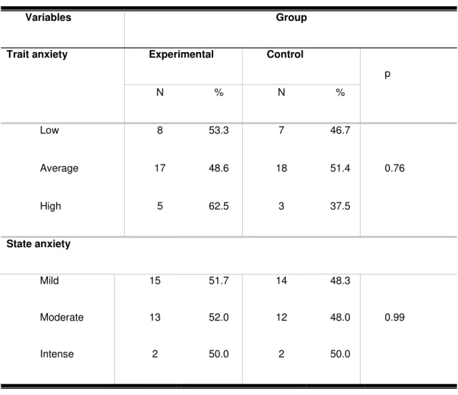
Table 1 describes the distribution of the results with respect to the
assessment of anxiety. Most of the participants exhibited low-to-average and
mild-to-moderate levels of trait and state anxiety, respectively. Significant association
Table 1 - Data corresponding to the participants’ trait and state anxiety levels
per group
Variables Group
Trait anxiety Experimental Control
p
N % N %
Low 8 53.3 7 46.7
0.76
Average 17 48.6 18 51.4
High 5 62.5 3 37.5
State anxiety
Mild 15 51.7 14 48.3
0.99
Moderate 13 52.0 12 48.0
Intense 2 50.0 2 50.0
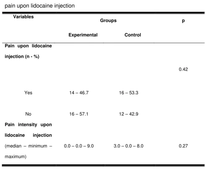
With respect to the assessment of pain as a function of lidocaine injection,
there was no association between the presence of pain and groups (p= 0.42), as
Table 2 shows. In addition, the median intensity of pain with respect to lidocaine
Table 2 - Sample characterization according to the presence and intensity of pain upon lidocaine injection
Variables
Groups p
Experimental Control Pain upon lidocaine
injection (n - %)
0.42
Yes 14 – 46.7 16 – 53.3
No 16 – 57.1 12 – 42.9
Pain intensity upon lidocaine injection (median – minimum –
maximum)
0.0 – 0.0 – 9.0 3.0 – 0.0 – 8.0 0.27
With respect to chest tube removal, no association was observed between
group and anxiety level (p= 0.94) or pain before the procedure (p= 0.67), as Table
3 demonstrates. The median intensity of pain upon removal of pleural or
Table 3 - Sample characterization according to anxiety level and pain intensity upon chest tube removal, Natal-RN, 2013.
Variables Groups p
Experimental (n=30)
Control (n=28)
Anxiety level before chest tube removal (n - %)
0.94
Low 19 – 51.4 18 – 48.6
Average 11 – 52.4 10 – 47.6
Pain before chest tube removal (n - %)
0.67
Yes 7 – 50.0 7 – 50.0
No 22 – 56.4 17 – 43.6
Pain intensity upon mediastinal chest tube
removal Mean (± SD) 4.0 (2.4) 3.7 (2.8) 0.56
Pain intensity upon pleural chest tube
DISCUSSION
There is evidence that adequate pain control is significantly beneficial for
patient comfort. Although multimodal analgesia combined with lidocaine is
considered an option for chest tube removal, this therapy was not effective in the
present study.
Although no association was observed between trait and state anxiety and
group, and despite the orientations and psychological support provided by the
nurses as a part of the routine care before the procedure, almost half of the
participants exhibited moderate anxiety.
Almost one-third of the participants reported the presence of pain before
chest tube removal. Both groups localized the majority of their pain to the
sternotomy site, as well as the site of chest tube insertion, particularly in the case
of the pleural drains, followed by the saphenectomy site. These findings agree with
previous studies, where pain occurred at the sternotomy site up to postoperative
day three [8,17].
There was no significant difference in pain associated with the
subcutaneous administration of lidocaine between the groups. In this regard, it is
worth noting that the intensity of pain reported was mild, which might be related to
the use of multimodal analgesia. According to the review by Cepeda et al [7],
discomfort and pain during the subcutaneous administration of lidocaine is
reported by patients as a whole. These symptoms might be related to the needle
gauge, anesthetic administration speed, injected solution volume or temperature,
patient profile, or the low pH of the anesthetic.
The groups did not exhibit significant differences in pain associated with the


