ABSTRACT
Immunohistochemical staining of Langerhans
cells in HPV-positive and HPV-negative cases of
oral squamous cells carcinoma
Karuza Maria Alves PEREIRA1, Rosilene Calazans SOARES2, Márcio Campos OLIVEIRA3, Leão Pereira PINTO4,
Antônio de Lisboa Lopes COSTA4
1- DDS, MSc, PhD, Professor, School of Dentistry, Federal University of Ceará - Campus Sobral, Sobral, CE, Brazil. 2- DDS, MSc, PhD, Professor, Department of Morphology, Federal University of Sergipe, Aracaju, SE, Brazil. 3- DDS, MSc, PhD, Professor, School of Dentistry, State University of Feira de Santana, Feira de Santana, BA, Brazil.
4- DDS, MSc, PhD, Professor, Graduate Program of Oral Pathology, Federal University of Rio Grande do Norte, Natal, RN, Brazil.
Corresponding address: Prof. Dr. Antônio de Lisboa Lopes Costa - Departamento de Odontologia da Universidade Federal do Rio Grande do Norte - Av: Senador Salgado Filho 1787 - Lagoa Nova - Natal, RN - 59056-000 - Brasil - Phone/Fax: +55-84-3215-4138 - e-mail: [email protected]/ [email protected]/
! "
T
he Human Papillomavirus (HPV) has been strongly implicated in development of some cases of oral squamous cell carcinoma (OSCC). However, the immunological system somehow reacts against the presence of this virus. Among the cells involved in such mechanism of defense Langerhans cells (LC) stand out, which are responsible for processing and presenting antigens. Objectives: The purposes of this study were to investigate the presence of HPV DNA and to evaluate the immunohistochemical reactivity for Langerhans cells between HPV-positive and HPV-negative OSCC. Twenty-seven cases of OSSC were !"#$!"#%&!' Viral typing was performed by dot blot hybridization. Immunohistochemistry was performed by the Streptavidin-biotin technique. Results: From the 27 cases, 9 (33.3%) were HPV-positive and 18 (66.0%) HPV-negative. HPV 18 was the most prevalent viral type (100% cases) and infection with HPV-16 (co-infection) was detected in only 1 case. In the OSCC specimens examined, immunoreactivity to S-100 antibody was detected in all cases, with a mean number of 49.48±30.89 Langerhans cells positive for immunostaining. The mean number of immunostained Langerhans cells was smaller in the HPV-positive cases (38 cells/case) than in the HPV-negative cases (42.5 cells/case), but this difference was not +$;< =>% "?@&!'BE"" indicates a possible participation of the virus in the development and progression of only a subgroup of these tumors. There was no association between the immunohistochemical +G+$EJ<<K%&!'BEE" ?+++ that the presence of HPV in such OSCC cases could not alter the immunological system, particularly the Langerhans cells.Key words: HPV. Carcinoma. Langerhans cells. Polymerase chain reaction. DNA probes.
INTRODUCTION
Oral squamous cell carcinoma (OSCC) is the most common type of cancer of the mouth, accounting for more than 90% of all malignant oral neoplasms. Smoking and alcohol are considered to be the main etiological factors of OSCC. However, some patients develop this neoplasm without
exposure to these risk factors, a fact suggesting the involvement of other etiological factors such as viral agents, particularly human papillomavirus (HPV)4,5,15,24,30.
+M+Q with the reported prevalence of HPV DNA in oral cancer tissue ranging from 0 to 100%3,12,15,22,25,30.
Cellular immunity seems to play an important role in the infection with HPV. The main cells involved in the elimination of altered cells are T lymphocytes. In the presence of an antigen, T cells need to be activated and this activation is mediated by antigen-presenting cells, including Langerhans cells13,14.
G+ epithelia of the epidermis and mucosa and their main function is to capture and transport protein antigens to the lymph nodes for effective presentation to T lymphocytes13. Thus, Langerhans
cells are able to induce either a cellular or a humoral immune response14,29.
Langerhans cells are believed to play an important role in tumor immunology, exerting a function against the emergence of new antigens expressing malignant transformation20. The
major cells involved in killing cancer are the T lymphocytes. However, T cells need to be activated upon antigen presentation, which is mediated by the antigen presenting cells, one of which is the Langerhans cell13,18.
Reduced immune surveillance may contribute to the severity of HPV-associated lesions8,12. Mota,
et al.21 (1999) found a progressive decrease in the
number of Langerhans cells with increasing severity of cervical intraepithelial lesions. In contrast, Levi, et al.16 (2005) observed an increase in the number
of these cells with the progression of cervical neoplasms. In cancer of the uterine cervix, which is strongly associated with HPV, the onset and progression of this neoplasm are related to changes in Langerhans cells inside the squamous epithelium of this mucosa8,12.
The aims of the present study were to investigate the presence of HPV DNA and to compare the immunohistochemical staining of Langerhans cells between HPV-positive and HPV-negative cases of OSCC.
MATERIAL AND METHODS
Twenty-seven cases of OSCC were used in this study. All specimens were taken from formalin-Q Oral Pathology Laboratory, School of Dentistry, Federal University of Rio Grande do Norte and from archives of Dr. Luis Antonio Hospital, Natal, RN, Brazil.
Of the 27 cases of OSSC evaluated, tumor primary sites included lip (7 cases), tongue (4 %Q M $Z % $[ % ? \ + 63 years (range 30-93), the sample comprised 19
men an 8 women.
Ten 10-μm thick histological sections were obtained for DNA extraction and two 3-μm sections were stained with hematoxylin/eosin for review by a pathologist.
DNA was extracted using the ammonium acetate-isopropanol technique which does not require the use of phenol-chloroform. This method in xylene heated to 65°C. Next, the tissues were
hydrated in a decreasing ethanol series (absolute, 95%, 70% and 50%). Then, 400 μL sterile lysis buffer (50 mM NaCl, 5 mM Tris-HCl, pH 8, 12.5 mM EDTA, pH 8, and 0.25% SDS) and proteinase K at a Z<<_+`G tissue pellet of each sample and the samples were incubated at 55°C for 3 to 5 days until complete
dissolution of the material. Next, 200 μL of a 4 M ammonium acetate solution was added to each sample for protein precipitation, followed by 600 μL 100% isopropanol for DNA precipitation. Finally, the DNA pellets obtained were washed with 70% ethanol, dissolved in 50 μL TE buffer and stored at -20°C.
Polymerase Chain Reaction (PCR) was carried out in an Eppendorf thermocycler in a reaction mixture containing the following components in a Z<_G< Z_!"B3 and PCO4 primers (Invitrogen, Life Technologies, Carlsbad, CA, USA), 1.0 U Taq DNA polymerase (Invitrogen, Life Technologies), 20 mM Tris-HCl, pH 8.4, 50 mM KCl, 1.5 mM MgCl2, 200 μM dNTP (GE Healthcare, Little Chalfont, Bucknghamshire, UK), and 0.7 or 7 _G ?!"# of the ß-globin gene were: initial denaturation at
95°"{Q{<
at 95°C for 1 min, 50°C for 1 min and 72°C for 1 min,
|}°C for 10 min. The
PCR products were analyzed by electrophoresis on 2.5% agarose gel (Nusieve, Cambrex Bio Science Rockland, Wokingham, Berkshire, United Kingdom) stained with ethidium bromide.
The samples positive for ß-globin were analyzed by PCR regarding the presence of HPV DNA using pair of generic primers GP5+ (5’TTTGTTACTGTGGTAGATACTAC3’) and GP6+ $Z?"???"???"=%QM a fragment of about 140 bp of the L1 gene, a highly conserved sequence in the genome of mucosal (genital and oral) HPVs. The use of this primer pair segment from at least 23 individual mucosal (genital and oral) HPV types, including high-risk HPV types.
Figure 1- DNA detection by PCR in samples from patients with OSCC. Lanes 1-7: samples from patients; CP: Positive control- HeLa cell DNA infected with HPV18; CN: Negative control (H2O2); PM: Molecular marker (100 pb)
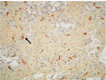
Figure 2- Immunostained Langerhans cells exhibiting characteristic dendritic shape. (SABC 400x)
Buckinghamshire, UK), and 0.7 to 7.0 μL DNA, in Z<_G ?!"# detection of HPV were: initial denaturation at 95°C
Z Q {< at 95°C for 1 min, 45°C for 2 min and 72°C for
J ZQ|}°C for 10
? !"# dot blot hybridization23 using radioactive probes
J[ &!' mucosal (genital and oral) infections. Each dot blot membrane included several negative and positive controls for HPV types, as well as PCR products from patients and controls. The membrane was hybridized overnight as 56°C in 2x standard saline
citrate (SSC), 0.5% sodium dodecyl sulfate (SDS) and 200 μg/mL DNA with 19 HPV types probes (6 ,11,16,18,31,33,34,35,39,40,42,43,44,45,51,52, 54,56,58) and label [32P]dATP. After hybridization,
the unbound probe was of 2x SSC and 0.5% SDS at room temperature and two 10-min washes of the same solution at 56°C. The membrane was exposed
}<°C for 24 h.
Immunohistochemical staining was performed on =_ sections. The sections were mounted on glass slides previously treated with organosilane as adhesive (3-aminopropyltrietoxi-silan, Sigma Chemical Co., St. Louis, MO, USA). The immunohistochemical technique used was a streptavidin-biotin method (streptavidin-biotin complex). The sections were incubated with antibody against S-100 protein (clone Cow S-100, Dako Corporation, Carpinteria, CA, USA), 1:200 dilution for 2 h. The reaction was developed with diaminobenzidine as chromogen, and the sections were counterstained with Mayer’s hematoxylin. Section from nervous tissue was used protein-positive control. In negative controls, the primary antibody was omitted.
After immunohistochemistry, the immunoreactive cells were analyzed taking into the presence or absence of immunostaining (positive, negative), following immunostaining density established by the determination of the number of cells
13,16. After the
immunohistochemical analysis, these data were correlated with a presence of HPV.
The results were analyzed statistically by the Fish and Mann-Whitney test. A level of statistical + Z This research was approved by Research Ethics Committee of the Federal University of Rio Grande do Norte (Process number 146/05).
RESULTS
With respect to HPV infection, of the 27 cases analyzed, 9 (33.3%) specimens were HPV-positive and 18 (66.7%) were HPV-negative. HPV-18 was
detected in all 9 HPV-positive cases (100%) and infection with HPV-16 (co-infection) was detected in only 1 case (Figure 1).
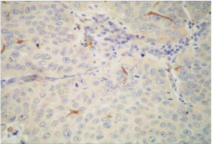
The presence of Langerhans cells in the tumors was demonstrated by immunohistochemical staining for the anti-S-100 antibody, which resulted in a brown staining of the cytoplasm of cells with a dendritic shape (Figures 2 and 3). In the OSCC specimens analyzed, a mean number of 49.48±30.89 Langerhans cells were stained for the anti-S-100 antibody, ranging from 6 to J=[ {<< +
The median number of immunostained cells was smaller in HPV-positive cases (38 cells) compared to HPV-negative cases (42.5 cells), but this difference +$;< =>}% of the presence or absence of HPV infection in relation to the number of S-100-stained Langerhans cells showed a wider variation in the number of immunostained cells in HPV-positive cases.
HPV Infection Low High p n (%) n (%)
Positive 7 (77.8%) 2 (22.2%)
Negative 15 (83.3%) 3 (16.7%) 1,000**
** Fisher test
Table 1- Cases categorized according with the median number of Langerhans cells immunostained, presence or DEVHQFHRI+39LQIHFWLRQDQGVWDWLVWLFDOVLJQL¿FDQFH Figure 3- Immunostained Langerhans cells in a HPV-positive case of oral squamous cell carcinoma (SABC 400x)
observed between the number of S-100-stained Langerhans cells and HPV-positive or HPV-negative cases of OSCC (p=1.000) (Table 1).
DISCUSSION
Oral cancer is a multifactorial disease, with smoking and regular alcohol consumption being the main etiological agents involved in oral carcinogenesis30. However, these habits do not
always explain the development of this type of neoplasm5,11.
Over the last few years, HPV has called the attention of researchers as a possible etiological agent of oral cancer1,10,15,24. This virus is strongly
associated with cancer of the uterine cervix and is detected in almost 100% of cases of this neoplasm, but its role in oral carcinogenesis is still inconclusive7,11,17,22,28,30.
Several techniques have been used for the detection of HPV, with PCR being considered the most sensitive method5. The most widely used
primers are the generic primer pairs GP5+/GP6+ and MY09/MY11, which are able to amplify various virus types. In the present study, the presence or absence of HPV !"# + !ZK`!K small fragment (140 bp). This is important when +Q done in the present study. Nine cases of the present
sample (33.3%) were HPV-positive and 18 (66.7%) were HPV-negative. This percentage is similar to data reported elsewhere15,26.
HPV-18 was detected in all HPV-positive cases, in agreement with previous studies4,19,26, in which
HPV-18 was the most frequent type. However, other investigators1,3,11,15,17,28 detected HPV-16 in 71%,
35%, 66.6%, 55.6%, 33.3% and 85% of cases, respectively, with this virus type being the most prevalent.
Local cellular immunity plays an important role in the response to infection with HPV and its progression in terms of neoplastic alterations6,8,9,12.
Impaired immune function results in an increase of the frequency of clinically detected HPV infections8.
Langerhans cells are antigen-presenting and -processing cells present in the epithelium of skin and mucosa and are the main cell population responsible for the capture of antigens close to the epithelium, presenting these antigens to T helper cells (CD4+ T lymphocytes) and thus inducing a
the evaluation of the patient’s immune system is the investigation of Langerhans cells in the epithelium14,18.
It has been suggested that HPV infection exerts an immunosuppressive effect, reducing the number of Langerhans cells in the epithelium of lesions infected with this virus6,9,12,21.
T h i s s u g g e s t i o n h a s e n c o u r a g e d t h e present investigation, which analyzed the immunohistochemical staining of Langerhans cells in cases of OSCC infected with HPV, a fact not described in the literature.
All 27 OSCC specimens analyzed presented positive staining for Langerhans cells (S-100+),
+ Q 21 (1999),
Sobhani, et al.27 (2002) and Karakök, et al.13
(2003), who investigated cases of OSCC, cervical intraepithelial lesions and laryngeal carcinoma, respectively.
In the present study, a mean number of 49.48 Langerhans cells were stained for the anti-S-100 antibody in each case. This result differs from those reported by Connor, et al.6 (1999), who
observed 6.0±2.3 immunostained Langerhans cells in high-grade cervical intraepithelial lesions, and Karakök, et al.13 (2003), who found 5.3±7.9 S-100+
Langerhans cells in squamous cell carcinoma of the larynx. Hubert, et al.9 (2005) detected about 3
Langerhans cells in cervical carcinomas, whereas Levi, et al.16 (2005) found a mean number of 6.1
Miyagi, et al.20 (2001) detected a larger number
of Langerhans cells in tumors infected with HPV (more than 100 cells per% not infected with this virus (fewer than 10 cells
per %
squamous cell carcinoma of the lung. In the present study, we found a wide variation in the number of Q+ was observed between the number of Langerhans cells and the presence of HPV in the OSCC cases B + + Uchimura, et al.29 (2004), who did not observe
differences in the density of Langerhans cells in cervical lesions between positive and HPV-negative groups. However, these authors noted a change in the morphology of these cells from their characteristic dendritic shape to a more round shape in HPV-positive cases. This fact was not observed in the present study. This divergence between the +M the lesions, in view of the fact that the oral mucosa markedly differs from the cervical and pulmonary epithelium.
++ between the number of Langerhans cells and the presence of HPV, the number of S-100+ cells tended to be smaller in HPV-positive cases. In agreement with this observation, Connor, et al.6
(1999) and Jimenez-Flores, et al.12 (2006) found
G++ cases of HPV-positive cervical neoplasms compared to HPV-negative lesions. We agree with Arany and Tyring2 (1998) who suggested that this depletion
of Langerhans cells is apparently associated with replication of HPV since oncoproteins of this virus may affect antigen presentation.
Jimenez-Flores, et al.12 (2006) suggest that
there is not a precise explanation for the decrease of dendritic cells found in the HPV-infected cervix, although several possibilities exist, including: reduction of the dendritic cells (or their immediate precursors) colonizing this area; increased exiting dendritic cell death. They believe that the last possibility, although feasible, is less likely to occur because HPV is known to induce proliferation rather than cell death, at least in epithelial cells.
When classifying the mean number of Langerhans cells into high and low, we found no association between the number of these cells and HPV-positive or HPV-negative cases. Though, we believe that HPV + microenvironment. According to Gianini, et al.8 (2002) and Jimenez-Flores, et al.12 (2006), a
consequence of this could be a more permissive microenvironment for the HPV to initiate and establish infection within the regional tissue, so "?
recruitment into this microenvironment.
So far, no study is available correlating Langerhans cells and HPV in OSCC, a fact impairing the comparison of the present data with the &!' may provoke changes in the immune system by interfering with Langerhans cell-associated antigen presentation, permitting the escape from this defense mechanism. However, the present results showed no interference of HPV with Langerhans cells in the OSCC cases analyzed.
CONCLUSION
In conclusion, we observed no association between the immunostaining of Langerhans cells (S-100+) and infection with HPV in the OSCC cases analyzed. Despite the reduced sample analyses, cases, which led to the exclusion these, our +++&!' cases of OSCC could not alter the immunological system, particularly the Langerhans cells. Further studies will be necessary to understand the real role between Langerhans cells and HPV in oral carcinogenesis.
REFERENCES
1- Anaya-Saavedra G, Ramirez-Amador V, Irigoyen-Camacho ME, García-Cuellar CM, Guido-Jiménez M, Méndez-Martínez. R, et al. High association of human papillomavirus infection with oral cancer: a case-control study. Arch Med Res. 2008;39:189-197. 2- Arany I, Tyring SK. Status of local cellular immunity in interferon-responsive and nonresponsive human papillomavirus-associated lesions. Sex Transm Dis. 1998;23:475-80.
3- Bouda M, Gorgoulis VG, Kastrinakis NG, Giannoudis A, Tsoli E, Danassi-Afentaki D, et al. “High risk” HPV types are frequently detected in potentially malignant and malignant oral lesions, but not in normal oral mucosa. Mod Pathol. 2000;13:644-53. 4- Boy S, Van Rensburg EJ, Engelbrecht S, Dreyer L, Van Heerden M, Van Heerden W. HPV detection in primary intra-oral squamous cell carcinomas – commensal, aetiological agent or contamination? J Oral Pathol Med. 2006;35:86-90.
5- Chaudhary AK, Singh M, Sundaram S, Mehrotra R. Role of human papillomavirus and its detection in potentially malignant and malignant head and neck lesions: updated review. Head Neck Oncol. 2009;1:22.
6- Connor JP, Ferrer K, Kane JP, Goldberg JM. Evaluation of Langerhans’ cells in the cervical epithelium of women with cervical intraepithelial neoplasia. Gynecol Oncol. 1999;75:130-5. 7- Doorbar J. The papillomavirus life cycle. J Clin Virol. 2005;32:S7-15.
>EGQ&!QQQ! M of the mucosal epithelium microenviroment on Langerhans cells: implications for the development of squamous intraepithelial lesions of the cervix. Int J Cancer. 2002;97:654-9.
10- Iamaroon A, Pattanaporn K, Pongsiriwet S, Wanachantararak S, Prapayasatok S, Jittidecharaks S, et al. Analysis of 587 cases of oral squamous cell carcinoma in northern Thailand with a focus on young people. Int J Oral Maxillofac Surg. 2004;33:84-8. 11- Ibieta BR, Lizano M, Frías-Mendivil M, Barrera JL, Carrillo A, Ruiz-Godoy LM, et al. Human papilloma virus in oral squamous cell carcinoma in a Mexican population. Oral Surg Oral Med Oral Pathol Oral Radiol Endod. 2005;99:311-5.
12- Jimenez-Flores R, Mendez-Cruz R, Ojeda-Ortiz J, Muñoz-Molina R, Balderas-Carrillo O, Diaz-Soberanes ML, et al. High-risk human papilloma virus infection decreases the frequency of dendritic G+\+ + 2006;117:220-8.
13- Karakök M, Bayazit YA, Ucak R, Özer E, Kanlikama M, Mumbuc EQ G+M+ squamous cell carcinoma. Auris Nasus Larynx. 2003;30:81-4. J{ # + disease. Immunol Lett. 2001;78:113-22.
15- Lee SY, Cho NH, Choi EC, Baek SJ, Kim WS, Shin DH, et al. Relevance of human papilloma virus (HPV) infection to carcinogenesis of oral tongue cancer. Int J Oral Maxillofac Surg. 2010;39:678-83.
16- Levi G, Feldman J, Holman S, Salarieh A, Strickler HD, Alter S, et al. Relationship between HIV viral load and Langerhans cells of the cervical epithelium. J Obstet Gynaecol Res. 2005;31:178-84. 17- Llamas-Martínez S, Esparza-Gómez G, Campo-Trapero J, Cancela-Rodríguez P, Bascones-Martínez A, Moreno-López LA, et al. Genotypic determination by PCR-RFLP of human papillomavirus in normal oral mucosa, oral leukoplakia and oral squamous cell carcinoma samples in Madrid (Spain). Anticancer Res. 2008;28(6A):3733-41.
18- Manickam A, Sivanandham M, Tourkova IL. Immunological role of dendritic cells in cervical cancer. Adv Exp Med Biol. 2007;601:155-62.
19- Mazon RC, Gerbelli TR, Benatti Neto C, Oliveira MRB, Donadi EA, Gonçalves MAG, et al. Abnormal cell-cycle expression of the proteins p27, mdm2 and cathepsin B in oral squamous-cell carcinoma infected with human papillomavirus. Acta Histochem. 2011;113(2):109-16.
20- Miyagi J, Kinjo T, Tsuhako K, Higa M, Iwamasa T, Kamada Y, +G+ favourable prognosis of HPV-infected squamous cell carcinoma and adenocarcinoma of the lung. Histopathology. 2001;38:355-67. 21- Mota F, Rayment N, Chong S, Singer A, Chain B. The antigen-presenting environment in normal and human papillomavirus (HPV)-related premalignant cervical epithelium. Clin Exp Immunol. 1999;116:33-40.
22- Rivero ERC, Nunes FD. HPV in oral squamous cell carcinomas !"# B # 2006;20:21-4.
23- Sambrook J, Russel, D. Molecular cloning. Philadelphia: CSHL; 2001.
24- Scully C. Oral cancer; evidence for sexual transmission. Br Dent J. 2005;199:203-7.
25- Siebers TJ, Merkx MA, Slootweg PJ, Melchers WJ, Van Cleef P, De Wilde PC. No high-risk HPV detected in SCC of the oral tongue in the absolute absence of tobacco and alcohol - a case study of seven patients. Oral Maxillofac Surg. 2008;12:185-8.
26- Soares RC, Oliveira MC, Souza LB, Costa ALL, Pinto LP. Detection of HPV DNA and immunohistochemical expression of cell cycle proteins in oral carcinoma in a population of Brazilian patients. J Appl Oral Sci. 2008;16:340-4.
27- Sobhani I, Walker F, Aparicio T, Abramowitz L, Henin D, Cremieux AC, et al. Effect of anal epidermoid cancer-related viruses on the dendritic (Langerhans’) cells of the human anal mucosa. Clin Cancer Res. 2002;8:2862-9.
28- Sugiyama M, Bhawal UK, Dohmen T, Ono S, Miyauchi M, Ishikawa T. Detection of human papillomavirus-16 and HPV-18 DNA in normal, dysplastic, and malignant oral epithelium. Oral Surg Oral Med Oral Pathol Oral Radiol Endod. 2003;95:594-600. 29- Uchimura NS, Ribalta JCL, Focchi J, Simões MJ, Uchimura TT, Silva ES. Evaluation of Langerhans’ cells in human papillomavirus-associated squamous intraepithelial lesions of the uterine cervix. Clin Exp Obstet Gynecol. 2004;31:260-2.