J C O L O P R O C T O L . 2 0 1 3 ;3 3 ( 4 ): 2 2 8 - 2 3 1
Case report
Recurrent aggressive angiomyxoma
*Suelene Suassuna Silvestre de Alencar
a,b,c,*, Romualdo da Silva Corrêa
b,c,
Emanuela Simone Cunha de Menezes
b,d, Antonio Luiz do Nascimento
b,d,
Davi Aragão Alves da Costa
b,d, Marcelo José Carlos Alencar
b,daDepartament of Integrated Medicine, Universidade Federal do Rio Grande do Norte (UFRN), Natal, RN, Brazil bService of Coloproctology, Hospital Universitário Onofre Lopes, Natal, RN, Brazil
cBrazilian Society of Coloproctology , Rio de Janeiro, RJ, Brazil
dMedical School, Universidade Federal do Rio Grande do Norte (UFRN), Natal, RN, Brazil
a r t i c l e i n f o
Article history:
Received 30 May 2013 Accepted 17 August 2013
Keywords:
Pelvic neoplasms
Neoplasms of connective tissues and soft tissues
Angiomyxoma
Local recurrence of neoplasm Laparotomy
a b s t r a c t
Introduction: aggressive angiomyxoma is a highly aggressive, rare neoplasm of the
mesen-chymal tissue with a high recurrence rate. It represents an important differential diagnosis of pelvic tumors in women of reproductive age. This study aims to describe a case of ag-gressive angiomyxoma.
Case report: woman, 37 years old, complained about a bulge on the right perianal region, and
anal itching and burning, bleeding, tenesmus and incontinence. The proctologic examina-tion conirmed the perianal bulge and extrinsic compression of the posterior wall of the rectum. Computed tomography (CT) of the pelvis showed a well-deined pelvic mass ex-tending to the right rectal area. Exploratory laparotomy showed a mass of ibro elastic con-sistency adjacent to the pelvic organs and closely attached to the distal rectum, and per-formed a resection of the pelvic tumor afterward. Anatomopathological analysis revealed an aggressive angiomyxoma. Magnetic resonance imaging (MRI) of the pelvis showed signs of recurrence in the pelvic cavity on the right side of the rectum. A surgical procedure was performed to resect the lesion. After an asymptomatic period, the MRI showed solid growths located in the right ischiorectal fossa. A new surgical procedure identiied only retention cysts in the pelvis and right ischiorectal fossa, only lysis of adhesions was per-formed. The patient is currently undergoing follow-up without disease recurrence.
© 2013 Elsevier Editora Ltda. All rights reserved.
*Study carried out at Departament of Integrated Medicine da Universidade Federal do Rio Grande do Norte, Natal, RN, Brazil.
* Corresponding author.
J C O L O P R O C T O L . 2 0 1 3 ;3 3 ( 4 ): 2 2 8 - 2 3 1
229
Palavras-chave: Neoplasias pélvicas
Neoplasias de tecido conjuntivo e de tecidos moles
Angiomixoma
Recidiva local de neoplasia Laparotomia
r e s u m o
Angiomixoma agressivo recidivante
Introdução: o angiomixoma agressivo é uma rara neoplasia do tecido mesenquimal de
gran-de agressividagran-de e alta taxa gran-de recorrência. Representa um importante diagnóstico diferen-cial de tumorações pélvicas de mulheres em idade reprodutiva. Este estudo objetiva relatar um caso de angiomixoma agressivo.
Relato de caso: mulher, 37 anos, com queixa de abaulamento em região perianal direita,
além de prurido e ardor anal, sangramento, tenesmo e incontinência anal. Exame procto-lógico conirmou o abaulamento perianal e compressão extrínseca da parede posterior do reto. Tomograia computadorizada (TC) de pelve evidenciou massa pélvica bem delimitada estendendo-se à região para-retal direita. Laparotomia exploradora demonstrou massa de consistência ibro-elástica adjacente aos órgãos pélvicos e intimamente aderida ao reto distal, sendo realizada ressecção do tumor pélvico. Anatomopatológico revelou angiomixo-ma agressivo. Ressonância nuclear angiomixo-magnética (RNM) de pelve demonstrou sinais de recidi-va na escarecidi-vação pélvica à direita do reto. Foi realizado procedimento cirúrgico para ressec-ção da lesão. Após período assintomática, RNM evidenciou processos expansivos sólidos localizados na fossa ísquio-retal direita. Novo procedimento cirúrgico identiicou apenas cistos de retenção na pelve e fossa ísquio-retal direita sendo feita apenas lise de aderên-cias. A paciente encontra-se em seguimento clínico sem recidiva da doença.
© 2013 Elsevier Editora Ltda. Todos os direitos reservados.
Introduction
Aggressive angiomyxoma (AA) is a neoplasm of mesenchy-mal connective tissue, which affects mainly women (6:1 ra-tio) of reproductive age, but there have been cases reported in perimenopausal women, men and children.1
Steeper and Rosai, in 1983, described the histological char-acteristics and the tendency for local iniltration and recur-rence.2 The adjective “aggressive” is due to its high recurrence
rate and iniltrative power, ranging from 33-72%.3,4 To date, AA
has approximately 250 cases reported in literature.1
Considering the rarity of cases of AA described in the lit-erature, this report aims to highlight the recurrent nature of this disease and include it in the differential diagnosis of pel-vic tumors, especially in women of reproductive age.
Case report
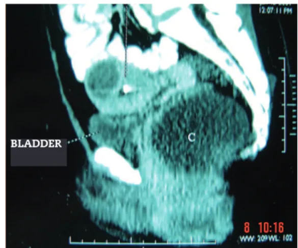
A 37-year-old female patient reported a bulge in the right perianal region, with progressive and painless growth for the past eight months. She also reported anal itching and burn-ing, bleedburn-ing, tenesmus and anal incontinence. The procto-logic examination disclosed a bulge in the right perianal re-gion and extrinsic compression of the right lateral posterior rectal wall. Endoanal ultrasonography (USG) revealed a large perineal collection of undeined nature. The computed to-mography (CT) revealed a well-deined pelvic mass extending to the right pararectal region (Figs. 1 and 2).
The patient underwent exploratory laparotomy, which re-vealed a mass of ibroelastic consistency adjacent to the pel-vic organs and closely adhered to the distal rectum. Resection of the pelvic tumor and a total hysterectomy were performed
(Fig. 3). The anatomopathological examination revealed an aggressive angiomyxoma and the presence of intrauterine leiomyoma. Immunohistochemical analysis showed spindle cells positive for desmin, vimentin, smooth muscle actin, HHF-35, as well as nuclear positivity for estrogen and progesterone and negative for CD34 and S-100 protein (Figs. 4, 5 and 6).
About one year after the surgery, the proctologic examina-tion revealed a small bulge in the right anterior anal region. Magnetic resonance imaging (MRI) of the pelvis showed signs of recurrence of the expansive process inside the pelvic cavity, right to the rectum, occupying the right ischiorectal fossa and conirmed by pelvis CT. A surgical procedure was performed to resect the recurrent lesion through perianal approach.
Fig. 1 - Pelvic CT (sagittal view) showing the presence of mass (C) in the pelvic region.
J C O L O P R O C T O L . 2 0 1 3 ;3 3 ( 4 ): 2 2 8 - 2 3 1
230
At the follow-up, after being asymptomatic for about three and a half years and displaying a normal proctologic examination, the MRI disclosed solid expansive processes located in the right ischiorectal fossa, adjacent to the left iliac vessels. A third laparotomy was performed and identiied only retention cysts in the pelvis and right ischiorectal fossa, and only lysis of adhesions was performed.
Currently, the patient is in clinical follow-up, with no recurrence of the disease for three years.
Discussion
Aggressive angiomyxoma (AA) is a rare, slow-growing and in-vasive myxoid mesenchymal neoplasm. Women of reproduc-tive age are affected in about 90% of cases, predominantly in the pelvic-perineal region, with a peak incidence at the fourth decade of life.3,5 It can also occur in menopausal women, in
men and in children.2
The clinical presentation of AA is variable. Initially, it may manifest as a perineal or vulvar asymptomatic polyp/nodule, perineal hernia or pelvic mass diagnosed incidentally on im-aging assessments.5
AA must be diagnosed considering age, clinical evolution, location, imaging assessment and anatomopathological and immunohistochemical evidence.6 However, due to the rarity
of cases and lack of typical characteristics, the preoperative diagnosis is very dificult, hindering the therapeutic planning.
Fig. 2 - Pelvic CT (cross-sectional view) showing the presence of mass (1) in the pelvic region.
Fig. 3 - Resected pelvic tumor.
Fig. 4 - HE01 (x20) – Myxoid stroma showing spindle cells and blood vessel proliferation.
Fig. 5 - Desm06 (x20) Spindle cells positive for desmin.
J C O L O P R O C T O L . 2 0 1 3 ;3 3 ( 4 ): 2 2 8 - 2 3 1
231
Conclusion
The present case depicts the characteristics of aggressive angiomyxoma, emphasizing the importance of the inclusion of this disease as a differential diagnosis in cases of locally invasive pelvic masses in women of reproductive age. Imag-ing tests are very important for the diagnosis of primary or recurrent AA, and immunohistochemical analysis should be considered whenever possible to conirm the diagnosis.
Conlicts of interest
The authors declare no conlicts of interest.
R e f e r e n c e s
1. Fuentea J, Zapardiela I, Herreroa S, Kazlauskasa SG, Vargasb J, Frutosa LS, et al. Angiomixoma agresivo vulvar. Prog Obstet Ginecol 2008; 51(2): p 99 - 103.
2. Akbulut M, Demirkan NÇ, Çolakoglu N, Düzcan E. Aggressive angiomyxoma of the vulva: a case report and review of the literature. Aegean Pathology Journal 2006; 3: p 1 – 4. 3. Danesh A, Sanei MH. Aggresive angiomixoma of the vulva:
dramatic response to gonadotropin-releasing hormone agonist therapy. Journal of research in medical sciences 2007; 12 (4): 217 – 221.
4. Dove S, Remoué P, Valo I, Ybarlucea LR, Panel N, Fondrinier E - Unusual female pelvic tumour: Aggressive angiomixoma. Letters to the editor/Eur J Obst and Reprod Biol 2008; 137, (1): 123 – 124.
5. Haldar K, Martinek IE, Kehoe S - Aggressive angiomyxoma: a case series and literature review. Eur J Surg Oncol 2010; 36(4): p 335 – 339.
6. Minagawa T, Matsushita K, Shimada R, Takayama H, Hiraga R, Uehara T, et al. Aggressive angiomyxoma mimicking inguinal hernia in a man. Int J Clin Oncol 2009; 14: p 365 – 368. 7. Bastian PJ, Fisang C, Schmidt ME, Biermann K, Textor J, Müller
SC. Aggressive angiomyxoma of the prostate mimcking benign prostatic hyperplasia. Eur J Med Res 2006; 11(4): p 167 – 169.
8. Jeyadevan NN, Sohaib SAA, Thomas JM, Jeyarajah A, Shepherd JH, Fisher C. Imaging features of aggressive angiomyxoma. Clinical Radiology 2003; 58(2): p 157 – 162.
9. Odashiro NA, Odashiro LN, Odashiro DN, Odashiro M, Campos JCP. Angiomixoma agressivo em paciente do sexo masculino acometendo cordão espermático: relato de caso e revisão da literatura. J Bras Patol Med Lab 2005; 41(2): p 131 – 134.
10. Morag R, Fridman E, Mor Y. Aggressive angiomyxoma of the scrotum mimicking huge hydrocele: case report and literature review. Case Reports in Medicine 2009; 2009 (ID 157624).
11. Wiser A, Korach J, Gotlieb WH, Fridman E, Apter S, Ben-Baruch G. Importance of accurate preoperative diagnosis in the management of aggressive angiomyxoma: report of three cases and review of the literature. Abdominal Imaging 2006; 31(3): p 383 – 386.
12. Velasco SM, Burgos AJC, López MR, Poveda IG, Santoyo JS. Resección laparoscópica de angiomixoma pélvico agresivo. Cirugía española 2010; 88(2), p 121 – 122.
Thus, most cases are conirmed histopathologically after the primary surgical resection.7
Imaging studies are of great importance for the diagnosis of AA. The USG can reveal a polypoid, hypoechoic mass of soft tissue, which may appear as a cyst. At the CT, the char-acteristics vary and may include a homogeneous well-de-ined and hypodense mass relative in relation to the muscle, a hypoattenuating solid mass with an internal spiral pattern after intravenous contrast agent or a predominantly cystic mass with solid components. The MNR image is isointense (less commonly hypointense) on T1 and hyperintense on T2 relative to the muscle. After intravenous contrast, a spiral component of lower intensity is seen inside. The recurrent tumor is also detected at the resonance with characteristic signs similar to the primary tumor.5,8
Macroscopically the AA consists of a homogeneous mass, partially or completely encapsulated, with a smooth surface, gelatinous appearance, of bluish gray color and small cysts or hemorrhagic areas can be found.1,5
The histopathological examination shows myxoid stroma, hypocellularity, small spindled and stellate mesenchymal cells with undeined cytoplasm. Pleomorphism and mitoses are not present and there is no evidence of coagulation necro-sis in the tumor cells. Characteristically, there is a prominent vascular component with vessels of various calibers.5,9
Immunohistochemical indings show that tumor cells have immunoreactivity for desmin, smooth muscle actin, vi-mentin and CD34. They can also be positive for estrogen and progesterone. They are immunonegative for S100 proteins, factor VIII, carcinoembryonic antigen and cytokeratin.
Cytogenetic analysis shows chromosomal translocations involving chromosomes 8 and 12, associated with the rear-rangement of HNGIC genes.10
The differential diagnosis of AA is based on other forms of soft tissue tumors (myxoma, myxoid lipoma, neuroibroma) and malignant tumors with metastatic potential (myxoid liposarcoma, myxoibrosarcoma, embryonal rhabdomyosar-coma).7 Additionally, it can occasionally mimic Bartholin’s
cyst, labial cyst, Gartner’s duct cyst and perineal herniation.3
Angiomyxomas are locally aggressive tumors, generally not capable of distant metastases, with only two cases re-ported in the scientiic literature.1,5
Because of its high rate of local recurrence, the irst line treatment for AA is complete surgical resection through ab-dominal, perineal or mixed approach, keeping margins free. However, the risk-beneit binomial should be considered, as most tumors occur in women of reproductive age and fertil-ity is a very important factor. In such situations this choice may not be the best option, as in cases of high surgical mor-bidity.1,5,11
Incomplete lesion resection occurs in 45-66% of cases and is directly associated with the possibility of recurrence, often requiring re-excision, such as the case reported here. In some free margin cases, the chances of recurrence are of 30-70%; the ischiorectal fossa is the most frequently affected site.1,5
Due to the low mitotic potential of this neoplasm, che-motherapy and radiotherapy are ineffective in their treat-ment.4 In cases of incomplete resection or recurrence, GnRH

