INTESTINAL OBSTRUCTION AFTER ROUX-EN-Y GASTRIC BYPASS
BY HIGA’S TECHNIQUE FOR TREATMENT OF MORBID OBESITY:
RADIOLOGICAL ASPECTS*
Ester Moraes Labrunie1, Edson Marchiori2
OBJECTIVE: The aim of this study is to describe the main radiological aspects of postoperative intestinal obstruction in patients submitted to Roux-en-Y gastric bypass by means of the Higa’s technique. MATERI-ALS AND METHODS: A total of 10 patients presenting with postoperative intestinal obstruction following a gastric reduction procedure were evaluated in the period between November 2001 and April 2006, in seven different medical centers. RESULTS: In the ten patients, the obstruction occurred in the small bowel, five of them because of internal hernias, three because of adhesions, one because of an umbilical hernia and one because of intussusception. Four patients presented obstruction early in the postoperative period (by the seventh post-op day), and six, late in the postoperative period (between the third month and the fifth year). CONCLUSION: All of the cases of intestinal obstruction were found in the small bowel. Internal hernia was the most frequent cause, followed by adhesion. Other causes included umbilical hernia and intussusception. Keywords: Morbid obesity; Bariatric surgery; Intestinal obstruction; Complication; Radiology; Computed tomo-graphy.
Obstrução intestinal pós-gastroplastia redutora pela técnica de Higa para tratamento da obesidade mórbida: aspectos por imagem.
OBJETIVO: O objetivo deste estudo foi descrever os principais aspectos radiológicos encontrados nas obs-truções intestinais pós-operatórias em pacientes submetidos a derivação gastrintestinal em Y de Roux pela técnica de Higa. MATERIAIS E MÉTODOS: Foram estudados 10 pacientes com obstrução intestinal no pós-operatório de gastroplastia redutora, examinados entre novembro de 2001 e abril de 2006. Os casos foram obtidos em sete instituições hospitalares distintas. RESULTADOS: Nos 10 pacientes, a obstrução ocorreu em alça de delgado, sendo cinco por hérnia interna, três por brida, um por hérnia umbilical e um por intus-suscepção gástrica. Em quatro pacientes a obstrução ocorreu precocemente (até o sétimo dia de pós-ope-ratório) e em seis aconteceu tardiamente (entre o terceiro mês e cinco anos de pós-opepós-ope-ratório). CONCLU-SÃO: Todos os casos de obstrução intestinal ocorreram ao nível do intestino delgado. A hérnia interna foi a causa mais freqüente, seguida de brida. Outras causas foram hérnia umbilical e intussuscepção gástrica. Unitermos: Obesidade mórbida; Cirurgia bariátrica; Obstrução intestinal; Complicação; Radiologia; Tomo-grafia computadorizada.
Abstract
Resumo
* Study developed in the Department of Radiology at Univer-sidade Federal do Rio de Janeiro (UFRJ), Rio de Janeiro, RJ, Brazil. 1. Assistant Professor of Radiology at Universidade Federal do Rio de Janeiro (UFRJ), Rio de Janeiro, RJ, Brazil.
2. Titular Professor, Department of Radiology at Universidade Federal Fluminense (UFF), Niterói, RJ, Adjunct Coordinator for the Course of Post-Graduation in Radiology at Universidade Fe-deral do Rio de Janeiro (UFRJ), Rio de Janeiro, RJ, Brazil.
Mailing address: Profa. Dra. Ester M. Labrunie. Avenida Epi-tácio Pessoa, 3490/502. Rio de Janeiro, RJ, Brazil, 22471-003. E-mail: [email protected]
Received September 8, 2006. Accepted after revision Octo-ber 18, 2006.
INTRODUCTION
Obesity is a chronic disease character-ized by excessive body fat, whose increase has achieved worldwide epidemic propor-tions. It is associated with an array of cor-related diseases which end up causing early mortality(1,2). Obesity is considered as a
morbid condition when the body mass
in-dex (BMI) is 40 kg/m² or greater, or 35 kg/ m²or greater in patients with some corre-lated disease (the so called co-morbid-ity)(2,3).
In response to the worldwide epidemic increase of morbid obesity, new treatments have been proposed and improved, with highlights particularly on the development of surgical techniques(4). Nowadays, the
surgical technique considered as the most effective for treating morbid obesity is the Roux-en-Y gastric bypass(4). The increase
in the frequency of bariatric surgeries de-mands the radiologist acquaintance with new techniques as well as with functional and anatomical changes resulting from these treatments(5).
The postoperative radiological study, the evaluation and early diagnosis of
com-plications pose more and more frequent problems for the radiologist in his/her daily practice. Technical limitations imposed by the biotype and clinical conditions of mor-bid obese patients may complicate the di-agnosis. A detailed and clear-sighted analy-sis of radiological studies represents a chal-lenge for both the radiologist and the sur-gical team. Incorrect evaluations or late diagnosis of complications may delay the treatment, and ultimately put the patient’s life at risk.
Small bowel obstruction occurs in about 1.3%–5% of cases, particularly in patients submitted to videolaparoscopy(4).
Lapa-roscopy results in less surgical trauma, with less formation of adhesions, although with higher incidence of internal hernias(4). On
adhesions in laparotomy seems to reduce the propensity to the formation of internal hernias(5).
The present study was aimed at discuss-ing radiological finddiscuss-ings in ten patients who developed intestinal obstruction as a complication of Roux-en-Y gastric bypass for treatment of morbid obesity.
MATERIALS AND METHODS
The present study included ten patients submitted to weight-reducing gastroplasty with Roux-en-Y gastric bypass, presenting with post-operative intestinal obstruction between November/2001 and April/2006. The cases originated from seven different hospital institutions, and the surgeries were performed by five different teams.
Four patients were men (40%), and six were women (60%), with ages ranging between 23 and 54 years (mean age, 37.2 years). Nine patients (90%) were submit-ted to videolaparoscopy surgery, and one (10%) to laparotomy. Fifteen studies were analyzed as follows: nine computed to-mographies, two seriographies of esopha-gus, stomach and jejunum, and four routine studies for acute abdomen.
All of the ten patients had surgical con-firmation of intestinal obstruction. The ra-diological findings were compared with the operative report. Indicators as well as pos-sible etiologies of intestinal obstruction were researched.
As regards the time period elapsed be-tween surgery and development of intesti-nal obstruction, the condition was classi-fied into early intestinal obstruction (those occurring up to the seventh post-operative day), and late intestinal obstruction (those occurring after the 30th post-operative day).
RESULTS
Intestinal obstruction occurred in small bowel loop in the ten patients, five of them by internal hernia, three by adhesion, one by umbilical hernia, and one by gastric in-tussusception. Four patients presented an early obstruction (up to the seventh post-operative day), two of them secondary to transmesocolic internal hernia, and two by adhesion in the distal ileum. The other six patients presented late intestinal obstruc-tion (between three months and five years after surgery), three of them related to in-ternal hernias (two transmesocolic and one caused by a mesenteric defect of the lower anastomosis), one caused by umbilical her-nia, on by adhesion associated with small bowel volvulus, and one by intussusception of the excluded stomach into the duodenal arch.
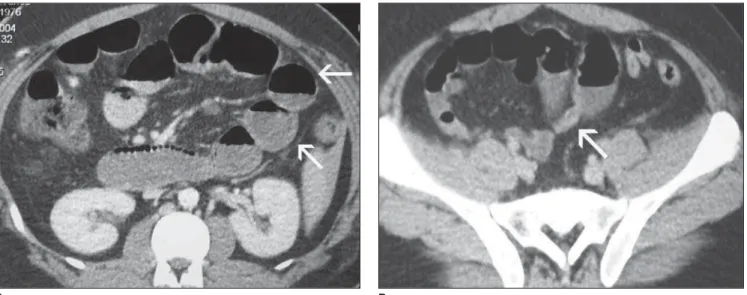
Surgical reintervention became neces-sary in all of the patients. In the three cases caused by adhesion, the obstruction oc-curred in the distal ileal loop (Figure 1). One case occurred in the site of insertion of the laparoscopic trocar, associated with
small bowel volvulus. In all of the cases, computed tomography (CT) could identify the zone of caliber transition from the dis-tended loops to the distal, non-disdis-tended segment. At surgery, two of these patients presented association with small bowel volvulus in the segment above the obstruc-tion. This finding could be suggested on the CT performed in one of the patients, be-cause of the anterior displacement of the surgical stapling line of the jejuno-jejunal anastomosis. Internal hernias occurred in five patients submitted to videolaparo-scopy, four of these hernias being trans-mesocolic (Figure 2), and one in the mesen-teric defect created in the construction of the Roux loop and jejuno-jejunal anasto-mosis. The four patients with transmeso-colic hernia presented distension of the alimentary loop through the transverse mesocolon. One of these patients also pre-sented migration of the lower anastomosis into the supramesocolic space, evidenced by a hyperdense line demonstrating the surgical stapling. The patients with hernia in the mesenteric defect (Figure 3), pre-sented distension of the biliopancreatic loop and excluded stomach. The case of umbilical hernia occurred later in the post-operative period, only in one patient in the group submitted to laparatomy.
One case of intussusception of the ex-cluded stomach into the duodenal arch was observed, with secondary displacement of the choledocus. This latest finding could be
Figure 1. Intestinal obstruction by adhesion. A: Small bowel loops distension (arrows). B: Zone of caliber transition of the distal ileal loop (site of obstruction – arrow).
more clearly observed on a magnetic reso-nance imaging cholangiography (Figure 4).
DISCUSSION
According to Blachar et al.(4), intestinal
obstructions occurring between three days and three months in the post-operative pe-riod, frequently are secondary to adhesions. Internal hernias, generally, occur later in the
post-operative period), as well as late ad-hesions (five years after). So, it seems that the time period elapsed between the bariatric surgery and the onset of symptoms should not be taken into consideration in the etiological evaluation of the intestinal obstruction.
Only one (secondary to umbilical her-nia) of the ten cases of obstruction occurred in patients submitted primarily to
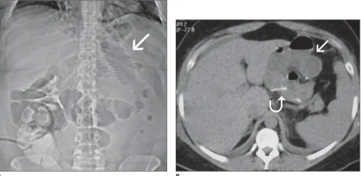
laparo-Figure 2. Transmesocolic internal hernia. A: Topogram demonstrating distension of supramesocolic, small bowel loops (arrow). B: Distension of supramesocolic alimentary loop (arrows). Dense suture threads of the lower anastomosis abnormally situated in supramesocolic floor (curved arrow).
B A
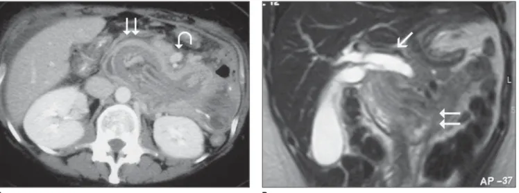
Figure 3. Internal hernia caused by mesenteric defect (surgical report). A: Contrast-enhanced small bowel loops (alimentary canal) distended and clustered on the supramesocolic floor (arrow). B: Mesenteric distension and vascular engorgement (two arrows). Clustered loops in the left upper abdominal quadrant, with slightly thickened walls (arrow).
A B
tomy. All of the cases of internal hernia and adhesion occurred after videolaparoscopy. This figure, however, is not statistically significant. The literature reports higher incidence of internal hernias in laparosco-pies(4) and adhesions in laparotomies(5).
According to Blachar et al.(3), the higher
incidence of post-laparoscopy internal her-nias, compared to laparotomy, would occur because a lower propensity to formation of adhesions as a result of this surgical method. Another predisposition factor may be related to the post-operative reduction in the amount of intraperitoneal fat, with a consequent enlargement of the mesenteric defect(3).
A delayed diagnosis, and, consequently, a late surgical reintervention in these pa-tients may lead to gangrene of intestinal segment, resulting in the need of na enter-ectomy of variable lengths of the small bowel, increasing the complication(6). Two
patients were found with intestinal disten-tion in the review of the routine studies for acute abdomen. Initially, these findings were interpreted as adynamic ileum, delay-ing both the diagnosis and the treatment. Retrospectively, in both cases, the CT per-formed at that time demonstrated a zone of caliber transition of small bowel loops, characterizing the obstruction. Both pa-tients underwent a quite difficult and com-plicated post-operative period. Also in both cases, the presence of an anastomosis fis-tula was observed, possibly secondary to
Figure 4. Gastric intussusception. A: Computed tomography at the level of the third duodenal portion. Heterogeneous contents, with fat and vessels with an aspect of “loop-within-loop” (two arrows), ectatic and ballotted mesenteric vessels (curved arrow). B: Coronal, T2-weighted magnetic resonance imaging dem-onstrating gall dilatation and choledocal distension (arrow) by invagination of the excluded stomach into the duodenal arch (two arrows).
the increased tension on the stapling line, and to the distention above the obstruction, according to the mechanism proposed by Luján et al.(5).
Small bowel obstruction may course with non-specific symptoms, such as long-lasting, intermittent abdominal pain, or acute abdominal pain associated with nau-sea, emesis and constipation(6). Both
pre-sentations occurred in our casuistic. A pa-tient who presented with tachycardia as a single clinical post-operative alteration, was submitted to pre-hospital discharge CT, and alterations compatible with early supramesocolic hernia were found. This patient was immediately operated with an excellent recovery.
CT has becoming the imaging method of choice for investigation of abdominal symptoms, especially in patients submitted to gastric bypass surgery(6). Patients with
non-specific and vague abdominal symp-toms should be promptly submitted to oral and intravenous contrast-enhanced CT(7).
Besides diagnosing the obstruction, CT allows to advance in the determination of the diagnostic etiology, indicating trans-mesocolic, mesenteric, umbilical hernias, and gastric intussusception. Also, CT al-lows the evaluation of intestinal loop dis-tress/thickening and correlated complica-tions, such as pneumoperitoneum, fistulas and collections. Nine of the ten patients with intestinal obstruction were submitted to CT, all of them with abnormal findings.
Even in the two cases were a delayed di-agnosis was observed, the retrospective review of the images had already indicated the abdominal distension with zone of cali-ber transition in the small bowel.
Blachar et al.(3,8)have discussed
tomo-graphic findings of internal hernias, and reported that some findings, if submitted to a clear-sighted analysis, may suggest a di-agnosis. The most significant alterations reported by them and also observed in our casuistic, were: small bowel distension suggesting obstruction; clustering of small bowel loops (especially in the left upper abdominal quadrant); engorgement and displacement of mesenteric vessels, ab-sence of omental fat covering the dis-tended/herniated loops and ascites. A find-ing that we have observed and not reported in this study, in a patient with transmeso-colic hernia, was the migration of the anas-tomosis stapling line towards to the supra-mesocolic floor of the abdomen visualized as a hyperdense, linear artifact in the inter-section between the alimentary and gall-bladder loops.
Oral contrast-enhanced radiography is also quite sensitive for internal hernias(9).
In our casuistic, abdominal plain or con-trast-enhanced radiographs of four patients were abnormal, indicating intestinal disten-sion and/or obstruction. The diagnosis of transmesocolic hernia may be suggested by the presence of supramesocolic clustering and distension of small bowel loops. The B
conventional radiological study may also demonstrate intestinal distension by ob-struction of other etiologies (in our casu-istic, by adhesion, with or without small bowel volvulus).
Intestinal intussusception is a rare com-plication of this surgery(7,10,11). One case of
late intussusception of the excluded stom-ach into the duodenal arch was observed. The CT finding was highly suggestive of invagination. The secondary complication was the development of jaundice by disten-tion and dilatadisten-tion of the choledocus, which is best visualized by magnetic reso-nance imaging. The diagnosis of this com-plication must be reached as early as pos-sible, considering the reported 10%-mor-tality in 48 hours, and 50%-mor10%-mor-tality after 72 hours(11).
REFERENCES
1. Moura LG Jr, Guimarães SB, Castro-Filho HF, Machado HF, Feijó FC, Vasconcelos PRL. Ca-pella’s gastroplasty: metabolites and acute phase proteins changes in midline and bilateral arciform approaches. Arq Gastroenterol 2004;41:215–219. 2. Blachar A, Federle MP. Gastrointestinal compli-cations of laparoscopic Roux-en-Y gastric bypass surgery in patients who are morbidly obese: find-ings on radiography and CT. AJR Am J Roentgenol 2002:179:1437–1442.
3. Blachar A, Federle MP, Pealer KM, Ikramuddin S, Schauer PR. Gastrointestinal complications of laparoscopic Roux-en-Y gastric bypass surgery: clinical and imaging findings. Radiology 2002; 223:625–632.
4. Blachar A, Federle MP, Pealer KM, Abu Abeid S, Graif M. Radiographic manifestations of nor-mal postoperative anatomy and gastrointestinal complications of bariatric surgery, with empha-sis on CT imaging findings. Semin Ultrasound CT MR 2004;25:239–251.
5. Luján JA, Frutos MD, Hernández Q, et al. Laparo-scopic versus open gastric bypass in the treatment
of morbid obesity: a randomized prospective study. Ann Surg 2004;239:433–437.
6. Onopchenko A. Radiological diagnosis of inter-nal hernia after Roux-en-Y gastric bypass. Obes Surg 2005;15:606–611.
7. Srikanth MS, Keskey T, Fox SR, Oh KH, Fox ER, Fox KM. Computed tomography patterns in small bowel obstruction after open distal gastric bypass. Obes Surg 2004;14:811–822. 8. Blachar A, Federle MP, Dodson SF. Internal
her-nia: clinical and imaging findings in 17 patients with emphasis on CT criteria. Radiology 2001; 218:68–74.
9. Carmody B, DeMaria EJ, Jamal M, et al. Internal hernia after laparoscopic Roux-en-Y gastric by-pass. Surg Obes Relat Dis 2005;1:543–548. 10. Duane TM, Wohlgemuth S, Ruffin K.
Intussus-ception after Roux-en-Y gastric bypass. Am Surg 2000;66:82–84.