Presynaptic Localization and Possible Function of
Calcium-Activated Chloride Channel Anoctamin 1 in the
Mammalian Retina
Ji Hyun Jeon1,2., Sun Sook Paik1,2., Myung-Hoon Chun1
, Uhtaek Oh3, In-Beom Kim1,2,4*
1Department of Anatomy, College of Medicine, The Catholic University of Korea, Seoul, Korea,2Catholic Neuroscience Institute, College of Medicine, The Catholic University of Korea, Seoul, Korea,3Channel Research Center, College of Pharmacy, Seoul National University, Seoul, Korea,4Catholic Institute for Applied Anatomy, College of Medicine, The Catholic University of Korea, Seoul, Korea
Abstract
Calcium (Ca2+)-activated chloride (Cl2) channels (CaCCs) play a role in the modulation of action potentials and synaptic
responses in the somatodendritic regions of central neurons. In the vertebrate retina, large Ca2+-activated Cl2 currents
(ICl(Ca)) regulate synaptic transmission at photoreceptor terminals; however, the molecular identity of CaCCs that mediate ICl(Ca)remains unclear. The transmembrane protein, TMEM16A, also called anoctamin 1 (ANO1), has been recently validated as a CaCC and is widely expressed in various secretory epithelia and nervous tissues. Despite the fact thattmem16awas first
cloned in the retina, there is little information on its cellular localization and function in the mammalian retina. In this study, we found that ANO1 was abundantly expressed as puncta in 2 synaptic layers. More specifically, ANO1 immunoreactivity was observed in the presynaptic terminals of various retinal neurons, including photoreceptors. ICl(Ca)was first detected in dissociated rod bipolar cells expressing ANO1. ICl(Ca)was abolished by treatment with the Ca2+channel blocker Co2+, the L-type Ca2+channel blocker nifedipine, and the Cl2channel blockers 5-nitro-2-(3-phenylpropylamino) benzoic acid (NPPB)
and niflumic acid (NFA). More specifically, a recently discovered ANO1-selective inhibitor, T16Ainh-A01, and a neutralizing antibody against ANO1 inhibited ICl(Ca)in rod bipolar cells. Under a current-clamping mode, the suppression of ICl(Ca)by using NPPB and T16Ainh-A01 caused a prolonged Ca2+spike-like depolarization evoked by current injection in dissociated rod bipolar cells. These results suggest that ANO1 confers ICl(Ca)in retinal neurons and acts as an intrinsic regulator of the presynaptic membrane potential during synaptic transmission.
Citation:Jeon JH, Paik SS, Chun M-H, Oh U, Kim I-B (2013) Presynaptic Localization and Possible Function of Calcium-Activated Chloride Channel Anoctamin 1 in the Mammalian Retina. PLoS ONE 8(6): e67989. doi:10.1371/journal.pone.0067989
Editor:Michael A. Fox, Virginia Tech Carilion Research Institute, United States of America
ReceivedFebruary 13, 2013;AcceptedMay 23, 2013;PublishedJune 26, 2013
Copyright:ß2013 Jeon et al. This is an open-access article distributed under the terms of the Creative Commons Attribution License, which permits unrestricted use, distribution, and reproduction in any medium, provided the original author and source are credited.
Funding:This study was supported by the Basic Science Research Program (2010-0022317) of the National Research Foundation (NRF) of Korea funded by the Ministry of Education, Science, and Technology. The funders had no role in study design, data collection and analysis, decision to publish, or preparation of the manuscript.
Competing Interests:The authors have declared that no competing interests exist.
* E-mail: [email protected]
.These authors contributed equally to this work.
Introduction
Calcium (Ca2+
)-activated chloride (Cl2) channels (CaCCs) are anion-selective channels that are activated by increased cytosolic Ca2+
. CaCCs have been implicated in many important physio-logical processes, such as the transepithelial transport of electro-lytes and water, control of vascular tone, and cardiac muscle and neuronal excitability [1–4]. In the nervous system, Ca2+-activated
Cl2 current (ICl(Ca)) is primarily observed in primary sensory neurons, such as olfactory receptor neurons (ORNs), taste receptor cells, somatosensory neurons of dorsal root ganglia (DRG), and photoreceptors of the retina, and is involved in corresponding sensory transduction. ICl(Ca)is also found in presynaptic terminals in the brain, where it is thought to modulate synaptic activity [1]. Anoctamin 1 (ANO1, also called TMEM16A) [5–7] is a CaCC because its biophysical and pharmacological characteristics correspond to those of endogenous CaCCs [8,9]. The identifica-tion of ANO1 as a CaCC has unveiled its significance in many physiological activities, including (1) Cl2 transport in airways [5,10,11], salivary glands [7,12], and gastrointestinal epithelial
cells [13,14], (2) rhythmic contraction in gastrointestinal tracts [13–15], and (3) heat sensation in DRG neurons [16].
The retina is a well-characterized model system that is used for the study of synaptic mechanisms, as it contains various neurotransmitters found in the central nervous system and its receptors; various types of synapses, such as conventional chemical, electrical, and distinct ribbon synapses; and several well-established synaptic circuits for visual processing. ICl(Ca)has been characterized in the photoreceptors of the vertebrate retina [17,18] and is thought to regulate synaptic transmission at photoreceptor terminals by stabilizing membrane potentials and Ca2+ channel modulation [19–24]. Recently, ANO1 [25] and
retinal neurons, even though its molecular identity and function remain unknown. Thus, we sought to examine the expression, localization, and function of ANO1 in the mouse retina.
Results
Expression and localization of ANO1 in the retina To investigate the expression and distribution pattern of the ANO1 protein in the mouse retina, western blotting and immunohistochemistry were performed. As shown in Fig. 1A, an ANO1 immunoreactive band (,130 kDa) was recognized in both
the retina and salivary gland (the latter was used as a control tissue in which ANO1 is expressed abundantly) [7,12]. Strong ANO1 immunoreactivity was observed as puncta in 2 synaptic layers, the outer plexiform layer (OPL) and the inner plexiform layer (IPL), whereas weak immunoreactivity was detected in some somata in the inner nuclear layer (INL) and ganglion cell layer (GCL). There was no immunoreactivity in the outer nuclear layer (ONL) (Fig. 1B).
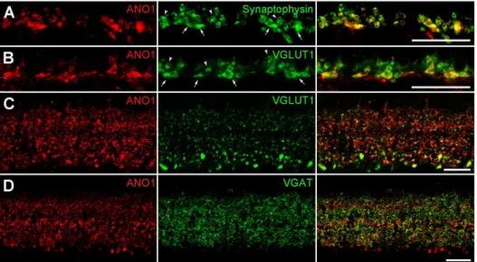
Next, we determined the cellular and subcellular localization of ANO1 in the retina via double-labeling experiments using various neuronal and synaptic markers. ANO1 puncta in the OPL, where photoreceptor terminals synapse onto bipolar and horizontal cell dendrites, showed immunoreactivities for synaptophysin and vesicular glutamate transporter 1 (VGLUT1), which are markers of photoreceptor terminals (Figs. 2A and B), but not for Goa, which is an ON bipolar cell marker (Fig. S1A), and calbindin, a horizontal cell marker (Fig. S1B). These results indicate that ANO1 is expressed in photoreceptor terminals, but not in bipolar and horizontal cell dendrites. In the IPL, where bipolar axon terminals synapse onto ganglion cell dendrites and amacrine processes, which in turn give synaptic inputs to ganglion dendrites and bipolar terminals, ANO1 was detected in bipolar cell terminals that were immunoreactive for VGLUT1, which is a bipolar terminal marker (Fig. 2C), and in amacrine cell processes that were immunoreactive for vesicular GABA transporter (VGAT), which is an amacrine terminal marker (Fig. 2D), but not in ganglion cell dendrites exhibiting immunoreactivity for SMI32, which is a ganglion cell marker (Fig. S1C).
In vertical retinal sections, large and strongly labeled ANO1 puncta exhibiting VGLUT1 immunoreactivity were observed in the IPL, close to the GCL (Fig. 2C). This is where stratification of
the axon terminals of rod bipolar cells, which are a subpopulation of second-order neurons involved in processing scotopic vision [29], occurs. Thus, we confirmed that ANO1 is expressed in rod bipolar axon terminals in retinal slices (Fig. 3A) and dissociated cells (Fig. 3B), as assessed via double-labeling of ANO1 and PKC, which is a marker for rod bipolar cells. Taken together, our results suggest that ANO1 is expressed in various retinal neurons, including rod bipolar cells, and that ANO1 is preferentially localized to the presynaptic region.
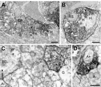
We used pre-embedding immunoelectron microscopy to con-firm the presynaptic localization of ANO1 in retinal neurons. In the OPL, cone pedicles and rod spherules were labeled for ANO1, whereas invaginating ON-bipolar and horizontal cell dendrites, which are postsynaptic elements located at the ribbon synapse of photoreceptor terminals, and OFF-bipolar cell dendrites, which make basal junctions with cone pedicles, were unlabeled (Figs. 4A and B). In the IPL, ANO1 labeling was observed in some bipolar terminals (Fig. 4C), including rod bipolar cells, which were identified by their synaptic ribbons, and amacrine processes, which were filled with synaptic vesicles and established conventional chemical output synapses (Fig. 4D). However, ganglion cell dendrites, which contain microtubules and microfilaments instead of synaptic vesicles, were not immunolabeled (Fig. 4C). These results were consistent with confocal microscopy findings, as shown in Figs. 2 and 3, and confirmed the preferential presynaptic localization of ANO1 in various retinal neurons.
Identification and characterization of ICl(Ca)in rod bipolar
cells expressing ANO1
Our anatomical findings strongly imply the presence of ANO1 in rod bipolar cells. However, the presence and functions of CaCCs and/or ICl(Ca)mediated by CaCCs have not been reported in rod bipolar cells in the mammalian retina (however, please refer to a previous report of ICl(Ca)in a bipolar cell type in the goldfish retina [28]). Thus, we aimed to test whether ICl(Ca)is present in mouse rod bipolar cells, and to characterize this current.
Whole-cell currents were recorded from somata (Fig. 5A; n = 20) and axon terminals (Fig. 5B; n = 7) of freshly dissociated rod bipolar cells, including long axons and terminals. The kinetics of the 2 currents were almost identical, as seen in Fig. 5. Upon depolarizing the cells to the voltage of+10 mV from a holding potential of285 mV, inward current was generated, followed by a
Figure 1. Expression of ANO1 in the mouse retina. A. Western blot analysis of ANO1 in mouse retina homogenates. Salivary gland extracts were used as a positive control. Preincubation of the anti-ANO1 antibody with a 10-fold excess (w/w) of the antigenic peptide led to an absence of bands. B. Confocal micrograph taken from a vertical vibratome section (50mm in thickness) of the mouse retina processed for ANO1 immunoreactivity. Strong ANO1-immunoreactive puncta are shown in the OPL and IPL. Many bipolar (arrowheads) and amacrine cells (asterisks) in the INL and some (arrows) cells in the GCL exhibit weak ANO1 immunoreactivity. The DIC image presented in the middle shows the retinal layers. In the control experiment shown on the right, preincubation of the anti-ANO1 antibody with a 10-fold excess (w/w) of the antigenic peptide led to an absence of labeling. OPL, outer plexiform layer; IPL, inner plexiform layer; INL, inner nuclear layer; GCL, ganglion cell layer; ONL, outer nuclear layer; DIC, differential interference contrast. Scale bar, 50mm.
slowly inactivating tail current (Itail) (Figs. 5A and B). This inward current was identified as L-type Ca2+
current (ICa), which was described previously in rod bipolar cells [30,31], because (1) potassium currents and h-current were suppressed by tetraethyl-ammonium chloride (TEA) and CsCl; (2) it was detectable at depolarizing potentials between about260 mV and+40 mV and had a peak amplitude of 2064 pA at a voltage of about230 mV; and (3) it was sensitive to nifedipine, which is L-type Ca2+
channel antagonist (Fig. S2). Itailwas also detectable; its activation started at about230 mV and peaked at about+10 mV. The maximum amplitude was,30 pA and Itaillasted for,1 s.
To test whether the activation of Itailwas dependent on ICa, a solution containing no Ca2+
and 3 mM Co2+
(Ca2+
channel blocker) were applied in series as shown in Fig. 5C (n = 13). In this series of recordings, both ICaand Itailwere not found in the absence of Ca
2+,
were recovered in control solution, and were suppressed again in the presence of 3 mM Co2+
. Next, we tested whether Ba2+
activated Itail as a carrier that permeates the Ca2+
channels (Fig. 5D; n = 10). Itail was abolished by the replacement of external Ca2+
with equimolar Ba2+
, whereas ICa was enhanced because of Ba2+
conductance. Moreover, the intracellular [Ca2+]
idependency of Itailwas tested by the application of Ca2+ chelators in the pipette solution. The
introduction of both BAPTA (.10 mM) (n = 8) and EGTA (5 mM) (n = 8) into bipolar cells via a recording pipette strongly suppressed Itail (Fig. S3). Furthermore, we applied BAPTA/AM (0.1 mM), which is a membrane-permeable Ca2+
chelator, extracellularly (Fig. 5E; n = 8). During the treatment with BAPTA/AM, the peak amplitude (40.368.1%) of Itail decreased, and this current disappeared almost completely after ,6 min (7.364.2%); in contrast, ICaremained unchanged. These experiments demonstrate that Itailof rod bipolar cells is activated by increased [Ca2+
]i. To identify the ionic component of Itail, we measured the reversal potential of Itailat 3 different [Cl2]i; 144 mM, 72 mM and 29 mM (Fig. 6A). As either standard or reduced [Cl2]isupplemented with methanesulfonate, the reversal potential of Itailfollowed the shift in Figure 2. Cellular and subcellular localization of ANO1 in the retina.(A, B) Outer plexiform layer (OPL).A. Large and small ANO1-labeled puncta (red) in the OPL demonstrate synaptophysin immunoreactivity (green). 2 types of synaptophysin-labeled puncta (smaller higher-positioned rod spherules (arrowheads) and larger lower-positioned cone pedicles (arrows)) are clearly seen.B. Similar toA, the anti-VGLUT1 antibody (green) labels rod and cone terminals. In the merged image, smaller puncta with a round shape, putative rod spherules (arrowheads), and 4 larger puncta with a linear shape (arrows; putative cone pedicles) show ANO1 (red) and VGLUT1 immunoreactivities. (C, D) Inner plexiform layer (IPL). Numerous ANO1-labeled puncta (red) of various sizes are observed in the IPL. InC, the anti-VGLUT1 antibody (green) labels numerous bipolar axon terminals in the IPL. Some ANO1-immunoreactive puncta show VGLUT1 immunoreactivity. InD, VGAT immunoreactivity (green) is seen as tiny puncta throughout the IPL. ANO1 is partially colocalized with VGAT. Scale bars, 20mm.
doi:10.1371/journal.pone.0067989.g002
Figure 3. ANO1 expression in the rod bipolar cell. Confocal micrographs taken from a vertical vibratome section of the mouse retina and a dissociated bipolar cell processed for ANO1 (red) and PKC (green) immunostaining.A. Many ANO1-labeled puncta of various sizes are observed throughout the IPL. Note the large puncta (arrows) located in the innermost part of the IPL. PKC-labeled rod bipolar (asterisks) and amacrine (arrowheads) cell somata are found in the INL, and PKC-labeled axon terminals are clearly seen in the IPL, close to the GCL. Large ANO1 puncta (arrows) are located in the PKC-labeled rod bipolar axon terminals. B. DIC image showing a dissociated retinal bipolar cell with a large axon terminal (arrow). The asterisk indicates its soma. The dissociated bipolar cell shows PKC immunoreactivity, and ANO1 immunoreactivity is seen strongly in the axon terminal (arrow) and weakly in the soma (asterisk). The merged image shows that ANO1 is expressed in a PKC-labeled rod bipolar cell, and that its expression is especially strong in the axon terminal. Scale bars, 20mm (A) and 5mm (B).
doi:10.1371/journal.pone.0067989.g003
the equilibrium potential of Cl2(ECl) (Fig. 6A, middle panel). The average reversal potentials, which were plotted as a function of log10 [Cl2]i, fitted to the values estimated from the Nernst equation with a slope of , 258 mV (Fig. 6A, right panel). Thus, the major
component of this Itailwas identified as a Cl2current. However, this Cl2conductance may have been due to the activation of glutamate transporters in the retina [32]. To rule out this possibility, we applied a glutamate transporter inhibitor,DL-threo-b -benzyloxyas-partate (DL-tBOA). Blocking the glutamate transporter did not induce a significant change in Itail(n = 8; data not shown), suggesting that the portion of Cl2 conductance that is mediated by the this transporter is negligible in the major component of this Itail.
Finally, we tested the effect of various Cl2channel blockers on Itail; 100mM NPPB, 100mM NFA, 1 mM 4,4’-diisothiocyanatos-tilbene-2,2’-disulfonic acid (DIDS), and 2 mM SITS (Fig. 6B; n = 10 for each blocker). Among the extracellular application of those blockers, NPPB (90.7610.0%) and NFA (79.867.3%) significantly suppressed the peak amplitude of Itail without a reduction in ICa. Although this Itailexhibited a lower sensitivity to DIDS (62.4612.2%) and SITS (61.2610.8%), these results suggest that Itaildetected in rod bipolar cells is ICl(Ca).
ANO1 is the CaCC that mediates ICl(Ca)in rod bipolar cells We performed 2 experiments to confirm that ANO1 is the molecular identity of ICl(Ca)observed in rod bipolar cell. First, we
applied a specific ANO1 inhibitor, T16Ainh-A01 [33], which is the most recent commercially available ANO1 inhibitor. As this was the first attempt to apply T16Ainh-A01 to retinal bipolar cells, we determined whether it acts as a specific blocker in these cells in a dose-dependent manner. The resulting dose-response curve showed that T16Ainh-A01 inhibited ICl(Ca) in response to depolarizing voltage steps in a dose-dependent manner, with a half-maximal dose (EC50) of 3mM. Upon applying the effective dose of 10mM, an apparent inhibition of ICl(Ca) was observed without any effects on ICa(Fig. 7A; n = 12).
Next, the neutralizing effects of ANO1 were assessed using a specific antibody against ANO1, via direct addition to the pipette solution (Fig. 7B). The anti-ANO1 antibody was developed against a peptide in the first intracellular loop between transmembrane domains 2 and 3 of mouse ANO1 as a target [7,12]. The first intracellular loop of ANO1 has recently been determined as being the critical region for voltage- and Ca2+
-dependent gating [34]. To rule out the possibility that the anti-ANO1 effect may have arisen from the antibody itself, we tested 2 other antibodies, as negative controls: an anti-glial fibrillary acidic protein (GFAP) antibody (Fig. S4; n = 11) and a peroxidase-conjugated donkey anti-rabbit antibody (n = 11; data not shown). The application of these negative-control antibodies elicited in no significant changes in either ICaor ICl(Ca). However, the application of the anti-ANO1 antibody had a strong blocking effect on ICl(Ca) (20.164.1%), without changing ICa(99.5614.6%), when current traces observed at,3 min were compared with those recorded at,10 min; the
black traces were control traces recorded prior to the full diffusion of the anti-ANO1 antibody into the cell (,3 min post-rupture),
and the colored traces depicted the neutralizing effect of ANO1 via the addition of the anti-ANO1 antibody to the pipette solution (,10 min post-rupture) (n = 12; data not shown). We further verified the blocking effect of the anti-ANO1 antibody on ICl(Ca) (14.066.7%) under 20 mM [Ca2+
]oconditions, which induced an increase in both ICa(146.8611.4%) and ICl(Ca)(Fig. 7B; n = 16).
ANO1 shortens Ca2+spike-like depolarization duration in rod bipolar cells
To investigate the possible function of ANO1 on rod bipolar cells, we recorded membrane potentials using the current-clamp mode in gramicidin-perforated whole cells. It has been demon-strated that rod bipolar cells dissociated from the mammalian retina exhibit membrane potentials of about245 mV [35–37] and EClmeasured in rod bipolar cell axon terminals is about260 mV [38]. The application of depolarizing currents (0.05,0.1 nA) to
cells resting near 245 mV (n = 14) led to the observation of a distinct Ca2+
spike-like depolarization in isolated rod bipolar cells (Figs. 8A and C). This waveform consisted of a first few huge spikes and subsequent small spikes riding atop a plateau potential that was similar to an excitatory postsynaptic potential. The application of 100mM NPPB and T16Ainh-A01 to the bath (Figs. 8A and C; n = 11 for each) revealed that the very first spike was maintained, whereas the depolarizing potential waveform was prolonged. The measurement of the width of the waveform at 1/2 total spike height showed that the application of NPPB and T16Ainh-A01 augmented the width of current-evoked response potential by
,60% compared with that observed in the absence of the drugs
(Figs. 8B and D). Interestingly, EC50of in increasing the width was similar to EC50of the 2 Cl2channel blockers in inhibiting ICl(Ca) (Figs. 8B and D; NPPB, 15.0 vs. 18.4mM; T16Ainh-A01, 2.6 vs. 3.0mM), indicating a close relationship between ICl(Ca) and current-evoked depolarization. These results suggest that ANO1 contributes to the shaping of the spike waveform in rod bipolar cells.
Figure 4. Presynaptic localization of ANO1 in the retina.
Electron micrographs taken from vertical ultrathin sections (90 nm in thickness) of the mouse retina processed for ANO1 immunostaining. (A, B) Outer plexiform layer. InA, ANO1 immunoreactivity is localized to a cone pedicle (CP). At each ribbon synapse (arrowheads) within the CP, 2 horizontal dendrites (H) and an invaginating dendrite of ON-cone bipolar cell (I) are unlabeled. The basal dendrites of OFF-cone bipolar cells (B) at the CP base are also unlabeled. InB, a rod spherule (RS) shows ANO1 immunoreactivity. Similar to A, the postsynaptic triad comprising 2 horizontal dendrites (H) and an invaginating rod bipolar dendrite (I) do not exhibit ANO1 immunoreactivity. (C, D) Inner plexiform layer. In right side ofC, a labeled cone bipolar terminal (BC+) establishes a ribbon synapse (arrowhead) onto a postsynaptic dyad composed of an unlabeled ganglion dendrite (G) and an unlabeled amarcrine process (A2). In addition, an unlabeled bipolar terminal (BC2) synapsing onto 2 unlabeled amacirne dendrites (A2) is seen in the left side. In D, a labeled amacrine process (A+) establishes a conventional chemical synapse onto an unlabeled amacrine process (A2). Scale bars, 0.5mm.
Figure 5. Itailrecorded in the rod bipolar cell is activated by Ca2+influx.(A, B) The cells that retained their axon terminals were filled with
Lucifer Yellow during recording and were morphologically identified under a fluorescence microscope after recording (left panel). The inwardly generating ICawas recorded from a voltage-clamped bipolar cell with a retained axon terminal at the voltage of+10 mV from a holding potential of
285 mV; subsequently, Itailwas activated when returned to the holding potential.C. A solution containing zero Ca2+, a control solution (5 mM Ca2+),
and a solution of 3 mM Co2+were applied serially (n = 13). Currents were recorded from a voltage-clamped bipolar cell with a retained axon terminal
at the voltage of+10 mV from a holding potential of285 mV.D. Ca2+
was replaced with Ba2+
in the extracellular solution (n = 10).E. BAPTA/AM (0.1 mM), which is a membrane-permeable Ca2+chelator, was applied extracellularly to show the time course of changes in I
tail. Scale bars, 5mm. doi:10.1371/journal.pone.0067989.g005
Discussion
This study documented the expression of ANO1, a CaCC, in the retina. This was the first demonstration of the presence of ICl(Ca) in rod bipolar cells, which are a specific type of retinal neurons that express ANO1, and its possible function. Our results showed that (1) various retinal neurons expressed ANO1; (2) this protein was exclusively localized in the presynaptic region; (3) rod bipolar cells expressing ANO1 conferred ICl(Ca), which was blocked by T16Ainh-A01 (a selective ANO1 inhibitor) and a neutralizing antibody; and (4) NPPB and T16Ainh-A01 prolonged the distinct Ca2+ spike-like depolarization evoked by current
injection in dissociated rod bipolar cells. These results suggest that ANO1 acts as an intrinsic regulator of the presynaptic membrane potential during synaptic transmission in retinal neurons.
In sensory neurons, CaCCs are localized in somatodendritic regions and are involved in signal transduction and amplification, ANO1 activation leads to depolarization in DRG neurons and acts as a heat sensor [16], whereas ANO2 contributes to signal amplification of olfactory sense in ORNs [39,40]. In addition, Huang et al. [41] have demonstrated that ANO2 is localized in the close vicinity of voltage-gated Ca2+
channels and NMDA receptors in the somatodendritic region in hippocampal neurons, where it
regulates action potentials and synaptic responses. Thus, CaCCs appear to play important functions in receptive and/or postsyn-aptic regions in neurons.
We showed that ANO1 was strongly expressed in 2 synaptic layers. Double-labeling experiments using various pre- and post-synaptic markers and immunoelectron microscopy clearly showed that ANO1 was localized to presynaptic terminals such as photoreceptor terminals, bipolar axon terminals, and amacrine cell processes. This preferential presynaptic localization of ANO1 was also observed in the cochlea, where ANO1 is exclusively localized at medial olivocochlear efferent nerve endings [42], and in the cerebellum where ANO1 is mainly found in mossy fibers (our unpublished data). Furthermore, when we performed whole-cell voltage clamp recordings on a rod bipolar whole-cell without the axon terminal, which is often lost during the procedure of enzymatic dissociation, ICl(Ca) was hardly elicited (Fig. S5), suggesting that ICl(Ca) may originate from the axon terminal and/or be linked with the Ca2+ channels localized at the axon
terminal. These findings suggest that CaCCs also have important functions in presynaptic terminals in neurons. In fact, the exclusive presynaptic localization of ANO1 in photoreceptor terminals observed in this study concurs with previous reports that showed that ICl(Ca)is elicited by depolarization-evoked Ca2+
influx, which Figure 6. Cl2ions are identified as the ionic component of I
tail. A. The reversal potential of Itailwas measured at 3 different [Cl2]i: 144 mM,
1.961.7 mV (n = 16); 72 mM,223.664.2 mV (n = 12); 29 mM,246.261.1 mV (n = 17). The peak amplitude of Itailwas measured 10 ms after the
command pulse, to avoid recording interference from the capacitative current. The average reversal potentials, which were plotted as a function of log10[Cl2]i, fitted to the values estimated from the Nernst equation with a slope of,258 mV.B. The NPPB (100mM), NFA (100mM), DIDS (1 mM) and SITS (2 mM) were applied. The results of statistical analyses are presented in the panel on the right as the normalized mean6S.D.. Student’st tests were used to compare the data from the 2 groups. Significance was set asP,0.05 (*) andP,0.01 (**).
doi:10.1371/journal.pone.0067989.g006
Figure 7. ANO1 is the molecular identity of ICl(Ca)in rod bipolar cells. A. The extracellular application of the specific ANO1 inhibitor T16Ainh
-A01 (10.0mM) to the rod bipolar cell (n = 10). The results of statistical analyses are presented in the panel on the right as the normalized mean6S.D..
B. Effect of the anti-ANO1 antibody on ICl(Ca)under the 20 mM [Ca2+]ocondition (n = 16). ICaand ICl(Ca)were recorded in the presence of the anti-ANO1
antibody in the pipette solution in response to depolarizing pulses,3 min after rupture and,10 min after rupture, respectively. The panel on the
right depicts the comparison of the amplitude changes of ICaand ICl(Ca)between,3 min after rupture and,10 min after rupture. The results of
statistical analyses are presented as the normalized mean6S.D.. Student’st-tests were used to compare the data from the 2 groups. Significance was set atP,0.05 (*) andP,0.01 (**).
activates CaCCs localized at photoreceptor terminals [17,18,25,38], and supports the proposed role of ICl(Ca) in membrane potential stabilization during synaptic activity [19,20,22,43] and presynaptic Ca2+
channel modulation [23–25]. In this study, the abundant expression of ANO1 detected in various retinal neurons, such as photoreceptors and bipolar and amacrine cells, exceeded our expectations. For example, the presence of ICl(Ca) in mouse rod bipolar cells has not been described previously, even though many studies on Ca2+channel
properties [30,31] and Ca2+ tail currents [44,45] have been
performed to understand visual signal processing and synaptic transmission in mammalian rod bipolar cells. The reasons for this may be that ICaamplitude that causes ICl(Ca)is too small in mouse rod bipolar cells and that the studies mentioned above focused on the detection and characterization of ICa itself. In this study, we found that ANO1 was expressed in rod bipolar cells; hence, we anticipated the presence of ICl(Ca) in these cells. To facilitate the identification of ICl(Ca), we lowered the concentration of the Ca
2+
chelator (0.5 mM EGTA) compared with that used in previous studies (5 mM EGTA) [30,31,46]. In these conditions, we were able to identify ICl(Ca)in rod bipolar cells easily and successfully. In fact, using this protocol (0.5 mM EGTA), ICl(Ca)was identified in
goldfish retinal bipolar cells [28] after ICaidentification [46,47]. Thus, to detect the presence and identify the function of CaCCs that are underestimated in the nervous system, further studies aimed at identifying CaCC conductance in other central neurons by using our protocol are needed.
Here, the suppression of CaCC using NPPB, which is a CaCC blocker, and T16Ainh-A01, which is a selective ANO1 blocker, prolonged the current-evoked depolarization that was experimen-tally induced by us. These results provide a very critical piece of information that explains the possible roles of ICl(Ca)mediated by ANO1 in rod bipolar cells. The dissociated preparation can be considered as a condition in which the synaptic network is free (i.e., synaptic events do not occur between presynaptic and postsynaptic neurons), but in which the intrinsic properties of the recording cells are preserved. The function of CaCCs may depend on their spatial distribution in cells and on local differences in ECl. Notably, to establish the function of ICl(Ca), it is crucial to know ECl. CaCCs promote membrane depolarization in rod photore-ceptor terminals, where ECl is <220 mV and the membrane potential is estimated at,245 mV [48]. Conversely, mouse rod bipolar cells, which receive synaptic inputs from rods, have ECl (<260 mV) that is more negative than the resting membrane Figure 8. The physiological role of ICl(Ca)during membrane depolarization was examined using NPPB and T16Ainh-A01.A. The bipolar
cell membrane potential was adjusted to close to245 mV with a steady hyperpolarizing current. A distinctive slow depolarizing potential that followed the Ca2+-dependent transient potentials was elicited by a current injection of 0.5 nA for 200 ms (n = 11). The extracellular application of
100mM NPPB enhanced the depolarizing membrane potential compared with the control experiment.B. The dose-dependent decrease elicited by NPPB on Itailamplitude obtained under the voltage-clamping mode (gray) was compared with the dose-dependent increase elicited by NPPB on the
width of current-evoked spikes (red). The half-maximal dose (EC50) of NPPB that was necessary to increase the width was similar to EC50of NPPB that
inhibited ICl(Ca)(15.0 vs. 18.4mM). C. In addition, T16Ainh-A01 application enhanced the depolarizing membrane potential (n = 8).D. The
dose-dependent decrease elicited by T16Ainh-A01 on Itailamplitude obtained under the voltage-clamping mode (gray) was compared with the
dose-dependent increase elicited by T16Ainh-A01 on the width of current-evoked spike (blue). The half-maximal dose (EC50) of T16Ainh-A01 that was
necessary to increase the width was similar to EC50of T16Ainh-A01 that inhibited ICl(Ca)(2.6 vs. 3.0mM). Significance was set atP,0.05 (*) andP,0.01 (**).
doi:10.1371/journal.pone.0067989.g008
potential (<245 mV) at its presynaptic axon terminal [38,49]. Under such conditions, activation of Ca2+
channels may open ANO1 in rod bipolar cells resulting in an outward current that shortens Ca2+
spike-like depolarizations or facilitates the repolar-ization of the cell membrane. Taken together, these results suggest that the physiologic function of ANO1 is dependent on Cl2 distribution and the establishment of ECl. ANO1 appears to confer an intrinsic electrical characteristic to retinal neurons and in the same way, may act as an important intrinsic regulator of the membrane potential in central neurons, including retinal neurons. In addition, during synaptic transmission in rod bipolar axon terminals in which ECl is more negative than the resting membrane potential, Cl2conductance stabilizes the presynaptic membrane potential, allowing an increase in glutamate release via an increase in [Ca2+]
i[19,24]. Vesicle release is regulated by L-type Ca2+channels, which in turn are regulated by Cl2moving
through CaCCs, as reported at photoreceptor ribbon synapses [17,18,25,38]. AII amacrine cells, which are located postsynapti-cally rod bipolar cells exhibit a mixture of transient and sustained components [50–52]. The transient component of the postsynaptic current is quite pronounced, whereas the sustained component is of relatively low amplitude [53–55]. This pattern is observed even in the presence of blockers of inhibitory GABAergic input and when only sustained Ca2+currents are activated in the rod bipolar
cell, which suggests that glutamate release from mammalian rod bipolar cells is inherently transient [53,56,57]. In this study, a CaCC blocker and a selective ANO1 blocker prolonged the current-evoked depolarization in an isolated cell preparation without the synaptic network, suggesting that ANO1 function may be attributed to inherently transient glutamate release in rod bipolar cells.
Materials and Methods
Ethical standards
This study was carried out in strict accordance with the recommendations provided in the Guide for the Care and Use of Laboratory Animals of the National Institutes of Health (NIH Publications No. 80-23; revised in 1996). The study protocol was approved by the Institutional Animal Care and Use Committee (IACUC) of the College of Medicine, The Catholic University of Korea (Approval Number: CUMS-2012-0087-01). All animal surgeries were performed under ketamine and xylazine anesthesia, and all efforts were made to minimize suffering.
Animals and tissue preparation
Three-month-old male C57BL/6 mice (Orient Bio, Seongnam, Korea) were used in this study. The mice were euthanized with 15% chloral hydrate.
For western blot analysis, the animals were transcardially perfused with saline, the eyeballs were enucleated, the anterior segments of the eyeballs were removed, and the retinal tissues were quickly dissected on an ice-cold plate, frozen on dry ice, and stored at270uC.
For immunohistochemistry, the eyecups were fixed by immer-sion in 4% paraformaldehyde in 0.1 M phosphate buffer (PB, pH 7.4). After fixation, the retinas were carefully dissected and transferred to 30% sucrose in 0.1 M PB. They were then frozen in liquid nitrogen, thawed, and stored at270uC.
For patch-clamp recordings, the retinas were quickly dissected and treated with a low-Ca2+ solution containing 4 mg/mL of
papain activated by 10 mM L110
cysteine. Subsequently, retinal cells were enzymatically isolated from retinas. The dissociated retinal preparations were kept in an oxygenated chamber during the recordings.
Antibodies
An anti-ANO1 polyclonal antibody (Cat. #LF-PA0208; Ab Frontier, Seoul, Korea) was raised in rabbits against a synthetic peptide with of amino acid sequence KDHPRAEYEARVLEKS (amino acids 451–466), which corresponded to a stretch located in the first intracellular loop between transmembrane domains 2 and 3 of mouse ANO1. The specificity of this antibody was tested by western blotting and immunocytochemical analyses in our previous study [7,42] and demonstrated by immunohistochemistry in knockout animals [12].
An anti-glial fibrillary acidic protein (GFAP) (Millipore, Temecula, CA) and peroxidase-conjugated donkey anti-rabbit (Molecular Probes, Eugene, OR) antibodies were used as controls in the patch-clamp recording experiment that was performed to test the neutralizing effect of the anti-ANO1 antibody. For double-labeling, various antibodies were used to label presynaptic or postsynaptic elements and specific neurons in the retina. Their target structures, together with dilution rates, sources, companies, and references, are listed in Table S1.
Western blotting
Western blot analysis was performed on extracts of the prepared tissues, which were homogenized in ice-cold RIPA buffer (50 mM Tris buffer, pH 8.0; 150 mM NaCl; 1% NP-40; 0.5% deoxycho-late; and 0.1% SDS). Samples from each tissue (corresponding to 50mg of total protein) were separated by SDS-PAGE, and the proteins were blotted onto a nitrocellulose membrane and probed with the anti-ANO1 antibody (dilution, 1:2000). The immunore-active bands were detected using an Enhanced Chemilumines-cence Detection Kit (Amersham, Arlington Heights, IL). Preincu-bation of the anti-ANO1 antibody with a 10-fold excess (w/w) of the antigenic peptide led to the absence of labeling in the subsequent western blot analysis (Fig. 1A); moreover, the reaction of the western blots with the secondary antibody alone produced no signal (data not shown).
Immunohistochemistry
Fifty-micrometer-thick vertical vibratome sections for the retina were used for immunohistochemistry. The sections were incubated in 10% normal donkey serum and incubated with the polyclonal antibody against ANO1 (dilution, 1:500) in 0.01 M phosphate-buffered saline (PBS, pH 7.4) containing 0.5% Triton X-100 for 1 day at 4uC. The sections were washed in PBS and incubated in the presence of biotin-labeled donkey anti-rabbit IgG (dilution, 1:100; Jackson Immuno Research, West Grove, PA) for 2 h. Subse-quently, the sections were washed and incubated with Cy3-conjugated streptavidin in PBS (dilution, 1:1000; Jackson Immuno Research) for 1 h. The fluorescent specimens were mounted using Vectashield mounting medium (Vector Laboratories, Burlingame, CA). The specificity of the immunostaining was evaluated in retinal sections by preincubating the anti-ANO1 antibody with a 10-fold excess of its antigenic peptide for 1 h at room temperature (Fig. 1B) and by omitting the incubation step with the primary antibody (data not shown); no staining was observed in these sections.
(Jackson Immuno Research) and Alexa Fluor 488 (Molecular Probes) at a dilution of 1:200.
Digital images (1,02461,024 pixels) were acquired using a Zeiss
LSM 510 Meta confocal microscope (Carl Zeiss Co. Ltd., Germany) and were imported into Photoshop (Adobe Systems, San Jose, CA). The brightness and contrast of the final images were adjusted.
Immunoelectron microscopy
Retinal sections were prepared as described above. After blocking, the sections were incubated in a solution containing the anti-ANO1 antibody at 4uC for 1 day, as described for light microscopy but without Triton X-100. The sections were washed in PBS for 45 min (3615 min), incubated with biotin-labeled donkey anti-rabbit IgG (dilution, 1:100; Jackson Immuno Research) for 2 h, and washed 3 times in PBS for 45 min (3615 min). The sections were then incubated in an avidin-biotin-peroxidase complex (ABC) solution (Vector Laboratories) for 1 h, washed in 0.1 M Tris buffer (TB, pH 7.6), and preincubated in 0.05% 3,3’-diaminobenzidine tetrahydrochloride (DAB) in TB for 10 min, followed by incubation in the same solution containing 0.05% hydrogen peroxide (H2O2) for an additional 10 min. The reaction was monitored using a low-power microscope and was stopped by replacing the DAB and H2O2solution with TB.
Stained sections were postfixed in 1% glutaraldehyde in PB for 1 h. After washing in PB containing 4.5% sucrose for 15 min (365
min), the sections were postfixed in 1% OsO4 in PB for 1 h. Subsequently, the sections were rewashed in PB containing 4.5% sucrose and dehydrated in a graded series of ethanol. During the dehydration procedure, the sections were staineden blocwith 1% uranyl acetate in 70% alcohol for 1 h, then transferred to propylene oxide, and flat embedded in Epon 812. After curing at 60uC for 3 days, well-stained areas were cut out and attached to an Epon support for further ultrathin sectioning (Reichert-Jung, Nubloch, Germany). Serial ultrathin sections (70–90 nm in thickness) were collected on single slot grids, and examined using a JEM 1010 electron microscope (JEOL, Tokyo, Japan).
Patch-clamp recordings
Whole-cell recordings were performed on rod bipolar cells by using an EPC-9 amplifier and the PULSE software (Heka Electronik, Germany). Electrodes were fabricated from borosili-cate microcapillary tubes (VWR Scientific, West Chester, PA) and fire-polished. The resistance of the electrode was 8–12 MV. Series resistance ranged from 25 to 40 MV. The extracellular solution contained (in mM): NaCl, 135; CsCl, 5; CaCl2, 10; MgCl2, 1; glucose, 10; and HEPES, 10. The pipette solution used for voltage-clamp recordings contained (in mM): CsCl, 120; MgCl2, 1; CaCl2, 0.5; EGTA, 0.5; tetraethylammonium (TEA)-Cl, 20; and Tris-ATP, 10 (pH adjusted to 7.2 with CsOH). The pipette solution used for current-clamp recordings in the perforated patch-clamp mode was (in mM): KCl, 140; MgCl2, 1; CaCl2, 0.5; EGTA, 0.5 and Tris-ATP, 10 and gramicidine D (14.8mg/mL) (pH adjusted to 7.2 with KOH). Dissociated rod bipolar cells were used in recordings for 4–5 h after isolation.
Current signals were filtered at 3 kHz and digitized at 5 kHz by using a data-acquisition interface (LIH 1600 A/D board, HEKA). Cell membrane capacitance and series resistance current were automatically compensated by the amplifier using the software. The calculated liquid junction potential was also subtracted using this software. Data were analyzed offline by using the PULSE-fit and ORIGIN programs. Statistical analysis was performed using analysis of variance (ANOVA) and Student’s t tests. Data are
represented as means 6 S.D.. The differences were considered significant atP,0.05.
Pharmacology
Test solutions were mostly dissolved in the Ringer solution and applied locally to a whole-cell clamped cell from a puffer pipette via Y-tube system. All chemicals were purchased from Sigma-Aldrich (St Louis, MO), with the exception of 2-(5-ethyl-4- hydroxy-6-methylpyrimidin-2-ylthio)-N-(4-(4-methoxyphenyl)thia-zol-2-yl)acetamide (T16Ainh-A01) (Tocris, Ellisville, MI),DL
-threo-b-benzyloxyaspartate (DL-tBOA) (Tocris), the anti-ANO1 antibody (Ab Frontier), and the anti-GFAP antibody (Millipore).
Some pharmacological agents were used for the manipulation of a specific ionic current: CoCl2for blocking Ca2+
current and CsCl and TEA in the pipette solution for blocking h-current and voltage- and Ca2+
-activated K+
currents. Nifedipine was used to block an L-type Ca2+
channel. The Ca2+
chelators 1,2-bis [o-aminophenoxy]ethane-N,N,N9,N9-tetraacetic acid (BAPTA) and ethylene glycol-bis [2-aminoethylether]-N,N,N9,N9-tetraacetic acid (EGTA), were applied in the pipette solution; when these agents were applied at higher concentration, the concentration of CsCl was appropriately reduced to adjust the osmolarity. 1,2-bis [2-aminophenoxy]ethane-N,N,N9,N9-tetraacetic acid tetrakis (BAPTA/AM), which is a cell-permeable Ca2+
chelator, was applied extracellularly at 0.1 mM. To suppress Cl2conductance, several agents were applied, i.e., 5-nitro-2-(3-phenylpropylamino) benzoic acid (NPPB), niflumic acid (NFA), 4,4’-diisothiocyanatos-tilbene-2,2’-disulfonic acid (DIDS), and 4-acetamido-4’-isothio-cyanatostilbene-2,2’-disulfonic acid (SITS), to block Cl2channels selectively. T16Ainh-A01 was tested as a specific ANO1 inhibitor.
DL-tBOA was used to suppress Cl2 current mediated by the glutamate transporter. In addition, we tested the effects of blocking ANO1 by adding the specific anti-ANO1 antibody directly to the pipette solution. Both the anti-ANO1 and anti-GFAP antibodies were used at dilutions of 1:500 in the pipette solution.
Supporting Information
Figure S1 Cellular and subcellular localization of ANO1 in the mouse retina.Confocal micrographs taken from vertical vibratome sections (50mm in thickness) processed for double labeling with antibodies against ANO1 (red) and Goa(A, green) or calbindin (B, green) or SMI32 (C, green) in the mouse retina. (A, B) Large and small ANO1-labeled puncta are visible in the OPL. InA, the anti-Goaantibody labels the somata of ON-bipolar cells (asterisks) in the outer INL and their dendrites extending into the OPL. In the merged image, ANO1 and Goaare not colocalized. In B, 2 calbindin-labeled horizontal cell somata (asterisks) and their dendrites located in the inner OPL are seen. In the merged image, ANO1-positive cells do not show calbindin immunoreac-tivity. C. Numerous ANO1-labeled puncta of various sizes are observed in the IPL. A SMI32-labeled ganglion cell soma (asterisk) and labeled dendrites are seen in the GCL and IPL, respectively. In the merged image, ANO1-immunoreactive puncta are not localized to SMI32-labeled ganglion cell dendrites in the IPL. Scale bars, 20mm.
(TIF)
Figure S2 Blocking effect of nifedipine on L-type Ca2+
channels in rod bipolar cells.Nifedipine (10mM) was applied to the rod bipolar cell. Nifedipine decreased the sustained component of ICa and Itail (n = 14). The results of statistical analyses are presented in the panel on the right as the normalized
mean6S.D.. Student’sttests were used to compare the data from the 2 groups.
(TIF)
Figure S3 Dependency of Itailon [Ca2+]i.The dependency of Itail on [Ca2+
]i was confirmed via the application of 2 Ca2+
chelators in the pipette solution. The introduction of both BAPTA (.10 mM) (n = 8) and EGTA (5 mM) (n = 8) into bipolar cells via a recording pipette strongly suppressed Itail. The results of statistical analyses are presented in the panel on the right as the normalized mean6S.D.. Student’sttests were used to compare the data from the 2 groups. Significance was set atP,0.01 (**) and
P,0.001 (***). (TIF)
Figure S4 A negative-control experiment of the blocking effect of the neutralizing antibody on ICl(Ca). A GFAP-conjugated donkey anti-rabbit antibody was introduced directly in the pipette solution to examine the blocking effect of the neutralizing antibody on ICl(Ca). In the presence of the anti-GFAP antibody, both ICaand ICl(Ca)were recorded at a holding potential of285 mV in response to depolarizing pulses of+10 mV,3 min
after rupture and,10 min after rupture (n = 11). The panel on the right depicts the comparison of the amplitude changes of ICaand
ICl(Ca)between,3 min after rupture and,10 min after rupture.
Student’sttests were used to compare the data from the 2 groups. (TIF)
Figure S5 Itail is absent in bipolar cells without axon terminals. A dissociated rod bipolar cell was filled with Lucifer Yellow during recording and was morphologically identified under a fluorescence microscope after recording (right panel). The representative trace recorded from a rod bipolar cell without axon terminals showed the presence of ICaand the absence of Itail. The currents were recorded at the voltage of+10 mV from a holding potential of285 mV. Scale bar, 5mm.
(TIF)
Table S1 Immunologic marker antibodies used in this study.
(DOC)
Author Contributions
Conceived and designed the experiments: JHJ SSP IBK. Performed the experiments: JHJ SSP. Analyzed the data: JHJ SSP MHC UO IBK. Wrote the paper: JHJ SSP IBK.
References
1. Frings S, Reuter D, Kleene SJ (2000) Neuronal Ca2+
-activated Cl2channels—
homing in on an elusive channel species. Prog Neurobiol 60: 247–289. 2. Eggermont J (2004) Calcium-activated chloride channels: (un)known, (un)loved?
Proc Am Thorac Soc 1: 22–27.
3. Hartzell C, Putzier I, Arreola J (2005) Calcium-activated chloride channels. Annu Rev Physiol 67: 719–758.
4. Duran C, Thompson CH, Xiao Q, Hartzell HC (2010) Chloride channels: often enigmatic, rarely predictable. Annu Rev Physiol 72: 95–121.
5. Caputo A, Caci E, Ferrera L, Pedemonte N, Barsanti C, et al. (2008) TMEM16A, a membrane protein associated with calcium-dependent chloride channel activity. Science 322: 590–594.
6. Schroeder BC, Cheng T, Jan YN, Jan LY (2008) Expression cloning of TMEM16A as a calcium-activated chloride channel subunit. Cell 134: 1019– 1029.
7. Yang YD, Cho H, Koo JY, Tak MH, Cho Y, et al. (2008) TMEM16A confers receptor-activated calcium-dependent chloride conductance. Nature 455: 1210– 1215.
8. Hartzell HC, Yu K, Xiao Q, Chien LT, Qu Z (2009) Anoctamin/TMEM16 family members are Ca2+
-activated Cl
-channels. J Physiol 587: 2127–2139. 9. Ferrera L, Caputo A, Galietta LJ (2010) TMEM16A protein: a new identity for
Ca2+-dependent Cl-channels. Physiology (Bethesda) 25: 357–363.
10. Rock JR, O’Neal WK, Gabriel SE, Randell SH, Harfe BD, et al. (2009) Transmembrane protein 16A (TMEM16A) is a Ca2+
-regulated Cl -secretory channel in mouse airways. J Biol Chem 284: 14875–14880.
11. Kunzelmann K, Tian Y, Martins JR, Faria D, Kongsuphol P, et al. (2011) Anoctamins. Pflugers Arch 462: 195–208.
12. Romanenko VG, Catala´n MA, Brown DA, Putzier I, Hartzell HC, et al. (2010) Tmem16A encodes the Ca2+
-activated Cl
-channel in mouse submandibular salivary gland acinar cells. J Biol Chem 285: 12990–13001.
13. Huang F, Rock JR, Harfe BD, Cheng T, Huang X, et al. (2009) Studies on expression and function of the TMEM16A calcium-activated chloride channel. Proc Natl Acad Sci U S A 106: 21413–21418.
14. Namkung W, Yao Z, Finkbeiner WE, Verkman AS (2011) Small-molecule activators of TMEM16A, a calcium-activated chloride channel, stimulate epithelial chloride secretion and intestinal contraction. FASEB J 25: 4048–4062. 15. Hwang SJ, Blair PJ, Britton FC, O’Driscoll KE, Hennig G, et al. (2009) Expression of anoctamin 1/TMEM16A by interstitial cells of Cajal is fundamental for slow wave activity in gastrointestinal muscles. J Physiol 587: 4887–4904.
16. Cho H, Yang YD, Lee J, Lee B, Kim T, et al. (2012) The calcium-activated chloride channel anoctamin 1 acts as a heat sensor in nociceptive neurons. Nat Neurosci 15: 1015–1021.
17. Barnes S, Hille B (1989) Ionic channels of the inner segment of tiger salamander cone photoreceptors. J Gen Physiol 94: 719–743.
18. MacLeish PR, Nurse CA (2007) Ion channel compartments in photoreceptors: evidence from salamander rods with intact and ablated terminals. J Neurophysiol 98: 86–95.
19. Maricq AV, Korenbrot JI (1988) Calcium and calcium-dependent chloride currents generate action potentials in solitary cone photoreceptors. Neuron 1: 503–515.
20. Barnes S, Descheˆnes MC (1992) Contribution of Ca and Ca-activated Cl channels to regenerative depolarization and membrane bistability of cone photoreceptors. J Neurophysiol 68: 745–755.
21. Thoreson WB, Miller RF (1996) Removal of extracellular chloride suppresses transmitter release from photoreceptor terminals in the mudpuppy retina. J Gen Physiol 107: 631–642.
22. Thoreson WB, Nitzan R, Miller RF (2000) Chloride efflux inhibits single calcium channel open probability in vertebrate photoreceptors: chloride imaging and cell-attached patch-clamp recordings. Vis Neurosci 17: 197–206. 23. Thoreson WB, Bryson EJ, Rabl K (2003) Reciprocal interactions between
calcium and chloride in rod photoreceptors. J Neurophysiol 90: 1747–1753. 24. Lalonde MR, Kelly ME, Barnes S (2008) Calcium-activated chloride channels in
the retina. Channels (Austin) 2: 252–260.
25. Mercer AJ, Rabl K, Riccardi GE, Brecha NC, Stella SL, Jr., et al. (2011) Location of release sites and calcium-activated chloride channels relative to calcium channels at the photoreceptor ribbon synapse. J Neurophysiol 105: 321– 335.
26. Sto¨hr H, Heisig JB, Benz PM, Scho¨berl S, Milenkovic VM, et al. (2009) TMEM16B, a novel protein with calcium-dependent chloride channel activity, associates with a presynaptic protein complex in photoreceptor terminals. J Neurosci 29: 6809–6818.
27. Pifferi S, Dibattista M, Menini A (2009) TMEM16B induces chloride currents activated by calcium in mammalian cells. Pflugers Arch 458: 1023–1038. 28. Okada T, Horiguchi H, Tachibana M (1995) Ca2+-dependent Cl-current at the
presynaptic terminals of goldfish retinal bipolar cells. Neurosci Res 23: 297–303. 29. Bloomfield SA, Dacheux RF (2001) Rod vision: pathways and processing in the
mammalian retina. Prog Retin Eye Res 20: 351–384.
30. Satoh H, Aoki K, Watanabe SI, Kaneko A (1998) L-type calcium channels in the axon terminal of mouse bipolar cells. Neuroreport 9: 2161–2165.
31. de la Villa P, Vaquero CF, Kaneko A (1998) Two types of calcium currents of the mouse bipolar cells recorded in the retinal slice preparation. Eur J Neurosci 10: 317–323.
32. Eliasof S, Werblin F (1993) Characterization of the glutamate transporter in retinal cones of the tiger salamander. J Neurosci 13: 402–411.
33. Namkung W, Phuan PW, Verkman AS (2011) TMEM16A inhibitors reveal TMEM16A as a minor component of calcium-activated chloride channel conductance in airway and intestinal epithelial cells. J Biol Chem 286: 2365– 2374.
34. Xiao Q, Yu K, Perez-Cornejo P, Cui Y, Arreola J, et al. (2011) Voltage- and calcium-dependent gating of TMEM16A/Ano1 chloride channels are physically coupled by the first intracellular loop. Proc Natl Acad Sci U S A 108: 8891– 8896.
35. Suzuki S, Kaneko A (1990) Identification of bipolar cell subtypes by protein kinase C-like immunoreactivity in the goldfish retina. Vis Neurosci 5: 223–230. 36. de la Villa P, Kurahashi T, Kaneko A (1995) L-glutamate-induced responses and cGMP-activated channels in three subtypes of retinal bipolar cells dissociated from the cat. J Neurosci 15: 3571–3582.
38. Varela C, Blanco R, De la Villa P (2005) Depolarizing effect of GABA in rod bipolar cells of the mouse retina. Vision Res 45: 2659–2667.
39. Stephan AB, Shum EY, Hirsh S, Cygnar KD, Reisert J, et al. (2009) ANO2 is the cilial calcium-activated chloride channel that may mediate olfactory amplification. Proc Natl Acad Sci U S A 106: 11776–11781.
40. Billig GM, Pa´l B, Fidzinski P, Jentsch TJ (2011) Ca2+
-activated Cl
-currents are dispensable for olfaction. Nat Neurosci 14: 763–769.
41. Huang WC, Xiao S, Huang F, Harfe BD, Jan YN, et al. (2012) Calcium-activated chloride channels (CaCCs) regulate action potential and synaptic response in hippocampal neurons. Neuron 74: 179–192.
42. Jeon JH, Park JW, Lee JW, Jeong SW, Yeo SW, et al. (2011) Expression and immunohistochemical localization of TMEM16A/anoctamin 1, a calcium-activated chloride channel in the mouse cochlea. Cell and Tissue Research 345: 223–230.
43. Babai N, Morgans CW, Thoreson WB (2010) Calcium-induced calcium release contributes to synaptic release from mouse rod photoreceptors. Neuroscience 165: 1447–1456.
44. Hartveit E (1999) Reciprocal synaptic interactions between rod bipolar cells and amacrine cells in the rat retina. J Neurophysiol 81: 2923–2936.
45. Pan ZH (2000) Differential expression of high- and two types of low-voltage-activated calcium currents in rod and cone bipolar cells of the rat retina. J Neurophysiol 83: 513–527.
46. Tachibana M, Okada T, Arimura T, Kobayashi K (1993) Dihydropyridine-sensitive calcium current mediates neurotransmitter release from retinal bipolar cells. Ann N Y Acad Sci 707: 359–361.
47. Kaneko A, Tachibana M (1985) A voltage-clamp analysis of membrane currents in solitary bipolar cells dissociated from Carassius auratus. J Physiol 358: 131– 152.
48. Thoreson WB, Stella SL, Jr., Bryson EI, Clements J, Witkovsky P (2002) D2-like dopamine receptors promote interactions between calcium and chloride channels that diminish rod synaptic transfer in the salamander retina. Vis Neurosci 19: 235–247.
49. Satoh H, Kaneda M, Kaneko A (2001) Intracellular chloride concentration is higher in rod bipolar cells than in cone bipolar cells of the mouse retina. Neurosci Lett 310: 161–164.
50. Nelson R (1982) AII amacrine cells quicken time course of rod signals in the cat retina. J Neurophysiol 47: 928–947.
51. Dacheux RF, Raviola E (1986) The rod pathway in the rabbit retina: a depolarizing bipolar and amacrine cell. J Neurosci 6: 331–345.
52. Bloomfield SA, Xin D (2000) Surround inhibition of mammalian AII amacrine cells is generated in the proximal retina. J Physiol 523 Pt 3: 771–783. 53. Singer JH, Diamond JS (2003) Sustained Ca2+
entry elicits transient postsynaptic currents at a retinal ribbon synapse. J Neurosci 23: 10923–10933.
54. Veruki ML, Mørkve SH, Hartveit E (2003) Functional properties of spontaneous EPSCs and non-NMDA receptors in rod amacrine (AII) cells in the rat retina. J Physiol 549: 759–774.
55. Trexler EB, Li W, Massey SC (2005) Simultaneous contribution of two rod pathways to AII amacrine and cone bipolar cell light responses. J Neurophysiol 93: 1476–1485.
56. Bieda MC, Copenhagen DR (2000) Inhibition is not required for the production of transient spiking responses from retinal ganglion cells. Vis Neurosci 17: 243– 254.
57. Wan QF, Heidelberger R (2011) Synaptic release at mammalian bipolar cell terminals. Vis Neurosci 28: 109–119.